INTRODUCTION
Ampulla of Vater cancer accounts for approximately 6%
of the tumors occurring in the region of the head of the pan- creas (1). The majority of tumors in this region are pancreatic adenocarcinomas and the remainders are divided among bile duct carcinomas, duodenal carcinomas, endocrine tumors and metastatic lesions. Patients with cancer of the ampulla of Vater account for up to 36% of those undergoing surgery for pancreato-duodenal malignacies and are the only patients among those affected by cancers of biliopancreatic origin who have upto a 50% chance of being cured by surgery alone (2-9).
In addition, these tumors grow more slowly than cholangio- carcinoma and pancreatic adenocarcinoma.
Ampulla of Vater cancer appears as a heterogenous disease from the pathophysiological and molecular point of view.
They can arise as a feature of a familial cancer syndrome or as a sporadic and quite uncommon event among gastroin- testinal malignancies. In the latter case, they can rise from a preexisting adenoma, closely resembling the colon adenoma- carcinoma sequence. This group of ampullary neoplasm has the histologic appearance of a tubular colon adenocarcinoma, shows an expansive pattern of growth and is characterized by
a K-ras mutational rate similar to that of colon cancer (10).
The existence of these types of ampullary tumors, which share some biological characteristics with colon carcinomas, may partially explain their striking difference from pancre- atic and biliary tract cancers in terms of growth behavior and patient survival.
Cyclooxygenase (COX), also referred to as prostaglandin endoperoxide synthase, is the rate-limiting enzyme for the metabolic conversion of arachidonic acid to prostaglandins (PGs) and related eicosanoids. There are at least two isoforms of cyclooxygenase, COX-1 and COX-2. COX-1 is constitu- tively expressed in many tissues and cell types (11), but in some cases is increased during differentiation (12). By con- trast, the expression of COX-2 is frequently up-regulated by mitogens, cytokines and tumor promoters.
Greater than 80% of colon cancers in humans have in- creased COX-2 levels when compared to adjacent normal tissue (11). The antitumor effect of COX-inhibitor (NSAIDs) in colorectal tumorigenesis is evidenced by abundant data from epidemiologic studies (12, 13), studies in patients with familial adenomatous polyposis (FAP) (14, 15), and multi- ple experiments in rodent models of colon cancer (16-19).
One potential mechanism for the antitumor effect of NSAIDs Hong Joo Kim, Tae Sung Sohn*, Kyu Taek Lee, Jong Kyun Lee, Seung Woon Paik, Jong Chul Rhee
Division of Gastroenterology and Department of Surgery*, Samsung Medical Center, Sungkyunkwan University School of Medicine, Seoul, Korea
Address for correspondence Tae Sung Sohn, M.D.
Department of Surgery, Samsung Medical Center, Sungkyunkwan University School of Medicine, 50 Ilwon-dong, Kangnam-gu, Seoul 135-710, Korea
Tel : +82.2-3410-3475, Fax : +82.2-3410-3849 E-mail : [email protected]
218
Expression of Cyclooxygenase-2 and Its Correlation with Clinicopathologic Factors of Ampulla of Vater Cancer
There has been no report for the expression of cyclooxygenase-2 (COX-2) and its clinicopathologic and biologic significance in ampulla of Vater cancer. This study was aimed for the clarification of COX-2 expression and its biologic roles in ampulla of Vater cancer. Fourty-six patients with ampulla of Vater cancer were enrolled and their COX-2 expression and clinicopathologic features were analyzed.
The median age of patients was 60 yr and the mean duration of follow-up was 35 months (range: 14-82 months). Immunohistochemical stainings for COX-2, Ki-67, CD34 and TUNEL staining were performed. The immunoreactive COX-2 expression was present in 24 (52.2%) patients of ampulla of Vater cancer and mainly localized in cytosolic and perinuclear region. There was no significant dif- ference in the length of survival between COX-2 postive and negative group (p=0.9420 by Log Rank test). Also, there were no significant differences of pro- liferation index (p=0.326), apoptotic index (p=0.764) and microvessel density (p=0.135) between COX-2 positive and negative group. Initial pTNM stage (p=
0.0028 by Log Rank test) and blood transfusion over 4 pints during operation (p=0.0254 by Log Rank test) were independent prognostic factor in patients with ampulla of Vater cancer. It is suggested that immunoreactivity of COX-2 is not correlated with clinicopathologic and biologic features of ampulla of Vater cancer.
Key Words : Vater’s Ampulla; Duodenal Neoplasms; Prostaglandin-Endoperoxide Synthase; Ki-67 antigen; Apoptosis; Antigen CD34
Received : 26 August 2002 Accepted : 28 November 2002
could involve inhibition of prostaglandin (PG) synthesis.
An important role for PGs in the pathogenesis of colon can- cer has been suggested by several lines of evidence: (a) the level of PGE2(20) and the mRNA for COX-2 (21) are ele- vated in human colon cancers; (b) PGs stimulate the prolif- eration of colon cancer cells (22); (c) PGE2 may interfere with host antitumor immunologic functions (23). Tsujii et al. (24) have shown that NSAIDs can inhibit angiogenesis by inhibiting COX-2 activity in colon carcinoma cells and downregulating the production of angiogenic factors. Another study has shown that NSAIDs could reduce the proliferation of HT-29 colon cancer cells in vitro, and they caused cell cycle quiescence and apoptosis, both of which could account for their anti-proliferative effect (25).
To our knowledge, there has been no report for the expres- sion of COX-2 in ampulla of Vater cancer. For the intestinal type of ampulla of Vater cancer, which is the most common among the ampullary cancers, an adenoma-carcinoma se- quence, which is similar to colorectal cancer, has been pos- tulated, and we hypothesized that biological study for COX-2 expression in ampulla of Vater cancer will demonstrate the similar clinicopathological significance with that of colorec- tal cancers. Hence, we have collected the surgical specimens of ampulla of Vater cancer and executed the immunohisto- chemical staining for COX-2, and correlated the results of immunohistochemical staining with the clinicopathologic parameters such as pTNM staging, cell type of tumor, in- volvement of resection margin and patient’s survival.
PATIENTS AND METHODS Patients
Except for one case of postoperative mortal course (within 30 days) and 8 cases of short-term follow-up (less than 13 months), 46 patients of ampulla of Vater cancer who under- went curative surgical resection between January 1995 and August 2000 at our institution were included in our study.
The median age of patients was 60 yr (range: 36-76 yr), and included 27 male (58.7%) and 19 female (41.3%) patients.
The mean duration of follow-up was 35 months (range: 14- 82 months). Twenty three (50%) patients were deceased during the follow-up period and 23 (50%) patients were still alive until the completion of follow-up.
Immunohistochemical Staining and Interpretation
The specimens obtained from surgical resection were fixed in 10%-buffered formalin, processed routinely, and embedded in paraffin. Five- m sections were stained with hematoxylin and eosin, and histopathological diagnosis was confirmed by at least two pathologists. In each case, accompanying normal mucosa was collected for comparison.
Tissue immunohistochemical staining was done with LSAB kit (DAKO, Carpinteria, CA, U.S.A.) with avidin-biotin peroxidase complex method. The deparaffinized slide was immersed for 45 min in 0.3% hydrogen peroxide in methanol solution to deplete endogenous peroxide. Nonspecific bind- ing sites were saturated with 0.3% bovine serum albumin and normal goat serum diluted to 1:66.7 in PBS for 20 min.
After they were washed with PBS, separate sections were incubated with primary antibodies against COX-2 (1 g/mL, Santa Cruz, Arbington, U.K.), Ki-67 (Signet Laboratories, Dedham, MA, U.S.A.) and CD34 (Immunotech, Marseille, France) at dilution of 1:50 in a humidified chamber at room temperature for 30 min. The sections were then washed with PBS for 10 min. Biotinylated goat antirabbit IgG were applied onto the tissue sections, and incubated at room temperature for 30 min. After washing with PBS for 10 min, these tis- sues were incubated with avidin-biotinylated peroxidase for 45 min. Finally, color was developed by the immersion of the sections in a peroxidase substrate solution with aminoethyl carbamazole. The sections were counterstained with Mayer’s hematoxylin and were examined under light microscope.
For each tissue specimen, the presence or absence of stain- ing with COX-2 was divided as positive or negative, and the observer assessed all tissues on the slide to assign the positive/negative status. Immunostaing for COX-2 appeared to be localized in cytosolic and perinuclear region (Fig. 1).
For evaluation of proliferation index (PI), the area with a high number of immunolabelled neoplastic nuclei were chosen. Only distinctly immunoreactive tumor cell nuclei were included, and the areas with necrosis and hemorrhage, section borders and vascular endothelium were omitted. In each specimen, approximately 1,000 tumor cells were counted in high-power fields using an eye grid. PI was defined as the ratio of labeled cells to total number of cells counted, repre- sented in percentages.
The number of apoptotic bodies was determined and was expressed in relation to the total number of cells within 20 random high-power fiels (area of each field 0.07 mm2). Strict criteria were used to define a cell as apoptotic on the H&E and TUNEL stained sections. These were: the presence of a condensed and often fragmented nucleus together with a sur- rounding halo. Cells containing weakly to moderately TUNEL positive nuclei in the absence of these additional morpho- logic features were not assessed as apoptotic. The apoptotic index (AI) was calculated as the percentage of tumor cell apoptotic bodies per total number of tumor cells counted for each case. Microvessel density was determined as mean number of CD34 positive microvessels in four ×200 fields.
Statistical Analysis
Comparison between the clinicopathological factors and immunohistochemical staining was performed using the chi- square test, Student t-test, one-way ANOVA and the linear
regression analysis. The survival time and the cumulative survival rate were estimated using the Kaplan-Meier method.
The significance level was set at 5% for each analysis, and the SPSS (version 10.0, SPSS Inc., U.S.A.) was used for sta- tistical analysis.
RESULTS
Clinicopathologic Features of Patients with Ampulla of Vater Cancer
No patient had a familiy history of malignant gastroin- testinal disease, including familial adenomatous polyposis (FAP) and hereditary non-polyposis colorectal cancer syn- drome. The 1, 2, 3, 4, and 5 yr cumulative survival rates of 46 patients with ampulla of Vater cancer were 97.8%, 77.9%, 57.0%, 42.2% and 35.2%, respectively, and median and mean survival were 39 and 48.9 months, respectively (Fig. 2).
Among 46 patients of ampullary cancer, 27 (58.7%) were male and 19 (41.3%) were female. There was no significant difference of cumulative survival rate between the group of male and female patients.
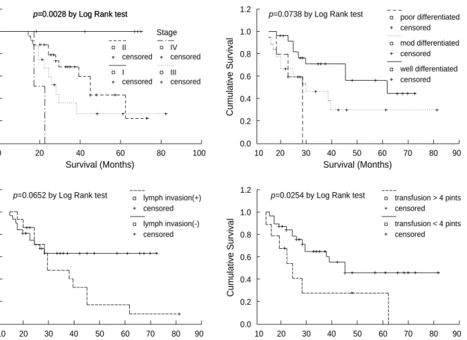
Among 46 patients of ampullary cancer, twenty four patients (52.2%) were pertained to pTNM stage II and fol- lowed by stage III (15 patients, 32.6%), stage I (5 patients, 7.8%) and stage IV (2 patients, 3.1%). There was no signif- icant difference in the distribution of pTNM stage with ref- erence to the presence or absence of COX-2 immunostaining (Table 1). However, there was significant difference of cumu-
lative survival rate between the groups of pTNM stage (Fig.
3). Subdividing the pTNM staging to pT and pN staging, there were significant differences of cumulative survival rate between the groups of pT and pN stage (Fig. 3).
Concerning the postoperative histopathology, well-differ- entiated tumor was most frequent (23 patients, 50.0%), fol- lowed by moderately differentiated (18 patients, 39.1%) and poorly differentiated tumor (5 patients, 10.9%). There was no significant difference in the distribution of tumor histo- pathology with reference to the presence or absence of COX-2 immunostaining (Table 1). There was a different tendency of cumulative survival rate between the groups of tumor histo- patholgy, however, did not reach the statistical significance (Fig. 3).
In our study, 31 patients (67.4%) of ampullary cancer had lymphatic tumor emboli in postoperative histopathology.
There was no significant difference of the presence or absence
Fig. 1.Marked expression of immunoreactive COX-2 staining was noticed mainly in cytosolic and perinuclear region of cancer cells (×400).
Age (yr) (mean±SD) 60.4±9.5 56.3±7.1 NS Sex (M/F) 18/6 9/13 0.035
pTNM stage NS
I 2 3
II 11 13
III 10 5
IV 1 1
Differentiation NS well differentiated 10 13
moderately differentiated 10 8
poorly differentiated 4 1 NS Complete resection (R0/R1) 24/0 22/0 NS Lymphatic tumor emboli (+/-) 9/15 6/16 NS
COX-2 (+) COX-2 (-)
(n=24) (n=22) p-value Table 1.Comparisons of clinicopathologic features between COX-2 positive and negative group of patients with ampulla of Vater cancer
NS: not significant.
Cumulative Survival
1.1 1.0 0.9 0.8 0.7 0.6 0.5 0.4 0.3 0.2 0.1 0.0
10 20 30 40 50 60 70 80 90
Survival (Months)
Fig. 2. The cumulative survival rate of 46 patients with ampulla of Vater cancer. The median and mean survival were 39 and 48.9 months, respectively.
censored
of lymphatic tumor emboli with reference to the COX-2 immunostaining (Table 1). There was a different tendency of cumulative survival rate between the positive and nega- tive group of lymphatic tumor emboli, however, did not reach the statistical significance (Fig. 3).
Among 46 patients of ampullary caner, 9 patients (19.6%) had more than 4 pints of blood transfusion during the oper- ation. There was significant difference of cumulative survival rate between the group of patients whether they had received more than 4 pints of blood transfusion or not (Fig. 3).
Immunohistochemical Staining of COX-2 and its Cor- relation with Proliferation Index (PI), Apoptotic Index (AI) and Microvessel Density
In the tissues of ampulla of Vater cancer, we found marked expression of immuno-reactive COX-2 in inflammatory mononuclear cells, vascular endothelial cells and cancer cells (Fig. 1). The cytosolic and perinuclear immunostaining for COX-2 was appeared in 24 (52.2%) patients with ampulla of Vater cancer. There was no significant difference of cumu- lative survival rate between the COX-2 positive and COX- 2 negative group (p=0.9420 by Log Rank test, Fig. 4).
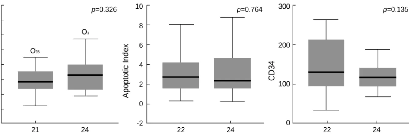
There were no significant differences of PI (p=0.326), AI (p=0.764), and microvessel density (p=0.135) in reference to the presence or absence of COX-2 immunostaining (Fig.
5). Also, there were no impacts on survival for PI, AI, and microvessel density in patients with ampulla of Vater cancer.
Cumulative Survival
1.2
1.0
0.8
0.6
0.4
0.2
0.0
0 20 40 60 80 100
Survival (Months)
IV censored Stage p=0.0028 by Log Rank test
p=0.0028 by Log Rank test
III censored II
censored I censored
Fig. 3. The comparisons of cumulative survival rate between the groups of pTNM stage, tumor histopathology, lymphatic tumor emboli, and blood transfusion.
Cumulative Survival
1.2
1.0
0.8
0.6
0.4
0.2
0.0
10 20 30 40 50 60 70 80 90
Survival (Months)
censored poor differentiated
mod differentiated censored
censored well differentiated
Cumulative Survival
1.2
1.0
0.8
0.6
0.4
0.2
0.0
10 20 30 40 50 60 70 80 90
Survival (Months)
p=0.0738 by Log Rank test
p=0.0652 by Log Rank test lymph invasion(+) censored
censored lymph invasion(-)
Cumulative Survival
1.2
1.0
0.8
0.6
0.4
0.2
0.0
10 20 30 40 50 60 70 80 90
Survival (Months)
p=0.0254 by Log Rank test transfusion > 4 pints censored
censored
transfusion < 4 pints
Cumulative Survival
1.2
1.0
0.8
0.6
0.4
0.2
10 20 30 40 50 60 70 80 90
Survival (Months)
Fig. 4.The comparison of cumulative survival rate between COX-2 positive and negative group of patients with ampulla of Vater can- cer.
p=0.9420 by Log-Rank test COX-2 (+) censored
censored COX-2 (-)
DISCUSSION
COX-2, a key enzyme required for the conversion of arachi- donic acid to prostaglandins, plays an important role in the promotion of intestinal tumorigenesis in animal models, but the underlying mechanism of action is still poorly understood.
Eberhart et al. (22) have reported that COX-2 expression is clearly up-regulated in the majority of colorectal carcinoma compared with levels of expression in accompanying normal mucosa. More recently, a subset of colorectal adenomas, the precursor lesions in colorectal carcinogenesis, show up-regu- lation of COX-2 mRNA.
Recently, several epidemiologic studies have suggested that NSAIDs decrease the incidence of colorectal cancer (12, 13).
COX and PGs may play a role in this mechanism because the main function of NSAIDs is in the inhibition of COX-1 and COX-2 enzymatic activity. Shiff et al. (26) reported that NSAIDS could inhibit the proliferation, alter morphological appearance, modify the cell cycle phase distribution and induce cell death by apoptosis of HT-29 colon adenocarcinoma cells.
There have been several immunocytochemical studies in localization of COX in the gastrointestinal tracts. The immu- nolocalization of COX in normal human colon and colorec- tal caner were reported by Mikkelson et al. (27) and Sano et al. (28), respectively. They detected the expression of immu- noreactive COX-2 in colorectal cancer cells, inflammatory mononuclear cells, vascular endothelial cells, and fibroblasts, but, immunoreactive COX-1 was very weak. In normal colonic tissues, immunoreactive COX-1 and -2 were weakly expressed in mucosal epithelial cells and vascular endothelial cells. In our study for ampulla of Vater cancer, the result is similar, and immunoreactive COX-2 staining was mainly localized in cytosolic and perinuclear region of cancer cells.
Kokawa et al. (29) have reported that marked COX-2 ex- pression was observed in 57% of pancreatic ductal adenocar- cinomas, 58% of adenomas, and in 70% of adenocarcinomas of intraductal pancreatic mucinous tumors (IPMTs) (38).
Likewise, COX-2 expression is known to be increased in human gastric cancers (30) and esophageal cancers (31). Over- expression of COX-2 in malignancies does not, however, seem to be a general phenomenon, since COX-2 protein was not found in human breast carcinoma (32).
In the current study, we found that immunoreactive COX-2 expression was noticed in 52.2% of patients with ampulla of Vater cancer. To our knowledge, there has been no report with a large number of patients with ampulla of Vater cancer focus- ing on COX-2 expression and its correlation with clinico- pathologic features. We found no significant difference in the cumulative survival rate between COX-2 positive and negative group, which was controversial points in previous reports for COX-2 and its relation with survival (33-37).
Also, we have correlated the COX-2 expression with prolif- erative index (PI), apoptotic index (AI), and microvessel density of ampulla of Vater cancer and could not find sig- nificant correlation between them. Our results of no correla- tion between COX-2 and proliferation, apoptosis, and angio- genesis for ampullary cancer were contradictory to the pre- vious report.
Cao et al. (38) have reviewed the many actions of COX-2 in cellular dynamics and in cancer. They indicate that the prostaglandin products of the COX-2 pathway enhance cell proliferation and growth in both normal and tumor cells.
Other important actions of COX-2 on cellular process which they reviewed, are inhibition of apoptosis, promotion of tumor cell migration, cell adhesion, tumor invasiveness and role of tumor angiogenesis. So far, it is not certain whether or not the dissension is due to the peculiar biologic properties of ampul- la of Vater cancer. Also, it is not certain whether or not the relatively small number of cases included in our study is the probable cause of statistical insignificance. Clinical studies of prospective design with a large number of samples for elucidating the biologic and therapeutic role of COX-2 in ampulla of Vater cancer will be warranted.
In our study, initial pTNM stage and blood transfusion
Fig. 5.The comparisons of PI, AI, and microvessel density between the COX-2 positive and negative group of patients with ampulla of Vater cancer. Each data contains median, quartiles and extreme values.
ProliferationIndex
80 70 60 50 40 30 20 10 0
COX-2(-) COX-2(+)
21 24
O25
p=0.326
O1
ApoptoticIndex
10
8
6
4
2
0
-2
COX-2(-) COX-2(+)
22 24
p=0.764
CD34
300
200
100
0
COX-2(-) COX-2(+)
22 24
p=0.135
more than 4 pints during operation were independent prog- nostic factors for patients with ampulla of Vater cancer. Al- though lymphatic tumor emboli and tumor differentiation in postoperative histopathology appeared as statistically in- significant prognostic factors, they showed distinctively dis- crepant tendency of survival.
In conclusion, our study suggests that COX-2 expression in ampulla of Vater cancer is not correlated with the clinico- pathologic and biologic features of tumor, such as length of survival, proliferation, apoptosis and angiogenesis of tumor cells.
REFERENCES
1. Howe JR, Klimstra DS, Moccia RD, Conlon KC, Brennan MF.
Factors predictive of survival in ampullary carcinoma. Ann Surg 1998; 228: 87-94.
2. Neoptolemos JP, Talbot IC, Shaw DC, Carr-Locke DL. Long-term survival after resection of ampullary carcinoma is associated inde- pendently with tumor grade and a new staging classification that assesses local invasiveness. Cancer 1998; 61: 1403-7.
3. Klempnauer J, Ridder GJ, Pichlmayr R. Prognostic factors after resection of ampullary carcinoma: multivariate survival analysis in comparison with ductal cancer of the pancreatic head. Br J Surg 1995; 82: 1686-91.
4. Roder JD, Schneider PM, Stein HJ, Siewert JR. Number of lymph node metastases is significantly associated with survival in patients with radically resected carcinoma of the ampulla of Vater. Br J Surg 1995; 82: 1693-6.
5. Allema JH, Reinders ME, van Gulik TM, van Leeuwen DJ, Ver- beek PC, de Wit LT, Gouma DJ. Results of pancreaticoduodenecto- my for ampullary carcinoma and analysis of prognostic factors for survival. Surgery 1995; 117: 247-53.
6. Shirai Y, Tsukada K, Ohtani T, Koyama G, Muto T, Watanabe H, Hatakeyama K. Carcinoma of the ampulla of Vater: histopathologic analysis of tumor spread in Whipple pancreatoduodenectomy speci- mens. World J Surg 1995; 19: 102-6.
7. Achille A, Biasi MO, Zamboni G, Bogina G, Iacono C, Talamini G, Capella G, Scarpa A. Cancers of the ampulla of Vater: mutator phe- notype is associated with good prognosis. Clin Cancer Res 1997; 3:
1841-7.
8. Talamini MA, Moesinger RC, Pitt HA, Sohn TA, Hruban RH, Lillemoe KD, Yeo CJ, Cameron JL. Adenocarcinoma of the ampul- la of Vater. A 28-year experience. Ann Surg 1997; 225: 590-9.
9. Dorandeu A, Raoul JL, Siriser F, Leclercq-Rioux N, Gosslin M, Martin ED, Ramee Mp, Raunois B. Carcinoma of the ampulla of Vater: prognostic factors after curative surgery: a series of 45 cases.
Gut 1997; 40: 350-5.
10. Matsubayashi H, Watanabe H, Yamaguchi T, Ajioka Y, Nishikura K, Kijima H, Saito T. Differences in mucus and K-ras mutation in relation to phenotypes of tumors of the ampulla of Vater. Cancer 1999; 86: 596-607.
11. Williams CS, Smalley W, DuBois RN. Aspirin use and potential
mechanisms for colorectal cancer prevention. J Clin Invest 1997;
100: 1325-9.
12. Smith CJ, Morrow JD, Roberts LJ, Marnett LJ. Differentiation of monocytoid THP-1 cells with phorbol ester induces expression of prostaglandin endoperoxide synthase-1 (COX-1). Biochem Biophys Res Commun 1993; 192: 787-93.
13. Smalley WE, DuBois RN. Colorectal cancer and non steroidal anti- inflammatory drugs. Adv Pharmacol 1997; 39: 1-20.
14. Thun MJ, Namboodiri MM, Heath CW Jr. Aspirin use and reduced risk of fatal colon cancer. N Engl J Med 1991; 325: 1593-6.
15. Khan KN, Masferrer JL, Woerner BM, Soslow R, Koki AT. Enhanced cyclooxygenase-2 expression in sporadic and familial adenomatous polyposis of the human colon. Scand J Gastroenterol 2001; 36: 865-9.
16. Wullen B, Muhlhofer A, Zoller WG. Effect of celecoxib, a COX-2 inhibitor, in familial adenomatous polyposis. Z Gastroenterol 2001;
39: 335-7.
17. Reddy BS, Hirose Y, Lubet R, Steele V, Kelloff G, Paulson S, Seib- ert K, Rao CV. Chemoprevention of colon cancer by specific cyclooxy- genase-2 inhibitor, celecoxib, administered during different stages of carcinogenesis. Cancer Res 2000; 60: 293-7.
18. Jacoby RF, Cole CE, Tutsch K, Newton MA, Kelloff G, Hawk ET, Lubet RA. Chemopreventive efficacy of combined piroxicam and difluoromethylornithine treatment of Apc mutant Min mouse adeno- mas, and selective toxicity against Apc mutant embryo. Cancer Res 2000; 60: 1864-70.
19. Oshima M, Dinchuk JE, Kargman SL, Oshima H, Hancock B, Kwong E, Trzaskos JM, Evans JF, Taketo MM. Suppression of intestinal polyposis in Apc delta 716 knockout mice by inhibition of cyclooxy- genase-2 (COX-2). Cell 1996; 87: 803-9.
20. Vogiagis D, Brown W, Glare EM, O’Brien PE. Rat colorectal tumours treated with a range of non steroidal anti-inflammatory drugs show altered cyclooxygenase-2 and cyclooxygenase-1 splice variant mRNA expression level. Carcinogenesis 2001; 22: 869-74.
21. Bennett A, Civier A, Hensby CN, Melhuish PB, Stamford IF. Mea- surement of arachidonate and its metabolites extracted from human normal and malignant gastrointestinal tissues. Gut 1987; 28: 315-8.
22. Eberhart CE, Coffey RJ, Radhika A, Giardiello FM, Ferrenbach S, DuBois RN. Up-regulation of cyclooxygenase 2 gene expression on human colorectal adenomas and adenocarcinomas. Gastroenterology 1994; 107: 1183-8.
23. Sheng H, Shao J, Morrow JD, Beauchamp RD, DuBois RN. Modu- lation of apoptosis and Bcl-2 expression by prostaglandin E2 in human colon cancer cells. Cancer Res 1998; 58: 362-6.
24. Tsujii M, Kawano S, Tsuji S, Sawaoka H, Hori M, DuBois RN.
Cyclooxygenase regulates angiogenesis induced by colon cancer cells. Cell 1998; 93: 705-16.
25. Marnett LJ. Aspirin and potential role of prostaglandins in colon cancer. Cancer Res 1992; 52: 5575-89.
26. Shiff SJ, Koutsos M, Qiao L, Rigas B. Nonsteroidal anti-inflamma- tory drugs inhibit the proliferarion of colon adenocarcinoma cells:
Effects on cell cycle and apoptosis. Exp Cell Res 1996; 222: 179-88.
27. Mikkelson HB, Rumessen JJ, Qvortrup K. Prostaglandin H synthase immunoreactivity in human gut: an immunohistochemical study. His- tochemistry 1991; 96: 295-9.
28. Sano H, Kawahito Y, Wilder RL, Hashiramoto A, Mukai S, Asai K, Kimura S, Kato H, Kondo M, Hla T. Expression of cyclooxygenase- 1 and -2 in human colorectal cancer. Cancer Res 1995; 55: 3785-9.
29. Kokawa A, Kondo H, Gotoda T, Ono H, Saito D, Nakadaira S, Kosuge T, Yoshida S. Increased expression of cyclooxygenase-2 in human pancreatic neoplasms and potential for chemoprevention by cyclooxy- genase inhibitors. Cancer 2001; 91: 333-8.
30. Ristimaki A, Honkanen N, Jankala H, Sipponen P, Harkonen M.
Expression of cyclooxygenase-2 in human gastric cancer. Cancer Res 1997; 57: 1276-80.
31. Wilson KT, Fu S, Ramanujam KS, Meltzer SJ. Increased expres- sion of inducible nitric oxide synthase and cyclooxygenase-2 in Barrett's esophagus and associated adenocarcinomas. Cancer Res 1998; 58: 2929-34.
32. Kargman SL, O’Neil GP, Vickers PJ, Evans JF, Mancini JA, Jothy S.
Expression of prostaglandin G/H synthase-1 and -2 protein in human colon cancer. Cancer Res 1995; 55: 2556-9.
33. Denkert C, Kobel M, Pest S, Koch I, Berger S, Schwabe M, Siegert A, Reles A, Klosterhalfen B, Hauptmann S. Expression of cyclooxy-
genase 2 is an independent prognostic factor in human ovarian car- cinoma. Am J Pathol 2002; 160: 893-903.
34. Zhang H, Sun XF. Overexpression of cyclooxygenase-2 correlates with advanced stages of colorectal cancer. Am J Gastroenterol 2002;
97: 1037-41.
35. Brabender J, Park J, Metzger R, Schneider PM, Lord RV, Holscher AH, Danenberg KD, Danenberg PV. Prognostic significance of cyclooxygenase 2 mRNA expression in non small cell lung cancer.
Ann Surg 2002; 235: 440-3.
36. Costa C, Soares R, Reis-Filho JS, Leitao D, Amendoeira I, Schmitt FC. Cyclooxygenase 2 expression is associated with angiogenesis and lymph node metastasis in human breast cancer. J Clin Pathol 2002; 55: 429-34.
37. Buskens CJ, van Rees BP, Sivula A, Reitsma JB, Haglund C, Bosma PJ, Offerhaus GJ, van Lanschot JJ, Ristimaki A. Prognostic signifi- cance of elevated cyclooxygenase 2 expression in patients with ade- nocarcinoma of the esophagus. Gastroenterology 2002; 122: 1800-7.
38. Cao Y, Prescott SM. Many actions of cyclooxygenase-2 in cellular dynamics and in cancer. J Cell Physiol 2002; 190: 279-86.