INTRODUCTION
The closure of cutaneous wounds involves complex tissue movements such as hemorrhage, inflammation, re-epitheliza- tion, granulation tissue formation, and the late remodeling phase of repair (1). These events involve coordination of dozens of types of cells and matrix proteins, which are all important to control stages of the repair process. Previous studies have demonstrated that endogenous growth factors, such as fibrob- last growth factors (FGF) (2), platelet derived growth factors (PDGF) (3), transforming growth factor-β(TGF-β) (1) and vascular endothelial growth factors (VEGF) (4) are the impor- tant regulatory polypeptides for coordinating the healing process. They are released from macrophages, fibroblasts, and keratinocytes at the site of injury and they participate in the regulation of re-epithelization, granulation tissue formation, collagen synthesis and neovascularization.
Ozone (O3) has been widely recognized as one of the best bactericidal, antiviral and antifungal agents (5, 6) and it has been used empirically as a clinical therapeutic agent for chron- ic wounds, such as trophic ulcers, ischemic ulcers and dia- betic wounds (7-9). The beneficial effects of O3on wound healing might be assumed to be due to decreased bacterial infection, ameliorated impaired dermal wound healing or
increased oxygen tension by O3exposure in the wound area (10, 11).
It was reported that O3exposure is associated with activa- tion of transcription factor NF-κB; this is important to reg- ulate inflammatory responses and eventually the entire pro- cess of wound healing (5, 12, 13). It was shown that huge amounts of PDGF and TGF-β1 were released from platelets in the heparinized plasma of a limb ischemia patient after ozonation (14, 15). It has been revealed that there were sub- stantial increases of steady-state mRNA levels of TGF-β1 in the fibroblasts that were co-cultured with bronchoepithelial cells after O3exposure (16). A recent study has shown that hydrogen peroxide (H2O2) potently induced the VEGF ex- pression in human keratinocytes which can stimulate wound healing (17). From these previous studies of O3, we hypothe- sized that O3might enhance acute cutaneous wound heal- ing, and this could be associated with growth factors such as FGF, PDGF, TGF-βand VEGF.
Nowadays, O3 is profitably and practically employed as ozonated olive oil; this contains the O3molecule stabilized as an ozonide between the double bonds of a monounsaturated fatty acid such as oleic acid, which is ideal for the topical use of O3to treat chronically infected cutaneous and mucosal areas of the body (5). Ozonated materials referred to as ozonides are
368
Hee Su Kim, Sun Up Noh, Ye Won Han, Kyoung Moon Kim, Hoon Kang, Hyung Ok Kim, and Young Min Park
Department of Dermatology, Seoul St. Mary’s Hospital, College of Medicine, The Catholic University of Korea, Seoul, Korea
Address for correspondence Young Min Park, M.D.
Department of Dermatology, Seoul St. Mary’s Hospital, College of Medicine, The Catholic University of Korea, 505 Banpo-dong, Seocho-gu, Seoul 137-040, Korea Tel : +82.2-2258-6223, Fax : +82.2-594-3255 E-mail : yymmpark6301@hotmail.com DOI: 10.3346/jkms.2009.24.3.368
Therapeutic Effects of Topical Application of Ozone on Acute Cutaneous Wound Healing
This study was undertaken to evaluate the therapeutic effects of topical ozonated olive oil on acute cutaneous wound healing in a guinea pig model and also to elu- cidate its therapeutic mechanism. After creating full-thickness skin wounds on the backs of guinea pigs by using a 6 mm punch biopsy, we examined the wound heal- ing effect of topically applied ozonated olive oil (ozone group), as compared to the pure olive oil (oil group) and non-treatment (control group). The ozone group of guinea pig had a significantly smaller wound size and a residual wound area than the oil group, on days 5 (P<0.05) and 7 (P<0.01 and P<0.05) after wound surgery, respectively. Both hematoxylin-eosin staining and Masson-trichrome staining reveal- ed an increased intensity of collagen fibers and a greater number of fibroblasts in the ozone group than that in the oil group on day 7. Immunohistochemical staining demonstrated upregulation of platelet derived growth factor (PDGF), transforming growth factor-β(TGF-β) and vascular endothelial growth factor (VEGF) expressions, but not fibroblast growth factor expression in the ozone group on day 7, as com- pared with the oil group. In conclusion, these results demonstrate that topical appli- cation of ozonated olive oil can accelerate acute cutaneous wound repair in a guinea pig in association with the increased expression of PDGF, TGF-β, and VEGF.
Key Words : Collagen; Ozone; Wound Healing
Received : 17 January 2008 Accepted : 7 July 2008
formed by the reaction of olefins with ozone. Any olefin can be treated with gaseous ozone to form an ozonide. The ozonide compositions have the capacity to deliver nascent oxygen deep within the lesion without causing primary skin irritation.
Ozonated oil has been used topically for the treatment of chronic wounds, but there have been few studies concerning with the therapeutic effects of ozonated olive oil on acute cuta- neous wound healing in animal models. The present study was designed to evaluate the therapeutic effect of topical ozo- nated olive oil on acute cutaneous wound healing in a guinea pig model, and to elucidate its therapeutic mechanisms that are associated with such growth factors as FGF, PDGF, TGF- β, and VEGF.
MATERIALS AND METHODS Animals
Sixteen female guinea pigs (400-450 g), aged 8-9 weeks, were placed in a room that was under a 12-hr light and 12- hr dark cycle. The animals were placed individually in sepa- rate cages with ad libitum access to food and water. The ani- mal care, handling, and experimental procedures were car- ried out in accordance with a protocol approved by the Ani- mal Care and Use Committee of the Catholic University of Korea.
Wound biopsy
After 7 days of acclimation, the guinea pigs were anesthe- sized with ketamine and their backs were shaved and then sterilized with normal saline. The skin was then pinched and folded, and a sterile biopsy punch (6 mm in diameter, Stiefel Co., Offenbach, Germany) was used to make a full-thickness hole in the skin. Two wounds were created on both sides of the back for a total 4 circular wounds per animal.
Application of topical ozonated olive oil
Two drops (about 0.1 mL) of ozonated olive oil (OZOO�, Aurora, Inc., Miami, FL, U.S.A.) were applied everyday to two sites of the four wounds (Ozone group). Olive oil (OLO) as a pure base was applied to a third wound (Oil group). As a control group, nothing was applied on the fourth wound.
The wounds were then dressed with Opsite�(Smith&Nephew, Hull, U.K.) to cover them without dryness. An elastic ban- dage was wrapped around the area to prevent further injury.
Measurement of wound closure
After each guinea pig was wounded as described above, the wounds were photographed at indicated times by using a digital camera (Canon 350D, Canon Inc., Tokyo, Japan) that
was placed at a 20 cm distance to help eliminate error in mea- suring the size of the wounds. The photographic images were analyzed by Canvas X10.0 software (ACDSEE Inc., Miami, FL, U.S.A.) and the area of wound closure was measured by using the same program. The parameters of the wounds, such as wound size and the residual wound area, were measured daily from the photographic images taken.
* Measured parameters 1) Wound size (mm2) 2) Residual wound area (%):
Area measured at the experiment day Initial wound area ×100
Analysis of clinical wound closure in each group of wounds was performed through digital processing on days 0, 3, 5, 7, and 11.
Processing and preparation of skin tissues
On days 3 (n=4), 7 (n=4), and 11 (n=8) after wounding, guinea pigs were euthanized using ketamine and the four wounded tissues from each guinea pig were excised. Then the tissues were cut into halves; one half was placed in formalin (10% formaldehyde in phosphate-buffered saline) for hema- toxylin-eosin and Masson-trichrome staining, and the second half was quickly kept as a frozen state (-80℃) for immuno- histochemistry.
Histology
The specimens for histological examination were collected from each group by the full-thickness excision. Intensity of the collagen fibers and the fibroblast proliferation were exam- ined under microscope in hematoxylin-eosin and Masson-tri- chrome staining. The staining intensity of the collagen fibers was graded under ×200 magnification as follows: -, com- pletely negative staining intensity; ±, lower staining inten- sity; +, moderate staining intensity; ++, slightly higher staining intensity; +++, considerably higher staining inten- sity. The number of fibroblasts was counted in the 5 random- ized fields per specimen under ×400 magnification. Three dermatologists, who were ‘‘blinded’’ to which groups the spec- imens were in, independently analyzed all the specimens and then the mean numbers and standard deviations of the fibrob- lasts in all groups of wound were calculated, respectively.
Immunohistochemical staining
All the cryostat sections (5 μm) of the healing wounds of guinea pigs were placed on coated micro slides (Muto-glass, Tokyo, Japan) for immunohistochemical staining procedures.
The samples for immunohistochemistry were washed sever- al times with 0.1 M phosphate buffered saline (PBS) and then
reacted for 15 min with 0.3% H2O2solution diluted with PBS to inactivate the endogenous peroxidase in the tissues.
After washing three times for 10 min with PBS, the nonspe- cific antigen-antibody reactions were inhibited by a 1 hr treat- ment with 2.5% normal horse serum (R.T.U. Vectastatin� Universal ABC Elite kit, Vector Laboratories, Inc., Burlin- game, CA, U.S.A.). The samples were reacted in 1:100 dilut- ed solutions of each of the primary antibodies such as rabbit anti-FGF serum (Santa Cruz Biotechnology Inc., Delaware, CA, U.S.A.), anti-PDGF serum (Santa Cruz Biotechnology Inc.), anti-TGF-βserum (Santa Cruz Biotechnology Inc.), and anti-VEGF serum (Santa Cruz Biotechnology Inc.) at 4℃ for 24 hr. After the primary antibody reaction, each sample was washed three times for 10 min with PBS, and then they were reacted at room temperature for 1 hr with treatment of the secondary antibody solution, biotinylated horse anti-IgG (R.T.U. Vectastatin�Universal ABC Elite kit). After wash- ing three times for 10 min each with PBS, the samples were reacted at room temperature for 1 hr with avidin-biotin-per- oxidase complex solution (R.T.U. Vectastatin�Universal ABC Elite kit). For the chromogen reactions, the Vector�Nova- REDTMsubstrate kit (Vector Laboratories, Inc., Burlingame, CA, U.S.A.) was used with reagents 1, 2, and 3 and H2O2at room temperature for 3-5 min. After the chromogen reac- tion, the sample slides were washed three times for 10 min each with PBS. The washed sample tissues were dried out on gelatin-coated slides for 2 hr at room temperature. They are made transparent with xylene treatment and next cov- ered with polymount. Three ‘‘blinded’’ dermatologists then
independently analyzed the relative intensity of the immuno- histochemical staining of the slides for FGF, PDGF, TGF-β, and VEGF on 5 randomized fields per specimen under ×200 magnification. The intensity was graded as follows: 0, no positive reaction; +, 1-25% of the cells were positively stained;
++, 26-50% of the cells were positively stained; +++, 51- 75% of the cells were positively stained; ++++, 76-100%
of the cells were positively stained. The mean values of the scores were analyzed for interpreting the histochemical reac- tions.
Statistical analysis
All statistical analyses were performed using the ANOVA followed by Turkey-Kramer’s multiple-comparison test, or Kruskal-Wallis test of variance with Bonferroni’s correction.
SPSS v10.0 (SPSS Inc., Chicago, IL, U.S.A.) was used for all calculations. All values were expressed as means and standard deviations. Differences were considered significant if P<0.05.
RESULTS
The ozonated OLO significantly enhances the acute cutaneous wound healing
As shown in Fig. 1, there was an enhanced wound closure in the ozone group of wounds, the left two of the four wounds, as compared to the oil group as well as the control group. On
Fig. 1.The effects of ozonated olive oil on clinical wound closure. The photomicrographs demonstrate the enhanced wound closure, in the ozone group, on the left two wounds (a and b) on the back of the guinea pig, as compared to the oil group (c) as well as the control group (d). (Bar=5 mm).
Day 0 Day 3 Day 5 Day 7 Day 11
a
b
c
d
Difference between the ozone group and the oil group. *P<0.05; �P<0.01.
Ozone group (n=16) Oil group (n=8)
Control (n=8) Post operation day
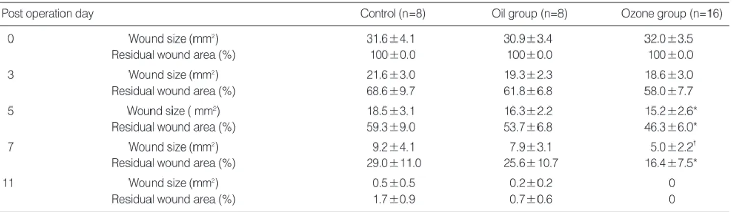
0 Wound size (mm2) 31.6±4.1 30.9±3.4 32.0±3.5
Residual wound area (%) 100±0.0 100±0.0 100±0.0
3 Wound size (mm2) 21.6±3.0 19.3±2.3 18.6±3.0
Residual wound area (%) 68.6±9.7 61.8±6.8 58.0±7.7
5 Wound size ( mm2) 18.5±3.1 16.3±2.2 15.2±2.6*
Residual wound area (%) 59.3±9.0 53.7±6.8 46.3±6.0*
7 Wound size (mm2) 9.2±4.1 7.9±3.1 5.0±2.2�
Residual wound area (%) 29.0±11.0 25.6±10.7 16.4±7.5*
11 Wound size (mm2) 0.5±0.5 0.2±0.2 0
Residual wound area (%) 1.7±0.9 0.7±0.6 0
Table 1.Comparison of the average wound size and residual wound area on post-operation days 0, 3, 5, 7, and 11
day 11, all of the wounds completely re-epithelized irrespec- tive of treatments. The ozone group showed a significantly smaller wound size than the oil group on days 5 (P<0.05) and 7 (P<0.01). The ozone group showed 58%, 46.3%, and 16.4% in residual wound area on days 3, 5 and 7, respective- ly. On the other hand, the oil group revealed 61.8%, 53.7%
and 25.6% in residual wound area, respectively (Table 1).
Thus, there was a significant difference in the residual wound area between the ozone group and the oil group on days 5 (P<0.05) and 7 (P<0.01). On repeated-measures of ANOVA, the ozone group showed a significantly decreased residual wound area as compared to the oil group, as well as the con-
trol group (P<0.05).
The ozonated OLO promotes collagen synthesis and fibroblast proliferation
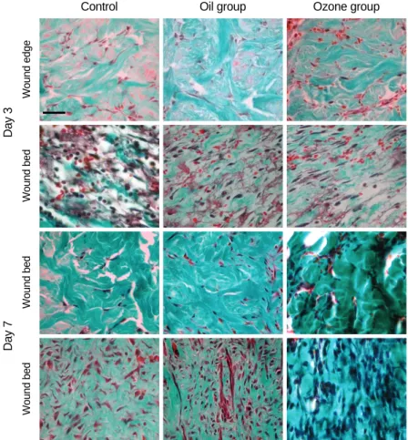
On the hematoxylin-eosin staining, epithelization, infil- tration of inflammatory cells and vascular proliferations were commonly seen on all of the wounds; but there were prolif- eration of fibroblasts and collagen fibers noted in the ozone group as well (data not shown). We performed the Masson- trichrome staining in order to determine whether ozonated OLO’s ability to accelerate wound closure was associated with
Fig. 2.Masson-trichrome staining of the wound bed and the edge of the injury site on days 3 and 7. The ozone group revealed the increas- ed staining intensity of collagen fibers and the number of fibroblasts at the wound bed and edge, in comparison to the oil group and the control group on day 7, but not on day 3 (original magnification ×400, Bar=50 μm).
Control Oil group Ozone group
Wound edgeWound bed
Day 3Day 7 Wound bedWound bed
B, wound bed; E, wound edge; -, completely negative staining intensity; ±, lower staining intensity; +, moderate staining intensity; ++, slightly higher staining intensity; +++, considerably higher staining intensity. *P<0.05.
Ozone group (n=8) Oil group (n=4)
Control (n=4) At post-operation day 3
B E B E B E
Staining intensity of collagen fibers ± + ± + ± +
Number of fibroblasts 30.3±1.3 16.3±1.4 29.7±1.1 17.7±1.7 30.1±1.8 19.3±2.1 At post-operation day 7
B E B E B E
Staining intensity of collagen fibers + ++ ++ ++ +++ +++
Number of fibroblasts 36.9±2.6 16.3±2.1 48.5±2.3 22.7±2.7 62.3±4.4* 35.6±3.1*
Table 2.Comparison of staining intensity of collagen fibers and the number of fibroblasts by the Masson-trichrome staining on days 3 and 7
collagen synthesis and fibroblast proliferation at the wound bed and at the edge of the injury site. The staining intensity of collagen fibers and the number of fibroblasts were evalu- ated on days 3 and 7. On day 3, the ozone group did not show a significant difference in the staining intensity of collagen fibers and the number of fibroblasts as compared to the oil group. In contrast, on day 7, the ozone group revealed an increased staining intensity of collagen fibers at the wound bed and at the edge of the entire dermis in comparison to the oil and control groups (Fig. 2). On day 7, the numbers of fibroblasts in the ozone group were 62.3 and 35.6 at the wound bed and edge, respectively; but, the numbers of fibrob-
lasts in the oil group were 48.5 and 22.7, respectively (Table 2). Thus, there was a significant difference in the staining intensity of collagen fibers and the number of fibroblasts bet- ween the ozone group and the oil group on day 7 (P<0.05), but not on day 3.
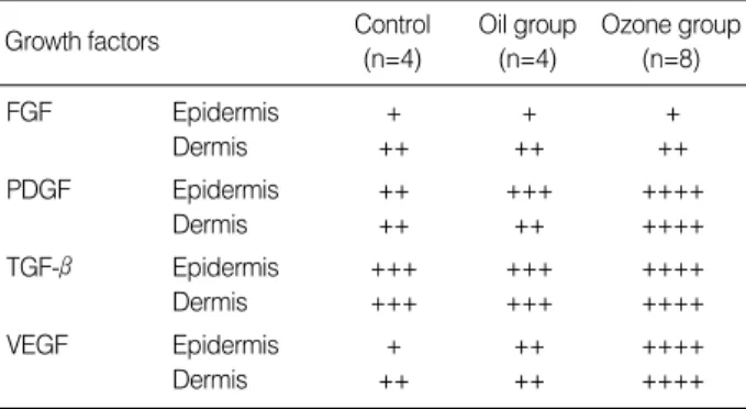
The ozone group revealed increased expressions of PDGF, TGF-ββ, and VEGF, but not FGF
In order to determine which growth factors play an impor- tant role for the accelerating wound closure associated with the proliferation of fibroblasts and collagen fibers by the ozo- nated OLO, we evaluated the immunohistochemical stain- ing intensity of FGF, PDGF, TGF-β, and VEGF on day 7 (Fig. 3). FGF expressions were identified in dermal fibrob- lasts and collagen fibers, but this was barely detected in the epidermis of the control group. There were little differences in the FGF expression between the ozone group and the oil group (Table 3). PDGF was expressed in dermal inflamma- tory cells, fibroblasts, epidermal cells and keratinocytes of hair follicles of the control group. The ozone group revealed a relatively higher PDGF expression, as compared to the oil group. There was a relatively distinct difference in the der- mis between the former and the latter. TGF-βexpressions were detected in the dermal fibroblasts, epidermal cells and keratinocytes of the hair follicles of the control group. Like- wise, the ozone group showed a relatively increased expres- sion of TGF-β, as compared to the oil group. VEGF expres-
Control
FGFPDGFTGF-βVEGF
Oil group Ozone group
Growth factors Control
(n=4)
Oil group (n=4)
Ozone group (n=8)
FGF Epidermis + + +
Dermis ++ ++ ++
PDGF Epidermis ++ +++ ++++
Dermis ++ ++ ++++
TGF-β Epidermis +++ +++ ++++
Dermis +++ +++ ++++
VEGF Epidermis + ++ ++++
Dermis ++ ++ ++++
Table 3.Comparison of the expression of growth factors on day 7
0, no positive reaction; 1+, the positively staining cells were 1-25%; 2+, the positively staining cells were 26-50%; 3+, the positively staining cells were 51-75%; 4+, the positively staining cells were 76-100%.
Fig. 3.Immunohistochemical sta- ining for FGF, PDGF, TGF-β‚ and VEGF on day 7. The ozone group revealed the relatively increased expressions of PDGF (B, F, J), TGF- β(C, G, K), and VEGF (D, H, L), but not FGF (A, E, I) as compared to the oil group on day 7 (×100, orig- inal magnification, inlet; ×400, Bar
=500 μm).
A E I
B F J
C G K
D H L
sion was identified in dermal fibroblasts, endothelial cells and collagen fibers, but it was barely detected in the epidermis of the control group. The ozone group revealed a relatively increased VEGF expression in both the dermis and the epi- dermis, as compared to the oil group. These findings demon- strated that the ozone group revealed relatively higher expres- sions of PDGF, TGF-β, and VEGF, but not FGF than the oil group on day 7.
DISCUSSION
Two different ozonized solutions, consisting of sunflower ozonized oil and ozonated OLO, have been usually used as a topical form of O3. Because of their antibacterial and anti- mycotic properties, these ozonated oils have been empirical- ly used for treating superficial bacterial and fungal infections (5, 18). Previous studies showed that O3could affect the ex- pression of pro-inflammatory cytokines, such as interleukin-1 (IL-1) and tumor necrosis factor-α(TNF-α), and the adaptive inflammatory responses including cyclooxygenase-2 (COX-2) gene activation in keratinocyte via activation of NF-κB (19, 20). It was also demonstrated that O3exposure increased the expression of proliferating cell nuclear antigen (PCNA) protein and K10, a keratin expressed in well differentiated suprabasal keratinocytes in skin tissues (20, 21). These find- ings suggested that O3could induce the keratinocyte prolif- eration and differentiation and it could affect skin biology.
OLO will react with O3, forming ozonide at room temper- ature in a mildly exothermic reaction. As a sample of OLO is ozonized, OLO will gain weight as the reaction proceeds.
OLO, high in essential fatty acids, is the only media that will allow O3to remain in an active or nascent state for a long length of time. Two drops of ozonated OLO, about 0.1 mL, were used in our experiment because it was thought that about 0.1 mL was sufficient to cover the 6 mm-sized punch wounds.
Although there are no reports about the toxicological study of ozonated OLO, it was reported that sunflower ozonized oil had no side effects in human-patients (18), except for slight irritation being noted at the epithelium of mice (22) from the previous toxicological studies. Because the O3molecule can be stabilized as an ozonide between the double bonds of a monounsaturated fatty acid such as oleic acid (5, 23), ozonat- ed OLO is an ideal preparation for the topical form of O3and it remains stable for 2 yr when stored at 4℃. Furthermore, ozonated OLO is easy to access as a commercialized form and to use for applying to cutaneous wound because of its liquid form. Thus, in this study, we used ozonated OLO as a topi- cal form of O3to treat cutaneous wound.
Full-thickness punch wounds used in this study, not includ- ing panniculectomy, are useful model to evaluate re-epithe- lization after topical application. Although pigs are often use- ful models since their cutaneous architecture is most similar
to that of human skin, guinea pigs are cheaper to obtain than pigs and the care of those is cheaper and easier as well (24).
Based on the results from the wound closure between the ozone group and the oil group on days 0, 3, 5, 7, and 11, the ozone group showed a significantly smaller wound size and residual wound area than the oil group in the guinea pig model on days 5 and 7. Thus, these results demonstrated that O3 could enhance acute cutaneous wound healing. Especially, on day 7, both the wound size and residual wound area of the ozone group were much more significantly decreased.
This implies that topical exposure of O3may affect granula- tion tissue formation of the wound healing process rather than affecting immediate formation of blood clot and recruit- ment of inflammatory cells during the inflammation phase.
On Masson-trichrome staining on day 3, there was not much difference in the staining intensity of collagen fibers and fibroblast proliferation between the ozone group and the oil group. However, on day 7, the ozone group revealed about one and half times increased staining intensity of collagen fibers and significantly increased fibroblasts at the wound edge as well as at the wound bed, compared to the oil group.
These findings indicated that O3 may act on acute wound healing directly or indirectly via collagen synthesis and fibrob- last proliferation during granulation tissue formation and the early tissue remodeling phase of wound healing.
Fibroblasts have been known to play important roles in re- epithelization, collagen fiber synthesis, extracellular matrix regeneration, remodeling of wounds, and for the release of such endogenous growth factors as FGF, PDGF, TGF-β, and VEGF (1, 25). In this study, increased expressions of PDGF and TGF-βwere seen in the ozone group on day 7, which is correlated with increased staining intensity of collagen fibers and fibroblast proliferation on the same day. There were also increased expressions of PDGF and TGF-βin the epidermal keratinocytes and the hair follicular cells adjacent to the injury.
These findings suggest that O3might induce expressions of PDGF and TGF-βfrom epidermal keratinocyte as well as from the dermal fibroblast at the injury site. In the present study, there was a relatively increased expression of VEGF in the epidermal keratinocytes of the ozone group on day 7; this was consistent with the previous study showing that VEGF was gradually increased from the 1st day to the 7th day of nor- mal wound healing process (26). On hematoxylin-eosin stain- ing, the relatively increased vascularity in the ozone group might be due to the increased expression of VEGF by ozo- nation. These findings may be associated with the genera- tion of H2O2through ozonation, which can directly induce a VEGF expression and/or indirectly induce it by the induc- tion of heme oxygenase-1 (27). Because VEGF is main cyto- kine of vascularization in the late phase of wound healing (28), further study will be needed to clarify the effect of O3 on the neovascularization of cutaneous wound healing. In contrast to the increased expressions of PDGF, TGF-β, and VEGF in the ozone group, there was little difference in the
FGF expressions between the ozone group and the oil group on day 7. One of the possible explanations for this result is that FGF had already been up-regulated within 24 hr after wounding (29).
In conclusion, these results demonstrate that application of ozonated OLO, the topical form of O3, can accelerate acute cutaneous wound repair in the guinea pig model by promot- ing collagen synthesis and fibroblast proliferation at the injury site and by increasing the expression of growth factors such as PDGF, TGF-β, and VEGF. Taken together, we can infer that topical O3may be regarded as an alternative therapeu- tic modality to enhance cutaneous wound healing.
REFERENCES
1. Werner S, Grose R. Regulation of wound healing by growth factors and cytokines. Physiol Rev 2003; 83: 835-70.
2. Ornitz DM, Itoh N. Fibroblast growth factors. Genome Biol 2001;
2: 3005.
3. Heldin CH, Westermark B. Mechanism of action and in vivo role of platelet-derived growth factor. Physiol Rev 1999; 79: 1283-316.
4. Lauer G, Sollberg S, Cole M, Flamme I, Sturzebecher J, Mann K, Krieg T, Eming SA. Expression and proteolysis of vascular endothe- lial growth factor is increased in chronic wounds. J Invest Dermatol 2000; 115: 12-8.
5. Valacchi G, Fortino V, Bocci V. The dual action of ozone on the skin.
Br J Dermatol 2005; 153: 1096-100.
6. Al-Dalain SM, Martinez G, Candelario-Jalil E, Menendez S, Re L, Giuliani A, Leon OS. Ozone treatment reduces markers of oxidative and endothelial damage in an experimental diabetes model in rats.
Pharmacol Res 2001; 44: 391-6.
7. Martinez-Sanchez G, Al-Dalain SM, Menendez S, Re L, Giuliani A, Candelario-Jalil E, Alvarez H, Fernandez-Montequin JI, Leon OS.
Therapeutic efficacy of ozone in patients with diabetic foot. Eur J Pharmacol 2005; 523: 151-61.
8. Bocci V. Ozone: a new medical drug. Dordrecht: Springer, 2005.
9. de Monte A, van der Zee H, Bocci V. Major ozonated autohemother- apy in chronic limb ischemia with ulcerations. J Altern Complement Med 2005; 11: 363-7.
10. Lim Y, Phung AD, Corbacho AM, Aung HH, Maioli E, Reznick AZ, Cross CE, Davis PA, Valacchi G. Modulation of cutaneous wound healing by ozone:differences between young and aged mice. Toxicol Lett 2006; 160: 127-34.
11. Gajendrareddy PK, Sen CK, Horan MP, Marucha PT. Hyperbaric oxygen therapy ameliorates stress-impaired dermal wound healing.
Brain Behav Immun 2005; 19: 217-22.
12. Janic B, Umstead TM, Phelps DS, Floros J. Modulatory effects of ozone on THP-1 cells in response to SP-A stimulation. Am J Physiol Lung Cell Mol Physiol 2005; 288: L317-25.
13. Valacchi G, van der Vliet A, Schock BC, Okamoto T, Obermuller- Jevic U, Cross CE, Packer L. Ozone exposure activates oxidative stress responses in murine skin. Toxicology 2002; 179: 163-70.
14. Bocci V. Biological and clinical effects of ozone. Has ozone therapy a future in medicine? Br J Biomed Sci 1999; 56: 270-9.
15. Valacchi G, Bocci V. Studies on the biological effects of ozone: 10.
Release of factors from ozonated human platelets. Mediat Inflamm 1999; 8: 205-9.
16. Lang DS, Jorres RA, Mucke M, Siegfried W, Magnussen H. Inter- actions between human bronchoepithelial cells and lung fibroblasts after ozone exposure in vitro. Toxicol Lett 1998; 96-97: 13-24.
17. Sen CK, Khanna S, Babior BM, Hunt TK, Ellison EC, Roy S. Oxi- dant-induced vascular endothelial growth factor expression in human keratinocytes and cutaneous wound healing. J Biol Chem 2002; 277:
33284-90.
18. Sechi LA, Lezcano I, Nunez N, Espim M, Dupre I, Pinna A, Molicot- ti P, Fadda G, Zanetti S. Antibacterial activity of ozonized sunflower oil (Oleozon). J Appl Microbiol 2001; 90: 279-84.
19. Fischer SM. Is cyclooxygenase-2 important in skin carcinogenesis?
J Environ Pathol Toxicol Oncol 2002; 21: 183-91.
20. Valacchi G, Pagnin E, Corbacho AM, Olano E, Davis PA, Packer L, Cross CE. In vivo ozone exposure induces antioxidant/stress-related responses in murine lung and skin. Free Radic Biol Med 2004; 36:
673-81.
21. Paramio JM, Casanova ML, Segrelles C, Mittnacht S, Lane EB, Jor- cano JL. Modulation of cell proliferation by cytokeratins K10 and K16. Mol Cell Biol 1999; 19: 3086-94.
22. Menendez S, Falcon L, Simon DR, Landa N. Efficacy of ozonized sunflower oil in the treatment of tinea pedis. Mycoses 2002; 45: 329- 32.
23. Bocci V. Oxygen-ozone therapy: a critical evaluation. Dordrecht:
Kluwer Academic Publisher, 2002.
24. FDA Wound Healing Clinical Focus Group. Guidance for industry:
chronic cutaneous ulcer and burn wounds-developing products for treatment. Wound Repair Regen 2001; 9: 258-68.
25. Vincent F. Mechanisms of cutaneous wound repair. In: Freedberg IM, Eisen AZ, Wolff K, Austen KF, Goldsmith LA, Katz SI, editors, Fitzpatrick’s dermatology in general medicine. 6th ed. New York:
McGraw Hill, 2003; 236-46.
26. Detmar M, Brown LF, Berse B, Jackman RW, Elicker BM, Dvorak HF, Claffey KP. Hypoxia regulates the expression of vascular per- meability factor/vascular endothelial growth factor (VPF/VEGF) and its receptors in human skin. J Invest Dermatol 1997; 108: 263-8.
27. Jazwa A, Loboda A, Golda S, Cisowski J, Szelag M, Zagorska A, Sroczynska P, Drukala J, Jozkowicz A, Dulak J. Effect of heme and heme oxygenase-1 on vascular endothelial growth factor synthesis and angiogenic potency of human keratinocytes. Free Radic Biol Med 2006; 40: 1250-63.
28. Nissen NN, Polverini PJ, Koch AE, Volin MV, Gamelli RL, DiPi- etro LA. Vascular endothelial growth factor mediates angiogenic activity during the proliferative phase of wound healing. Am J Pathol 1998; 152: 1445-52.
29. Werner S, Peters KG, Longaker MT, Fuller-Pace F, Banda MJ, Williams LT. Large induction of keratinocyte growth factor expres- sion in the dermis during wound healing. Proc Natl Acad Sci USA 1992; 89: 6896-900.