1)
Introduction
Hepatoblastoma is a primary tumor of the liver occur- ring predominantly in children
1). The usual site of metasta- sis in hepatoblastoma is the lung. Metastasis of hepato- blastoma to brain and/or heart is quite rarely reported and we could not find a prior case of hepatoblastoma invading the left atrium of the heart. We report a case of hepato- blastoma in a 6-year-old orphan girl who had recurrent disease at multiple sites including brain, left atrium of the heart and lung after the initial treatment 4 years earlier.
Preoperative chemotherapy and tumor resection was per- formed initially; however, postoperative chemotherapy was refused by the legal guardian of the child. She remained alive after the cessation of the treatment for over 6 months.
Case Report
A 6-year-old orphan girl was admitted for the evalu- ation of an abnormal mass-like lesion noted on a plain
접수 : 2005년 11월 14일, 승인 : 2006년 1월 20일 책임저자 : 김흥식, 계명의대 동산의료원 소아과 Correspondence : Heung Sik Kim, M.D.
Tel : 053)250-7516 Fax : 053)250-7783 E-mail : [email protected]
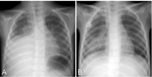
chest film. The patient had coughing for about 10 days, and had no improvement of symptoms despite treatment at a private clinic. A chest X-ray revealed a mass-like lesion occupying the entire right lower lung field (Fig. 1A). The patient was immediately transferred to Keimyung Univer- sity Dongsan Medical Center. On admission, she was pale with ill-looking appearance. Her body temperature was 37.4
℃, pulse rate was 80/min, and respiratory rate was 36/min.
Her body weight was 18 kg (10-25th percentile) and her height was 108 cm (3-10th percentile). Breathing sound was markedly decreased on right lower chest. There was no palpable mass on her abdomen. She had a history of treatment of hepatoblastoma diagnosed about four years prior to presentation. At that time, four cycles of preopera- tive chemotherapy consisting of cisplatin, 5-fluorouracil, and vincristine were administered and right lobectomy of the liver was performed. Alpha fetoprotein level was de- creased from over 65,000 ng/mL on admission to 6,810 ng/
mL after chemotherapy. The patient missed the final 2 cy- cles of chemotherapy. The patient returned to our hospital with another problem one year after the completion of the surgery, but again, the remaining two cycles of chemother- apy was refused. Alpha-fetoprotein was measured at that point and was found to be normal. The patient remained stable for the following 4 years, until cough developed and a large mass was identified in the right chest on standard
A case of recurrent hepatoblastoma : lung, heart and brain metastasis
Sun Mi Park, M.D., Byung Kyu Choe, M.D., Yeo Hyang Kim, M.D.
Heung Sik Kim, M.D., Tae Chan Kwon, M.D. and Hee Jung Lee, M.D.
*Department of Pediatrics and Radiology
*, Keimyung University School of Medicine, Daegu, Korea
Hepatoblastoma is a hepatic tumor predominantly occurring in children. The usual site of metastasis is the lung. There are only several reports worldwide on the distant metastasis of hepatoblastoma to the central nervous system in children. Only one reported case showed survival of a patient after multiple resections of a recurrent brain lesion. Involvement of the cardiovascular system has been reported in the medical literature. Lesions almost always involve the right-side of the heart. We re- port a case of recurrent hepatoblastoma at multiple sites, including brain, left atrium of the heart and lung in a 6-year-old girl who was partially treated in the past at the age of 1.5 years; the pa- tient had been event-free for four and a half years. (Korean J Pediatr 2006;49:691-695)
Key Words : Hepatoblastoma, Metastasis, Left atrium, Brain
X-ray evaluation. A recurrence of the hepatoblastoma was immediately suspected. A chest CT was performed and showed a well defined heterogeneous mass measuring 63×
72×49 mm in the right middle lobe of the lung with direct invasion into the left atrium through the right pulmonary artery (Fig. 2A, 2B). There was no abnormal lesion iden- tified in the liver. The Alpha fetoprotein was elevated over 65,000 ng/mL. A needle aspiration of the mass revealed an
epithelial histology consistent with the diagnosis of hepato- blastoma; which was different from the prior report that showed a mixed type of tumor (both epithelial and mesen- chymal components). The results of the whole body bone scan and the bone marrow aspiration were unremarkable.
Chemotherapy was started with CCG 8881B protocol con- sisting of cisplatin and continuous infusion of adriamycin.
An echocardiogram was performed and the left ventricular Fig. 1. Chest PA on admission revealed a large mass-like lesion in the lower half of
right lung (A). Follow up chest PA taken after 4th cycle of chemotherapy showed im- provement (B).
Fig. 2. Chest CT on admission showed 63×72×
49 mm sized well defined heterogeneous mass in right middle lung. Invasion of RPA and LA by tumor mass was seen (A, B). After 4th cycle of chemotherapy, pulmonary mass size was de- creased to 48×35×42 mm and intracardiac mass size also was decreased (C). Abbreviations : IVS, interventricular septum; LA, left atrium; LV, left ventricle; PV, pulmonary vein; RA, right atrium;
RPA, right pulmonary artery; RV, right ventricle;
RVOT, right ventricular outflow tract.
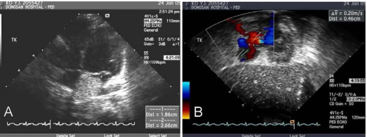
function was found to be normal. An atrial septal defect of 4.6 mm and a mass measuring 2.6×1.8 cm in the left atrium were identified (Fig. 3). Surgical biopsy of the in- tracardiac mass was not performed. At the 6th day of chemotherapy, the patient had 2 episodes of generalized tonic-clonic convulsions each lasting less than 1 minute. A brain CT performed immediately revealed multiple het- erogeneous lesions bilaterally in each hemisphere (Fig. 4A).
There were no further seizures, and the EEG remained normal. The patient underwent four cycles of chemotherapy with febrile neutropenia and mucositis but without serious complications. The serum alpha-fetoprotein gradually de- creased from over 60,500 ng/mL at the start of chemother- apy to 28,000 ng/mL at the completion of the 4th cycle.
Follow up chest PA (Fig. 1B) and chest CT (Fig. 2C) showed a decreased size of the mass in the lung mea-
suring 48×35×42 mm. A small residual mass but mark- edly decreased size measuring 1×1 cm, in the left atrium was observed in the follow up chest CT (Fig. 2C); echo- cardiogram also showed decreased size of the intracardiac mass. The brain CT, after the 4th cycle of chemotherapy, showed decreased size and number of multiple heterogene- ous metastases in both cerebral hemispheres (Fig. 4B). Ac- cording to the treatment protocol, surgical resection of the lung mass was the next step. However, at that point, the legal guardian of the child declined further treatment. The patient remained alive in fair condition 6 months after the interruption of the treatment.
Discussion
The usual site of metastasis of hepatoblastoma is the Fig. 4. Brain CT on 6th day of the chemotherapy showed multiple, variable sized heterogeneous, highly enhancing masses in the gray-white matter junction sites of both cerebral hemispheres (A). Follow up brain CT after 4th cycle of chemotherapy showed decreased size and numbers of multiple variable sized hemorrhagic metastases in both cerebral hemispheres (B).
Fig. 3. Echocardiogram on admission showed left atrial mass of 2.6×1.8 cm (A) and atrial septal defect
of 4.6 mm (B).
lung. Lung metastases identified on diagnosis is one of the most reliable prognostic indicators of an unfavorable out- come for patients diagnosed with hepatoblastoma
2). Extra- pulmonary metastatic sites in patients with hepatoblastoma, including brain as well as abdomen and bone, are uncom- mon. The prognosis for children with hepatoblastoma with distant metastasis is poor at diagnosis and even worse with relapse
3). In cases with multiple site metastases, sur- gical treatment of pulmonary lesions has not been effective
4)