87 Corresponding Author :
, Paralichthys olivaceus Philasterides
dicentrarchi
The Pathogenicity of Scuticocilate Philasterides dicentrarchi Isolat- ed from Cultured Olive Flounder, Paralichthys olivaceus
Chang-Nam Jin , Hyun-Sil Kang, Chang-Hoon Lee , Sun kyoung Kang, Young-Don Lee, Jehee Lee and Moon-Soo Heo
Jeju Regional Maritime Affairs and Fisheries Office, Jeju 690-704 Jeju Fisheries Resource Institute NFRKE, Jeju 690-102 Faculty of Marine Science, Cheju National University, Jeju 690-756
In previous study, scuticociliate P. dicentrachi was isolated from farmed flounder during the december 2004 to april 2005. Pathogenicity, mortality and infection symptom were studied using 3 cm and 5 cm groups of juvenile flounder.
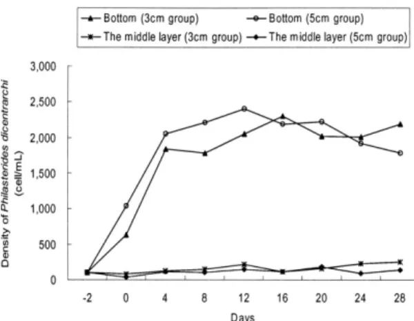
The ciliates were exponentially increased from bottom layer of the experimental tanks, which propagated within 2000 cells/ml (1.4 103cell -1~ 2.5 103cell -1) after 6 days of inoculation. The middle layer was maintained within 300 cells/ml.
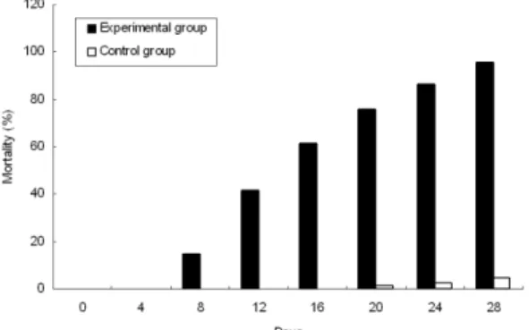
Both 3 cm and 5 cm groups were infected with ciliates, mainly 3 cm group showed high mortality during the experimental period. The death of 3 cm group was started from 5 days and showed 95.6% of mortality after 28 days of first inoculation. The control group showed 4.4% of mortality however, we could not observed any ciliates.
The death of 5 cm group was started later than 3 cm group after 18 days of first inoculation. The total mortality was 71% during 28 days. No mortality and infection symptoms were observed in the control.
We also studied SSU rRNA gene of ciliates which, re-isolated from infected flounder of experimental groups. When SSU rRNA in this study compared with previous data showed that the identified strain of both previous and present study was same.
Key words: Philasterides dicentrarchi, Scuticociliate, Paralichthys olivaceus.
(Ototake and Mat- susato, 1986; Mizuno, 1993; Lee et al., 2001),
(Lee et al., 2001; Jin et al., 2003).
, ,
(Mizuno, 1993;
Lee et al., 2001; Jin et al., 2003).
southern bluefin
tuna, , ,
Corresponding Author : Moon-Soo Heo, Tel : 064-754-3473, Fax : 064-756-3493, E-mail : [email protected]
, Uronema (Cheung et al.
1980, Bassleer 1983, Gill and Callinan 1997, Mun- day et al. 1997) Miamiensis (Thompson and Moewus 1964), Tetrahymena (Ferguson et al.
1987), Philasterides (Dragesco et al. 1995, Igle-
sias et al. 2001) .
Philasterides dicentrarchi , ,
(Iglesias et al., 2001),
(Kang et al., 2005).
,
. ,
Pseudocohnilembus persalinus, Miamiensis avidus, Uronema marinum, P. dicentrarchi
(Jee et al., 2001; Kim et al., 2001; Kim et al., 2004; Jung et al., 2005).
.
P.
dicentrarchi
.
P. dicentrarchi .
Philasterides dicentrarchi
(14 ) P. dicentrarchi
.
. 3
( 3.2 , : 2.7 3.6 ) 5 ( 5.4 , : 4.5 6.4 )
.
Fig. 1 43
63 25 4 FRP
. 43 43
25 , 43 20 25
, 20 (Fig. 1).
37 L, 50 L
, 80 20
.
6 8 , 4 6
Fig. 1. Schematic diagram of experimental tank
, .
1.5 .
3 5
. 5
100 , 3 160
.
5
1/2 , 3 1/3 .
P. dicentrarchi
P. dicentrarchi
, cell culture
flask (20 ) 20 H
0.5 0.6 (Table 1) 1
mg/ml, penicillin streptomycin 100 IU
. CHSE-214 (chinook
salmon embryo)
3.0 103cell/ml 15 ,
7 9.8 104cell/ml
.
5 ,
2 g .
ml 100 cell
3.7 106cell
. 2
, 2 0.2
g
.
2 10%
.
6 pipetting counting chamber
glutaraldehyde 1%
(18 18 ) (Olympus BX 50, 100) .
100 pipetting
counting chamber glutaralde-
hyde 1%
(Olympus BX 50, 100) .
. Bouin's
5 Haematoxylin-
Eosin .
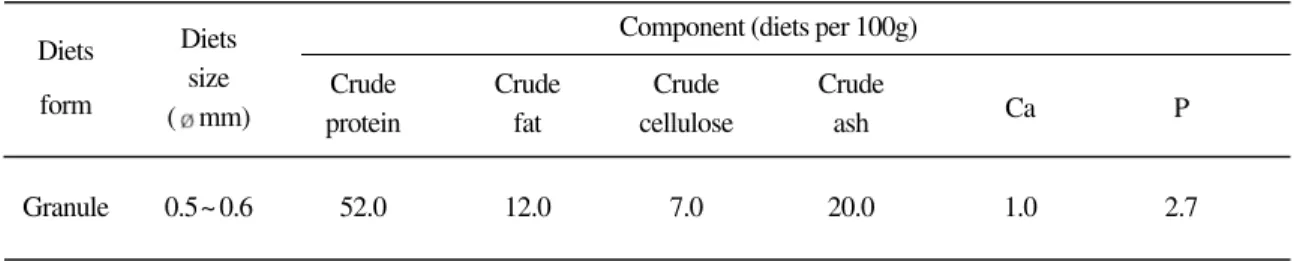
Table 1. Composition of the experimental diets
Diets Diets Component (diets per 100g)
form size Crude Crude Crude Crude
( mm) protein fat cellulose ash Ca P
Granule 0.5~ 0.6 52.0 12.0 7.0 20.0 1.0 2.7
(Unit : g)
.
70% 10
washing .
, genomic DNA
CHSE-214 15 , 5
.
1.5 ml 1,500 rpm, 10
pellet .
pellet proteinase K lysis buffer
genomic DNA (Qiagen, Germany).
small subunit
(SSU) rRNA Scu18F 5'-
AACCTGGTTGATCYTGCCAGTA-3', Scu18R 5'-GATCYWTCTGCAGGTTCACCTAC-3'
, PCR 100 ng genomic DNA, dNTPs, 10 Ex taq polymerase buffer, 0.5 unit Ex taq polymerase (Takara, Japan)
50 Takara
PCR thermal cycler (Takara, Japan)
. SSU rRNA 94 , 3
predenaturation, 94 30 denaturation,
55 35 annealing, 72 2
extension 30 , 72 5
extension . PCR
1% agarose gel
. PCR pBluescript II
SK(-) plasmid DNA
( ) SSU rRNA
.
4
.
.
, 2
TSA Agar, BHI Agar 25 , 24
.
. 2
6.3 102cell/
1.0 103cell/
, 6 (2 )
1.4 103cell/ 2.5 103cell/
2,000 cell .
100 200 cell (Fig. 2), .
Fig. 2. Ciliate density at the bottom layer and middle layer of experimental tank according to incubation days.
3 P. dicentrarchi . 3 cm
4 (1 )
5 2
(5 8 ) 15%
. 3 (9 12 )
40%
(Fig. 3). 28 95.6%
. 5
, 14
(Fig. 9).
3 cm 28
4.4%
.
5 3
16 (4 ) 5
(17 20 ) .
3
3 28 (4 )
71% 3
(Fig. 4).
.
Fig. 3. Accumulative mortality on experimental infection and control groups of 3 cm flounder by P. dicentrarchi.
Fig. 4. Accumulative mortality on experimental infection of 5 cm flounder by P. dicentrarchi.
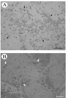
Fig. 5. A number of ciliates are infected to fin of flounder. (A) Photograph of ciliates (arrows) infiltrated into the fin ray of dorsal fin in flounder. Scale bar = 100 . (B) Light micrograph of the caudal fin in the infected flounder showing numerous ciliates (arrows) that have invaded this fin tissue. Note that the areas of severe tissue necrosis surrounding the invading cili- ates, Fm: fin membrane, Nf: necrosis fin membrane. HE staining. Scale bar = 50 .
3 5 .
(Fig. 5), (Fig. 6), (Fig. 7), (Fig. 8), (Fig.
9) . 3
(Fig.
10A). 5
, ,
(Fig. 10B).
Fig. 6. Light micrograph of the muscle in the infected floun- der showing ciliates (arrows) that have invaded this muscle tissue. Note that the areas of severe tissue lysis surrounding the invading ciliates, M: muscle. HE staining. Scale bar = 50
.
Fig. 9. Photograph of scuticociliates, P. dicentrarchi (arrows) are infected to the brain of flounder. (A) Micro- scopic observation, (B) Light microscopic observation, HE staining. Scale bar = 100 .
Fig. 7. Light micrograph of the gill in the infected flounder showing ciliates (arrows) that have invaded into the gill fila- ment tissue. HE staining. Scale bar = 50 .
Fig. 8. Light micrograph of eye in the infected flounder showing ciliates (arrows) that have invaded into the vitreous cavity of eye tissue, Eb; eye ball. HE staining. Scale bar = 50
.
ribosome SSU rRNA
.
3 cm 5 cm
genomic DNA SSU
rRNA PCR 1760
bp .
,
SSU rRNA PCR forward
reverse primer 1759 bp
. SSU rRNA
SSU rRNA ( )
99.8 100% identity
. ,
.
, , Vibrio ichthyoenteri Vibrio. sp.
.
1986 Ototake Matsusato
(Mizuno, 1993; Lee et al., 2001), 1990
(Lee et al. 2001). Jin (2003)
7 53.8%
87.5%
. P. dicentrarchi
,
100% (Iglesias et al.,
2001). , , ,
(Paramá et al., 2003)
.
,
P. dicen- trarchi Kim (2004)
. P. dicentrarchi
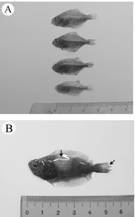
Fig. 10. External features of infected flounder infected with ciliates. (A) The flounder of 3 cm in size. (B) The flounder of 5 cm in size.
P.
dicentrarchi .
6 2,000 cell
300 cell
. Chi (1997)
7.3 102cell/ , 4.1 cell/
. in vitro
105cell/ ,
.
2,000 cell
. 5
3
. 5
3
4.4% .
3 5%
.
3
. 5
. 3
5
.
, ,
Vibrio ichthyoenteri Vibrio sp.
,
. Jin (2003) 2
2 .
P. dicentrarchi
(Iglesias et al., 2003)
. P. dicentrarchi
2
. scuticociliate
, taxa
(Shang et al., 2003).
SSU rRNA .
SSU
rRNA ,
.
2004 12 2005 4
P. dicentrarchi
3 5
.
P. dicentrarchi
6 1.4 103cell/ 2.5
103cell/ 2,000 cell
. 300 cell
. P. dicentrarchi
3 5
,
3 .
5 , 28
95.6% .
4.4%
.
5 18
3 .
28 71% 3
. 5
.
SSU rRNA
, P. dicentrachi
SSU rRNA
.
. ,
2005 Brain
Korea 21 .
Bassleer, G.: Uronema marinum, a new and com-
mon parasite on tropical salt-water fishes.
Freshw. Mar. Aquar., 6: 78-79, 1983.
Cheung, P. J., Nigrelli, R. F. and Ruggieri, G. D.:
Studies on the morphology of Uronema marinum Dujardin (Ciliatea: Uronematidae) with a description of the histopathology of the infection in marine fishes. J. Fish Dis., 3:
295-303, 1980.
Choi, S. D., Kim, J. M., Kim, S. Y., Jo, Y. C., Choi, K. K. and Yang, H. C.: Study on distribution and extermination of scuticociliatids para- sitizing to Japanese flounder, Paralichthys olivaceus in southern Korea. J. Fish Pathol., 10(1): 21-29, 1997.
Dragesco, A., Dragesco, J., Coste F., Gasc, C., Romestand, B., Raymond, J. C. and Bouix G.: Philasterides dicentrarchi, n. sp. (Cilio- phora, Scuticociliatida), a histophagous opportunistic parasite of Dicentrarchus labrax (Linnaeus, 1758), a reared marine fish. Europ. J. Protistol., 31: 327 340, 1995.
Ferugson, H. W., Hicks, B. D., Lynn, D. H. and Ost- land, V. E.: Cranial ulceration in Atlantic salmon Salmo salar associated with Tetrahy- mena sp. Dis. Aquat. Org., 2: 191-195, 1987 Gill, P. A. and Callinan, R. B.: Ulcerative dermatitis
associated with Uronema sp. infection of farmed sand whiting sillago ciliata. Aust.
Vet. J., 75: 357, 1997.
Iglesias, R., Paramá, A., Alvarez, M. F., Leiro, J., Aja, C. and Sanmartín, M. L.: In vitro growth requirements for the fish pathogen Philasterides dicentrarchi (Ciliophora, Scu- ticociliatida). Vet. Parasitol., 111: 19-30, 2003.
Iglesias, R., Paramá, A., Alvarez, M. F., Leiro, J., Fernández, J. and Sanmartín, M. L.: Philas- terides dicentrarchi (Ciliophora, Scuticocili-
atida) as the causative agent of scuticocil- iatosis in farmed turbot Scophthalmus max- imus in Galicia (NW Spain). Dis. Aquat.
Org., 46: 47-55, 2001.
Jee, B. Y., Kim, Y. C. and Park, M. S.: Morphology and biology of parasite responsible for scu- ticociliatosis of cultured olive flounder Par- alichthys olivaceus. Dis. Aquat. Org., 47:
49-55, 2001.
Jin, C. N., Lee, C. H., Oh, S. P., Jung, Y. U., Song, C. B., Lee, J. and Heo, M. S.: Scuticociliato- sis in Flounder Farms of Jeju Island. J. Fish Pathol., 16(2): 135-138, 2003.
Jin, C. N., Lee, C. H., Oh, S. P., Na, O. S. and Heo, M. S.: Infection Route of Scuticociliates in the Juvenile of the cultured Flounder, Par- alichthys olivaceus. J. Fish Pathol., 16(1):
13-21, 2003.
Jung, S. J., Kitamura, S. I., Song, J. Y., Joung, I. Y.
and Oh M. J.: Complete small subunet rRNA gene sequence of the scuticociliate Miamiensis avidus pathogenic to olive flounder Paralichthys olivaceus. Dis. Aquat.
Org., 64: 159-162, 2005.
Kang, B. S., Go, H. B., Kim, S. J. Na, O. S., Lee, C.
H., Kim, S. Y., Lee, J. and Lee, Y. D.: Exter- nal Symptoms of Tiger Puffer, Takifugu rubripes Infected with Scuticociliates and Distribution of the Scuticociliates in the Skin, Gill and Blood Vessel. J. Fish Pathol., 18(1): 29-37, 2005.
Kim, S. M., Cho, J. B., Kim, S. K., Nam, Y. K. and Kim, K. H.: Occurrence of scuticociliatosis in olive flounder Paralichthys olivaceus by Philasterides dicentrarchi (Ciliophora: Scu- ticociliatida). Dis. Aquat. Org., 62: 233-238, 2004.
Kim, S. M., Cho, J. B., Lee, E. H., Kwon, S. R., Kim, S. K., Nam Y. K. and Kim, K. H.:
Pseudocohnilembus persalinus (Ciliophora:
Scuticociliatida) is an additional species causing scuticociliatosis in olive flounder Paralichthys olivaceus. Dis. Aquat. Org., 62:
239-244, 2004.
Lee, C. H., Kang, Y. J., Ha, D. S. and Lee, Y. D.:
Occurrence and Histopathological Observa- tion of Scuticociliatosis in the Cultured Olive Flounder, Paralichthys Olivaceus.
Bull. Natl. Fish. Res. Dev. Inst. Korea., 59:
68-73, 2001.
Mizuno, Y.: Control methods of diseased Japanese flounder, Paralichthy olivaceus, used in fish farm in Japan. J. Fish Pathol., 6: 219 231, 1993.
Munday, B. L., O'Donoghue, P. J., Watts, M., Rough, K. and Hawkesford, T.: Fetal encephalitis due to the scuticociliate Urone- ma nigricans in sea-cage, sounthern bluefin tuna Thunnus maccoyii. Dis. Aquat. Org., 30: 17-25, 1997.
Ototake, M. and Matsusato, T.: Notes on Scuticocil- iata infection of cultured juvenile flounder Paralichthys olivaceus. Bull. Natl. Res.
Aquacult., 9: 65-68, 1986.
Paramá, A., Iglesias, R., Álvarez, M.F., Leiro, J., Aja C. and Sanmartín M. L.: Philasterides dicentrarchi (Ciliophora, Scuticociliatida):
experimental infection and possible routes of entry in farmed turbot (Scophthalmus max- imus). Aquaculture 217: 73-80, 2003.
Shang, H., Song, W. and Warren, A.: Phylogenetic positions of two ciliates, Paranophrys magna and Mesanophys carcini (Ciliophora:
Oligohemenophorea), within the subclass scuticociliatia inferred from complete small subunit rRNA gene sequences. Acta Proto- zool. 42; 171-181, 2003.
Thompson, J. C. and Moewus, L.: Miamiensis
avidus n, g., n. sp., a Marine Facultative Par- asite in the Ciliate Order Hymenostomatida.
J. Protozool., 11(3): 378-381, 1964.
Manuscript Received : March 14, 2006 Revision Accepted : May 5, 2006 Responsible Editorial Member : Ki-Hong Kim (Pukyong Univ.)