Tuberc Respir Dis 2012;72:77-81
CopyrightⒸ2012. The Korean Academy of Tuberculosis and Respiratory Diseases. All rights reserved.
폐결핵으로 오인된 폐분아균증 1예
성균관대학교 의과대학 삼성서울병원
1내과학교실,
2흉부외과학교실,
3병리학교실
전병우1, 김다민1, 박지현1, 유홍석1, 심훈보2, 김진국2, 한정호3, 권오정1A Case of Pulmonary Blastomycosis Mimicking Pulmonary Tuberculosis
Byung Woo Jhun, M.D.
1, Da Min Kim, M.D.
1, Ji Hyeon Park, M.D.
1, Hong Seok Yoo, M.D.
1, Hunbo Shim, M.D.
2, Jhin Gook Kim, M.D.
2, Joungho Han, M.D., Ph.D.
3, O Jung Kwon, M.D., Ph.D.
1Departments of
1Medicine,
2Thoracic and Cardiovascular Surgery, and
3Pathology, Samsung Medical Center, Sungkyunkwan University School of Medicine, Seoul, Korea
Blastomyces dermatitidis is a dimorphic fungus that causes the systemic pyogranulomatous disease known as blastomycosis. Blastomycosis most often involves the lungs, skin, and may involve nearly every organ in the body.
It is difficult, however, to diagnose blastomycosis in the early stage of pulmonary disease because clinical manifestations are varied from subclinical infection to acute respiratory distress syndrome. Since blastomycosis is often accompanied by granulomatous inflammation in histopathologic findings, differentiation from other etiologic diseases is important. We report a case of a 45-year-old male with pulmonary blastomycosis who had been misdiagnosed with tuberculosis for 3 months.
Key Words: Blastomycosis; Granuloma; Tuberculosis
Address for correspondence: O Jung Kwon, M.D., Ph.D.
Division of Pulmonary and Critical Care Medicine, Depart- ment of Medicine, Samsung Medical Center, Sungkyunkwan University School of Medicine, 50, Irwon-dong, Kangnam- gu, Seoul 135-710, Korea
Phone: 82-2-3410-3421, Fax: 82-2-3410-6956 E-mail: [email protected]
Received: Aug. 12, 2011 Revised: Aug. 25, 2011 Accepted: Sep. 7, 2011
서 론
분아균증(blastomycosis)은
Blastomyces dermatitidis
에 의한 전신성 농성 육아종성 질환으로 폐감염증을 비롯 한 전신적인 감염증의 형태로 나타날 수 있다1. 임상증상 이 비특이적이고 방사선학적 소견이 다양하게 나타나기 때문에 초기에 진단이 쉽지 않고, 조직병리학적으로 흔히 만성 육아종성 염증을 동반하기 때문에 다른 병인과의 감 별이 중요하다2,3. 저자들은 육아종성 염증의 침윤성 폐병 변을 보이는 환자가 폐결핵으로 오인되어 3개월 동안 항결핵제를 복용하던 중, 조직병리 검사 및 DNA 염기서열 분석을 통해 폐분아균증으로 진단된 1예를 경험하였기에 보고하는 바이다.
증 례
환 자: 채○○, 남자, 45세
주 소: 기침, 가래, 흉벽의 고름 양상 분비물 현병력: 환자는 내원 3개월 전 기침으로 3차병원을 방 문하였고, 흉부 전산화 단층촬영에서 우폐상엽에 4 cm 크 기의 종괴양상의 침윤 소견이 보였다. 경피적 세침흡인술 에서 만성 육아종성 염증 소견이 관찰되어, 폐결핵 의증 하에 1차 항결핵제(isoniazid, rifampin, ethambutol, pyr- azinamide) 복용을 시작하였다. 항결핵제 복용 1개월 후 에도 증상의 호전이 없고 약물로 인한 간독성이 의심되어 다른 3차병원을 방문하였고, 재시행한 경피적 세침흡인술 에서도 만성 육아종성 염증 소견이 확인되어 isoniazid, rifampin, ethambutol, levofloxacin으로 약제조정 후 항결
Figure 1. Posteroanterior chest radiograph showed air- space consolidation with irregular margin in the right up- per lung zone.
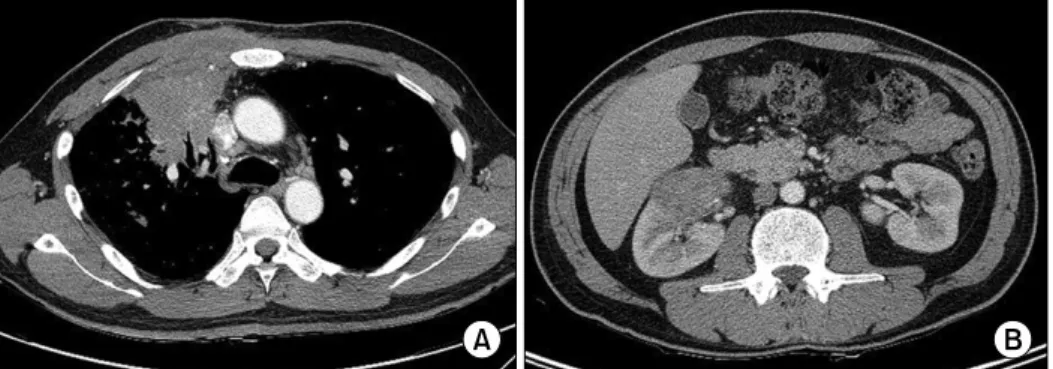
Figure 2. (A) Chest computed tomography revealed a low attenuation lesion communicating with the right anterior chest wall, suggestive of empyema necessitatis formation. (B) In the right kidney area a 5 cm size wedge shaped low attenuated lesion was observed.
핵제 복용을 지속하였다. 항결핵제 복용 2개월째, 경피적 세침흡인술 시행부위에 고름 양상의 분비물이 발생하여 2차 항결핵제(streptomycin, cycloserine, para-amino- salicylic acid, prothionamide, levofloxacin)로 변경하여 치료받았으나 증상이 지속되어 본원으로 전원되었다. 그 동안 두 곳의 3차병원에서 객담과 흉벽분비물로 시행한 항산균 염색과 결핵균 및 세균 배양 검사는 모두 음성이었 다.
과거력: 23년 전 폐결핵으로 치료받은 병력이 있었다.
사회력: 환자는 26갑년의 과거흡연가였다. 증권업에 종 사하였고, 내원 4년 전부터 2년 동안 미국 Tennessee주에 거주했던 적이 있었다.
가족력: 특이사항은 없었다.
이학적 소견: 입원 당시 혈압 120/82 mm Hg, 맥박 99 회/분, 호흡수 20회/분, 체온 36.4oC였고, 산소 투여 없이 측정한 산소포화도는 97%였다. 급성 병색의 소견은 없었 고 의식은 명료한 상태였다. 두경부 진찰에서 오른쪽 이 마부위에 1 cm 크기의 결절모양의 피부병변이 있었다. 흉 부 청진상 심잡음이나 수포음은 들리지 않았고, 우상부 전면의 경피적 세침흡인 검사부위에 고름 양상의 분비물 이 배출되고 있었다. 복부진찰과 사지진찰에서 특이 소견 은 없었다.
검사실 소견: 입원 당시 말초혈액 검사에서 백혈구 13,650/μL (호중구 71.0%, 호산구 3.4%), 혈색소 12.0 g/dL, 혈소판 451,000/μL, 적혈구 침강속도 120 mm/hr, C-반응 단백은 6.8 mg/dL였고, 혈액화학 검사에서 총단백 6.7 g/dL, 알부민 3.6 g/dL였고 간기능과 신장기능에 이상 소견은 없었다. 객담과 흉벽분비물로 시행한 항산균 염색, 배양 검사 및 결핵에 대한 중합효소 연쇄반응 검사 모두 음성이었으며 그람 염색과 배양 검사 결과도 음성이었다.
Aspergillus galactomannan 항원 검사 결과 음성이었고 Anti-HIV 항체 검사 결과도 음성이었다.
방사선 검사: 단순 흉부촬영에서 우측 폐상엽에 경계가 불분명한 기강경화(air-space consolidation) 소견이 관찰 되었다(Figure 1). 흉부 컴퓨터 단층촬영에서 우폐상엽에 종괴양상의 침윤 소견과 주변에 다발성 결절이 관찰되었 고, 우중엽과 우하엽 상분절에 다발성 결절 소견이 관찰되 었다. 흉벽의 우상부 전면에는 흉벽천공성 농흉(empye- ma necessitates)을 시사하는 피하조직과 연결되는 경계 가 분명한 저음영 병변 소견이 관찰되었고(Figure 2A), 우 측 신장부위에는 쐐기모양의 경계가 분명한 저음영의 병
Figure 3. Microscopic findings of video-assisted thor- acopscopic surgery biopsy showed granulomatous in- flammation with a multinucleated giant cell (hematoxylin and eosin stain, ×400).
Figure 4. (A) Yeast form fungal organisms with a thick cell wall and a single broad-based bud were stained with Gomori methenamine silver stain (Gomori methenamine silver stain, ×400). (B) No highlighting organism was identified with muci- carmine stain (Mucicarmine, ×400).
Figure 5. Posteroanterior chest radiograph indicated an improved consolidative lesion in the right lung zone after right upper lobectomy and 2 months of itraconazole treatment.
변 소견이 관찰되었다(Figure 2B). 이 외 종격동이나 폐문 부에 병적으로 커져있는 림프절은 없었다.
기관지내시경 검사: 기관지 내 협착이나 종괴의 소견은 보이지 않았고, 우상엽 전분절에서 기관지세척 검사를 시 행하였다. 기관지세척액으로 시행한 항산균 염색과 배양 검사 결과는 음성이었고 그람 염색과 배양 검사 결과도 음성이었다. Sabouraud dextrose agar 배지로 시행한 진 균 배양 검사에서는 원인균을 확인할 수 없었다. 거대세 포 바이러스에 대한 배양 검사는 음성이었고, 아데노 바이 러스, 인플루엔자 바이러스, 파라인플루엔자 바이러스, 호 흡기세포융합 바이러스에 대한 중합효소 연쇄반응 검사 도 음성이었다.
치료 및 경과: 환자는 본원에서 시행한 항산균 염색 검
사에서 음성이었지만, 타 병원에서 3개월 동안 항결핵제 를 복용하고 있었고 두 번의 경피적 세침흡인 검사에서 모두 만성 육아종성 염증이 확인되어, 항결핵제를 지속적 으로 투여하였다. 환자는 흉벽천공성 농흉이 있었고 3개 월간의 결핵치료에도 불구하고 임상증상이 좋아지지 않 았기 때문에 우상엽절제술, 우중엽 쐐기절제술 및 농흉부 위 괴사조직 제거술을 시행하였다. 조직병리 검사에서, 다핵거대세포를 동반한 만성 육아종성 염증 소견이 보였 고, 건락괴사 소견은 관찰되지 않았다(Figure 3). Gomori
methenamine sliver 염색에서 효모형태의 진균이 관찰되 었고 Mucicarmine 염색에서는 특이 소견이 없었다(Figure 4). 이후 Internal transcribed spacer (ITS) 및 D1/D2 ribo- somal DNA에 대한 염기서열분석을 시행하였고
Blasto- myces dermatitidis
에 의한 폐분아균증으로 진단하였다.또한 오른쪽 이마부위의 결절모양의 피부병변에서도 조 직 검사를 시행하였고 육아종성 염증 소견과 효모형태의 진균이 확인되었다. 컴퓨터 단층촬영상에서 관찰되었던 우측 신장의 쐐기모양의 저음영 병변의 소견은 blastomy- cosis의 신장 침범 소견으로 판단하였다. 수술 후, 하루 itraconazole 400 mg을 경구투약 하기 시작했고 임상적으 로 호전되어 퇴원하였다. 이후 치료 2개월 뒤에 시행한 단순 흉부촬영에서 호전된 소견을 확인하였고(Figure 5) 지속적으로 외래 추적관찰 중이다.
고 찰
분아균증(blastomycosis)은
Blastomyces dermatitidis
에 의한 전신성 농성 육아종성 질환으로, 1894년 Gilchrist 에 의해 처음 보고되었다. 초기에는 원생동물이 원인일 것으로 여겨졌으나 후에 Stokes 등에 의해 진균감염에 의 한 것으로 확인되었고, 일차적인 폐감염 후에 이차적인 피부, 비뇨생식계, 중추신경계 등의 전신적인 감염을 일으 키는 것으로 알려져 있다4,5. 대개 강이나 습기가 많은 지 역에서 발생률이 높고, 진균에 오염된 토양이나 부패된 식물에의 노출과 관련성이 높으며, 북아메리카지역이 유 행지역으로 알려져 있다6. 국내에서의 분아균증은 주로 골이나 피부를 침범하는 증례가 드물게 보고되고 있으나7,8 폐분아균증의 사례는 극히 드물고 비유행지역 여행 후에 폐증상을 보이는 1예만 보고된 상태이다9.폐분아균증은 급성과 만성 혹은 재발성 폐렴의 형태로 나타날 수 있다. 감염된 환자의 절반 정도에서는 급성 폐 렴의 형태로 나타나는데 발열, 기침, 가래 등의 비전형적 인 증상을 보이기 때문에 바이러스나 박테리아에 의한 폐 렴과 임상적으로 감별이 어렵다1. 그리고 만성 혹은 재발 성 폐렴의 형태로도 나타나는데10, 지속되는 호흡기증상과 함께 폐외감염증(extrapulmonary disease)을 동반하는 경 우가 많다. 방사선학적 소견 또한 다양하게 나타나며 흔 히 종괴나 결절양상으로 나타나기 때문에 종종 악성 질환 으로 오인되기도 한다2,5.
본 증례는 분아균증 유행지역에 2년간 거주했던 환자에 서 폐분아균증과 피부와 신장을 침범한 폐외감염증이 동
반된 경우로, 환자의 과거력에 대한 충분한 조사와 이학적 검사가 진단에 있어서 중요한 역할을 할 수 있음을 확인할 수 있었다. 또한 두 곳의 3차병원에서 시행한 세침흡인 검사 시술부위에 흉벽천공성 농흉이 발생하였고 항결핵 제에 의한 간독성의 합병증이 발생했다는 점을 볼 때, 정 확한 진단의 중요성을 보여주는 예라고 하겠다.
폐분아균증은 조직병리학적으로 흔히 육아종성 염증 소견이 동반되는데 진균 감염 이외에도 마이코박테리아 감염, 과민성 폐렴, 유육종증 등의 다양한 질환에서도 나 타날 수 있어 감별진단에 주의를 요하게 된다3. 증례의 환 자에서 진단이 늦어진 주된 원인으로는 이와 같은 육아종 성 염증의 병변을 보이는 환자에서 감별해야 할 질환들에 대한 접근이 부족했던 점과, 결핵치료로도 경과가 좋아지 지 않았는데 좀 더 적극적인 검사를 시행하지 않았다는 점이다. 조직병리 검사에서 진균의 확인은 염색법을 이용 하게 되고, 주로 Gomori methenamine silver염색, peri- odic acid-schiff염색이 이용된다11. 특히 Mucicarmine염 색에서는
Blastomyces dermatitidis
가 거의 염색되지 않기 때문에Cryptococcus neoformans
등의 다른 진균을 감별 하는데 도움을 줄 수 있다. 증례의 경우에서는 Gomori methenamine silver염색에서는 염색이 되지만, Mucicar- mine염색에서는 염색되지 않는 효모양상의 진균을 확인 하여 blastomycosis에 의한 감염증을 추정할 수 있었고, 추가적인 DNA 염기서열분석법을 사용하여Blastomyces dermatitidis
를 확인할 수 있었다12.분아균증의 주된 치료로는 항진균제를 이용한 약물치 료로, 생명을 위협하는 정도의 심한 감염증이나 중추신경 계감염증에는 amphotericin계열의 항진균제를 사용하고, 이외 중등도 이하의 감염증의 경우에는 azole계열의 항진 균제를 사용하게 된다. Itraconazole이 우선적으로 권고되 는데, 대개 하루 200∼400 mg을 6개월 정도 사용한다13. 증례의 환자는 동반된 다른 악성 질환이 없고 정상면역기 능을 가진 성인이었기 때문에, 병변부위 엽절제술 및 농흉 부위 괴사조직 제거술을 받은 후 하루 itraconazole 400 mg을 경구투약 하였고 임상적으로 호전되어 외래 추적관 찰 중이다.
참 고 문 헌
1. Baumgardner DJ, Halsmer SE, Egan G. Symptoms of pulmonary blastomycosis: northern Wisconsin, United States. Wilderness Environ Med 2004;15:250-6.
2. Sheflin JR, Campbell JA, Thompson GP. Pulmonary blastomycosis: findings on chest radiographs in 63 patients. AJR Am J Roentgenol 1990;154:1177-80.
3. Mukhopadhyay S. Role of histology in the diagnosis of infectious causes of granulomatous lung disease. Curr Opin Pulm Med 2011;17:189-96.
4. Gilchrist TC, Stokes WR. A case of pseudo-lupus vulga- ris caused by a blastomyces. J Exp Med 1898;3:53-78.
5. Smith JA, Kauffman CA. Blastomycosis. Proc Am Thorac Soc 2010;7:173-80.
6. Dworkin MS, Duckro AN, Proia L, Semel JD, Huhn G.
The epidemiology of blastomycosis in Illinois and fac- tors associated with death. Clin Infect Dis 2005;41:
e107-11.
7. Koh JK. Clinicohistopathologic findings and their differ- ential diagnoses of pathogenic fungal infections of cu- taneoua deep mycoses. Korean J Med Mycol 1997;2:
101-9.
8. Cho JH, Suh JS, Kim JH. Systemic blastomycosis with osseous involvement of the foot: a case report. J
Korean Foot Ankle Soc 2005;9:216-9.
9. Seo CG, Seo YW, Park HP, Choi WI, Beom HS, Kwon KY, et al. A case of blastomycosis after traveling around non-endemic area. Tuberc Respir Dis 2005;58:
619-23.
10. Pappas PG. Blastomycosis. Semin Respir Crit Care Med 2004;25:113-21.
11. Lee YB. Studies on the systemic mycosis in Korea and the special stainings for fungi. Korean J Med 1964;7:
523-43.
12. Bialek R, Cirera AC, Herrmann T, Aepinus C, Shearn- Bochsler VI, Legendre AM. Nested PCR assays for de- tection of Blastomyces dermatitidis DNA in paraffin- embedded canine tissue. J Clin Microbiol 2003;41:
205-8.
13. Chapman SW, Dismukes WE, Proia LA, Bradsher RW, Pappas PG, Threlkeld MG, et al. Clinical practice guidelines for the management of blastomycosis: 2008 update by the Infectious Diseases Society of America.
Clin Infect Dis 2008;46:1801-12.