∙Received: October 10, 2013. Accepted: November 2, 2013.
∙Corresponding author : Dong Chan Han
Department of Nuclear Medicine, Samsung Medical Center, 50 Il won-dong, Gangnam-gu, Seoul 135-701, Korea
Tel: +82-2-3410-6286, Mobile: +82-2-3410-2639 E-mail: [email protected]
Original Article PET/CT 검사에서 열소 주변 인공물 감소를 위한 DFOV
위치 변화 재구성 방법의 유용성 평가
삼성서울병원 핵의학과
한동찬⋅홍건철⋅최춘기⋅이 혁⋅최성욱
The Effectiveness Evaluation of Reconstruction Method Using DFOV Position Changes for Reduction of Artifact Around Hotspot in PET/CT Images
Dong Chan Han, Gun Chul Hong, Choon ki Choi, Hyeok Lee and Seong Wook Choi Department of Nuclear Medicine, Samsung Seoul Medical Center, Seoul, Korea
Purpose: In the PET/CT images, various artifacts cause degradation of the quantitative assessment. Most hotspot generated by radiopharmaceutical injection errors cause an artifact and degrade the quality of the images as well as the accuracy of the quantitative evaluation. The purpose of this study is to assess effectiveness of the elimination of the hotspot at the injection sites using shifting the center of DFOV (Display Field of View, DFOV) method and evaluate the quantitative evaluation of result. Materials and Methods: GE Discovery STE 16 (GE Healthcare, Milwaukee, USA) and 1994 NEMA phantom were used for imaging acquisition. Phantom was filled with 0.005 MBq/mL of
18F-FDG. A hotspot was artificially placed on the outside of the phantom. The ratio of hotspot area activity to background area activity was regulated as 200:1. After image acquisition with routine protocol, all of the images were reconstructed using the shifting the center of DFOV method that wasn't overlapped with hotspot. Those images obtained before and after applying the shifting reconstruction method were compared. ROIs (Region Of Interests) were set in the hotspot areas, meanSUVs and standard deviations were calculated. Percentage differences were calculated with those meanSUVs and standard deviations. The evaluation on the effects of the shifting reconstruction method was done by comparison of the meanSUVs and the standard deviations, which were calculated for background areas unaffected by hotspot. Results: In the areas of unaffected by hotspot, meanSUVs before and after applying the shifting of center of DFOV method were 0.67±0.06 g/mL and 0.65±0.06 g/mL, respectively. In the artifact areas affected by hotspot, meanSUVs before and after applying the shifting of center of DFOV method were 0.32±0.08 g/mL and 0.56±0.12 g/mL, respectively. The percentage differences of the area adjacent to the hotspot and the area distant from the hotspot were 65.3% and 97.4%, respectively. Conclusion: In the PET/CT images, meanSUV was improved by 32.1%
when the effect of artifact was removed with application of the shifting the center of DFOV methode. In other areas unaffected by artifacts, meanSUVs were not significantly different after applying DFOV center shift method. As shown in the result, adverse effects of hotspot made by swelling in the injection site can be reduced by applying DFOV center shift method. Therefore, DFOV center shift method can be applied for the more precise quantitative evaluation, and contribute to the increase of the diagnostic value of the images. (Korean J Nucl Med Technol 2013;17(2):90-94)
Key Words : *,
서 론
양전자 방출 단층영상(Positron Emission Tomography, PET) 은 종양의 치료반응과 심장 및 신경 등의 진단에 사용되는 비 침습적인 영상기법이다. 또한 PET은 매우 높은 민감도를
Fig. 1. 10 cc syringe was attached to NEMA 1994 PET Phantom.
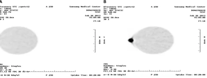
Fig. 2. (A) The image was reconstructed after phantom scan. (B) The image was reconstructed after moving the center of DFOV not to overlap with hotspot.
가져 해부학적인 수술 없이 쉽게 병변과 악성결정 등을 발견 하는 유용한 도구로 사용되며 특히 종양학 분야에서 임상적 으로 응용되는 횟수가 지속적으로 증가해왔다. 이와 병행하 여 PET/CT (Computed Tomography, CT)의 개발 등 PET의 기 술적인 발전으로 이어져 진단적으로 더 나은 서비스를 제공 하기 위한 지속적인 성장을 해왔다.1-3) 엑스선 일반촬영, 초 음파촬영, CT, MRI (Magnetic Resonance Imaging), SPECT (Single Photon Emission Computed Tomography)와 마찬가지로 PET/CT에서도 보통 변형체(Normal Variants), 인공물(Artifact), 가양성(false-positive)를 야기하는 원인으로 부터 판독오류를 범하지 않기 위한 영상에 대한 정확한 인지가 필요하다.4) 현 재 진행되고 있는 PET/CT 검사는 다양한 원인에 의해 인공 물이 발생되고 있으며, 이러한 인공물의 발생원인과 영상에 서 미치는 영향 등 판독오류로 이어질 수 있는 요소들에 대 한 이해를 토대로 영상의 질을 높이도록 해야 한다. 판독오 류의 원인 중 인공물은 영상에서 불필요한 구성요소로 나타 나게 되며 표준섭취계수(Standard Uptake Value, SUV)에 영향 을 주어 판독 시 정량평가에 영향을 주게 된다. 그 중 방사선 의약품 정맥주사 시 주사오류로 인해 방사성의약품이 주사 부위 주변부에 남게 되면 종창이 발생되어 영상에서 열소로 나타나 인공물을 발생시키게 된다. 다량의 방사성의약품으 로 이루어진 종창에 의한 인공물은 주변 조직의 표준섭취계 수를 변화시켜 판독결과에 영향을 줄 수 있으므로 인공물의 감소를 위한 노력이 필요하다. 본 논문에서는 열소가 포함되 지 않도록 표시시야(Display Field Of View, DFOV)의 중심을 이동하여 영상을 재구성함으로써 인공물의 감소에 대한 유 용성을 평가해 보았다.
실험재료 및 방법 1. 장비 및 실험재료
GE Discovery STE 16 (GE Healthcare, Milwankee, USA) 장 비를 이용하여 실험을 진행하였다. 장비의 재원은 BGO 크리 스탈(Crystal), 장비의 민감도 8.5 CPS/kBq, 공간분해능 5.1 mm, 축 방향 시야는 Frame 당 15.7 cm, 16 slice의 나선형 CT를 사용 하였다. 실험에 사용된 모형은 NEMA (National Electrical Manufacturers Association) 1994 PET 모형이었으며, 재원으로 는 배후용적 4,558 mL, 구체용적 304 mL, 외곽직경 20.3 mm, 외곽길이 22.8 mm 총 용적은 5,640 mL이다.
2. 모형 실험 방법
배후방사능을 구현하기 위해 NEMA 1994 PET 모형에 총
A B
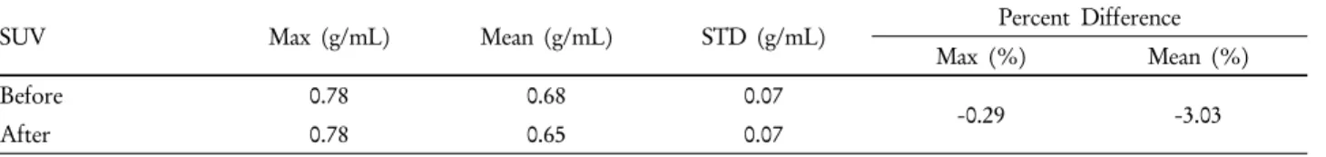
Table 1. SUV and Percentage Difference in other areas unaffected by artifacts
SUV Max (g/mL) Mean (g/mL) STD (g/mL) Percent Difference
Max (%) Mean (%)
Before 0.78 0.68 0.07
-0.29 -3.03
After 0.78 0.65 0.07
*P>0.05.
Table 2. SUV and Percentage Difference in the artifact areas affected by hotspots
SUV Max (g/mL) Mean (g/mL) STD (g/mL) Percent Difference
Max (%) Mean (%)
Before 0.51 0.32 0.08
48.61 74.26
After 0.76 0.56 0.13
*P<0.05.
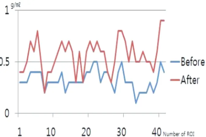
Fig. 3. This graph was composed with meanSUV of areas that were unaffected by artifacts.
체적 5,640 mL를 배후방사능이 0.005 MBq/mL가 되도록 28.2 MBq의 18F-FDG와 물을 함께 모형에 주입하였고. 열소는 1 MBq/mL가 되도록 10 cc주사기에 10 MBq의 18F-FDG와 물로 채워 열소 당 배후방사능의 방사능 비는 1:200이 되도록 하 였다.
이 후 NEMA 1994 PET 모형에 10 cc주사기를 외곽에 인위 적으로 부착하였다(Fig. 1).
CT를 이용한 투과촬영을 한 후 Frame 당 2분 30초의 방출 영상을 3차원(3D)모드로 획득하였다.
얻어진 데이터에 적용 된 재구성방법으로는 Z축 필터 stand- ard, post필터 4.29 반치폭(FWHM) mm, 반복횟수(iterations) 2회와 부분집합 수(subset) 20회였다. 영상 획득 후 재구성을 통해 DFOV 50 cm로 하여 위치 이동 없이 열소가 포함된 영 상을 얻었으며 DFOV의 중심 위치를 이동하여 열소부위가 DFOV로부터 벗어나도록 영상을 추가로 재구성하였다(Fig. 2).
3. 영상분석
DFOV 중심이동 전, 후의 재구성 영상이 얻어지면 열소로 부터 영향을 받지 않은 배후방사능 부위의 5곳과 열소 주변 인공물이 발생한 3곳에 관심영역(Region Of Interest, ROI)을 설정하여 DFOV 중심이동전, 후의 meanSUV, 표준편차, 백분 율 오차를 각각 산출해 비교, 분석하였다.
4. 통계분석
모든 실험 결과를 SPSS (Statistical Package for the Social Sciences) 18 version을 이용하여 통계분석 하였다. 각각의 ROI를 비소수 검정인 Wilcoxon signed rank test를 사용하여
분석하였으며 모든 결과의 유의수준은 5% (P<0.05)로 설정 하여 통계분석하였다.
결 과
1. 인공물의 영향을 받지 않은 배후방사능 부위
모형영상에서 열소의 영향을 받지 않은 배후방사능 부위 의 DFOV 중심이동 적용 전, 후 5개의 ROI를 분석한 결과 DFOV 변경 전 meanSUV와 표준편차는 0.67±0.06 g/mL, DFOV 변경 후의 meanSUV와 표준편차는 0.65±0.06 g/mL로 나타났으 며 백분율 오차는 maxSUV 0.29%, meanSUV –3.03%로 나타나 통계적으로 유의한 차이가 없었다(Table 1, Fig. 3).
2. 인공물의 영향을 받은 Cold Artifact 부위
열소에 의해 발생한 인공물의 영향을 받은 Cold Artifact 부
FIg. 4. This graph was composed with meanSUV of artifact areas that were affected by hotspots.
위의 DFOV 변경 전, 후 3개의 ROI를 분석한 결과 DFOV 변 경 전의 meanSUV와 표준편차는 0.32±0.08 g/mL, DFOV 변경 후의 meanSUV와 표준편차는 0.56±0.12 g/mL로 나타났으며 백 분율 오차는 maxSUV 48.61%, meanSUV 74.26%로 나타나 통계 적으로 유의한 차이를 나타냈다(Table 2, Fig. 4).
결 론
열소에 의한 인공물의 발생은 현재 진단적으로 사용되는 PET/CT 장비에서 재구성 시 이용되는 신틸레이터(Scintillator) 성분, 검출기의 구조와 전기적인 요소에 의해 4-7 mm의 반치 폭을 사용함으로 써 공간분해능과 해상도가 제한되어 나타 난 것으로 이때 부분체적효과(Partial Volume Effect, PVE)나 부분체적오류(Partial Volume Error)가 발생되어 복셀 내의 count의 크기가 평균이 되어 실제 보다 영상이 부드럽게 나 타나 열소와 배후방사능의 경계의 count의 측정에 오류가 발 생한 것이다.5,6) 본 연구에서는 DFOV의 중심 이동법을 통해 부분체적효과에 의해 발생 된 복셀 내의 count가 평균이 되 는 영향을 감소시켜 열소 주변부의 감소된 meanSUV를 32.1%
향상시킬 수 있었다. 또한 DFOV의 중심 이동 전, 후의 열소 의 영향이 없는 배후방사능을 측정하여 mean SUV의 변화를 측정한 결과로 DFOV에 이동에 의한 mean SUV가 통계적으 로 유의한 변화가 없다는 결과를 얻게 되었다. 이러한 결과 로 볼 때 주사오류에 의해 발생한 종창으로 열소가 발생 시 DFOV의 중심 이동법은 인공물의 영향을 적게 하는데 유용 하며 임상에서 적용하였을 시 정확한 정량평가를 위한 방법 으로 이용하여 영상의 진단적 가치를 높이는데 기여할 수 있 을 것으로 생각된다.
고 찰
본원에서 시행하고 있는 kg 당 5 MBq의 비율로 열소와 배 후방사능을 구성하였으며 열소 대 배후방사능 비는 200:1로 실제 주사오류가 발생하여 인공물의 영향이 나타난 검사를 기초하여 동일 비율로 실험을 진행 하였다. 인공물을 구현하 기 위한 200:1 미만의 열소 대 배후방사능 비에서는 뚜렷한 인공물의 발생이 나타나지 않은 사례가 많았다. 따라서 비율 이나 주입량에 따라 결과에 미치는 영향이 달라 질 수 있다.
오직 본원에서 사용되는 GE STE Discovery 16 장비의 실험 진행으로 이루어져 장비의 공간분해능과 민감도에 따른 차 이를 관찰할 수 없었다. 장비의 공간분해능과 민감도에 따라 인공물의 발생 정도는 차이를 보일 수 있으며 검사 시 적용 되는 복셀의 크기에 따라 달라질 수 있다. 또한 본 실험은 모 형에 의해서만 진행되어 실제 임상에서 적용 시 인체의 조직 이나 골격, 신체조건에 의한 차이가 발생할 수 있어 추가적 인 연구가 필요하다.
요 약
PET/CT 영상에서 인공물은 정량성을 저하시키는 원인이 된다. 여러 인공물 중 방사성의약품 주사 시 주사오류로 인 해 발생할 수 있는 열소는 그 주변부에 인공물을 발생시켜 영상의 질을 저하시킬 뿐 아니라 정량평가의 정확도를 저하 시킨다. 본 연구에서는 영상의 재구성시 표시시야(Display Field of View, DFOV)의 중심이동법을 이용하여 주사부위에 발생한 열소부위를 제거하고 정량평가에 미치는 영향을 평 가해 보고자 한다. GE Discovery STE 16 (GE Healthcare, Milwaukee, USA) 장비에 1994 NEMA 모형을 이용하였다. 모 형에 0.005 MBq/mL의 18F-FDG를 채우고 모형주변에 열소 대 배후방사능의 농도비가 200:1이 되도록 열소를 만들어 모 형외곽에 인위적으로 두었다. 영상획득 후 DFOV의 중심 위 치를 이동하여 열소부위가 DFOV로부터 벗어나도록 영상을 재구성한 후 적용 전, 후를 비교하였다. 영상에 대한 평가는 열소의 영향을 받지 않은 부위에서 DFOV 중심이동 전, 후의 배후방사능의 평균 표준섭취계수와 표준편차를 산출하여 재 구성에 의한 영향을 비교, 평가하였다. 또한 인공물이 발생한 부위에 관심영역을 설정하고 인공물의 발생 전, 후의 평균 표준섭취계수와 표준편차를 산출하여 백분율 오차를 각각 비교하였다. 모형영상 내 열소로 인한 인공물의 영향을 받지 않은 부위에서 DFOV 중심이동 법을 적용하기 전 평균 표준 섭취계수는 0.67±0.06 g/mL이었고, 적용 후에는 0.65±0.06
g/mL로 나타났다. 또한 영상에서 열소에 의해 발생한 인공물 이 있는 부위의 평균 표준섭취계수와 표준편차는 0.32±0.08 g/mL였으며, DFOV 중심이동을 적용한 경우는 0.56±0.12 g/mL로 나타났다. 이 때 열소의 영향을 받은 열소 인접부위 와 상대적으로 영향을 받지 않은 부위에 대한 백분율 오차는 65.3%와 97.4%로 각각 나타났다. PET/CT 영상에서 열소에 의해 발생 된 인공물은 DFOV의 중심이동법 적용 시 평균 표 준섭취계수를 32.1% 향상시킬 수 있으며, 이 때 중심이동 법 에 의한 다른 부위의 영향은 유의한 차이가 없음을 알 수 있 다. 결과에서와 같이 방사성의약품의 주사오류 시 발생한 종 창 등으로 발생되는 인공물의 영향은 DFOV 중심이동법을 적용할 경우 보다 정확한 정량평가가 가능해지고 그로 인하 여 영상의 진단적 가치를 높일 수 있을 것이다.
REFERENCES
1. Lardinois D, Weder W, Hany TF, et al. Staging of non-small- cell lung cancer with integrated positron-emission tomography and computed tomography. N Engl J Med 2003;348:2500-2507.
2. Kresnik E, Mikosch P, Gallowitsch HJ, et al. Evaluation of head and neck cancer with 18F-FDG PET: a comparison with con- ventional methods. Eur J Nucl Med 2001;28:816-821.
3. Weber WA, Avril N, Schwaiger M. Relevance of positron emis- sion tomography (PET) in oncology. Strahlenther Onkol 1999;
175:356-373.
4. Gary JR Cook. Artifacts and normal variants in whole-body PET and PET/CT imaging. Springer London 10.1007/1-84628- 187-3_5
5. Paul E Kinahan, James W Fletcher. PET/CT standardized up- take values (SUVs) in clinical practice and assessing response to therapy. J Sult 2010;31:496-505.
6. Lars Åkesson. Partial volume correction in PET/CT. Master Science Thesis Med Radiation Physics 2008.