Invasive cribriform carcinoma of the breast is a dis- tinct histologic type of invasive carcinoma that was first described by Page et al. in 1983 (1) and is characterized by a cribriform histologic pattern in the majority of its invasive component (1). Cribriform carcinoma is a well- differentiated variant of invasive ductal carcinoma and has a relatively favorable prognosis and low frequency of axillary nodal metastases (2). To our knowledge, the radiologic findings of invasive cribriform carcinoma of the breast are limited. MR imaging findings have not been previously described and there were highly suspi- cious findings of the masses on mammography and sonography in previous reports (3, 4). We report a case of invasive cribriform carcinoma of the breast with sonographic findings indicating a low suspicion for ma-
lignancy and MR imaging findings.
Case Report
A 48-year-old woman visited our hospital because of left breast cancer which was detected by screening. She underwent a mammography and breast sonography for screening at an outside clinic. A sonographically-guided core biopsy was performed at an outside clinic and the biopsy result was ductal carcinoma in situ. The patient was referred to our hospital for surgery.
Upon physical examination, there was no palpable mass or axillary lymphadenopathy. Moreover, skin changes or nipple retraction were not found. On mam- mography, there was no notable finding except for het- erogeneously dense breast (not shown). On ultrasonog- raphy, an approximately 0.9-cm microlobulated isoe- choic mass was detected in the left breast at the 1- o’clock position (Fig. 1A). Breast conservation surgery was planned and breast MR imaging was performed to evaluate the extent of the disease. MRI was performed with a 1.5T system (GE Signa Excite GE Healthcare, Milwaukee, WI, U.S.A.) using a dedicated breast coil.
The mass showed iso-signal intensity on a fat-saturated
J Korean Soc Radiol 2011;64:83-86
─ 83 ─
Invasive Cribriform Carcinoma of the Breast:
A Case Report
1Tae Wook Heo, M.D., Hyo Soon Lim, M.D.
2, Ji Shin Lee, M.D.
3, Min Ho Park, M.D.
4, Su Jin Jeong, M.D.
2, Jin Woong Kim, M.D.
2, Jin Gyoon Park, M.D., Heoung Keun Kang, M.D.
21Department of Radiology, Chonnam National University Hospital
2Department of Radiology, Chonnam National University Hwasun Hospital
3Department of Pathology, Chonnam National University Hwasun Hospital
4Department of Surgery, Chonnam National University Hwasun Hospital Received July 28, 2010 ; Accepted September 7, 2010
Address reprint requests to : Hyo Soon Lim, M.D., Department of Radiology, Chonnam National University Hwasun Hospital, 160 Ilsim-ri, Hwasun-eup, Hwasun-gun, Jeollanam-do 519-763, Korea.
Tel. 82-61-379-7112 Fax. 82-61-379-7133 E-mail: [email protected]
Invasive cribriform carcinoma is a rare type of invasive breast carcinoma with an ex- cellent prognosis and is characterized by a cribriform histological pattern. However, there is scant information about the radiologic features of invasive cribriform carcino- ma of the breast. We report a case of invasive cribriform carcinoma of the breast with radiologic features including MR imaging findings.
Index words : Breast Neoplasms Ultrasonography
Magnetic Resonance Imaging
Adenocarcinoma
T1 weighted image and iso- to slightly high signal inten- sity on a fat-saturated T2 weighted image (Figs. 1B, C).
After contrast (Gadopentetate dimeglumine) enhance- ment, the mass showed an oval, smooth, marginated, and heterogenously early enhancement with a delayed washout kinetic pattern (Figs. 1D, E). In addition, there was no evidence of multiple or bilateral breast cancer on breast MR images and the breast conservation surgery was performed. Macroscopically, the main tumor, which had a long axis of 0.9 cm, was well-demarcated in some regions, but had irregular invasive margins in oth-
er regions. Upon microscopic examination, the invasive component exhibited a cribriform pattern with well dif- ferentiated nuclei. Cribriform ductal carcinoma in situ was also present (Fig. 1F). The lymphovascular inva- sion, tumor necrosis, or microcalcifications were absent.
There was no lymph node metastasis detected on sen- tinel lymph node biopsy. Immunohistochemical stain- ing indicated a positive finding for the estrogen and progesterone receptors. The final histopathological diag-
Tae Wook Heo,
et al : Invasive Cribriform Carcinoma of the Breast
─ 84 ─
A B
C D
Fig. 1. Invasive cribriform carcinoma of the breast in a 48-year-old woman.
A. Sonogram shows a 0.9-cm, microlobulated isoechoic mass (arrows) in the left breast at a 1-o’clock position.
B, C. The mass (arrow) has an iso-signal intensity on a fat-saturated T1 weighted image (B) and an iso- to slightly high signal intensi- ty on a fat-saturated T2 weighted image (C).
D, E. Images reveal oval, smooth early enhancement (arrow) on fat-saturated T1 weighted subtraction images after contrast infu-
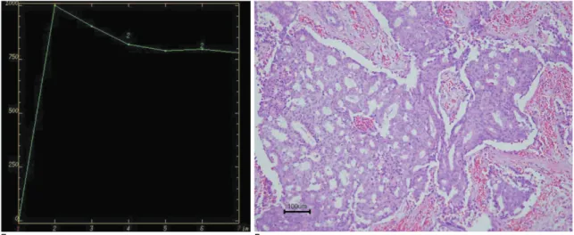
sion (D) and show an initial rapid rise and delayed washout pattern in the kinetic curve (E).
nosis was invasive cribriform carcinoma. Following di- agnosis, the patient has received radiation therapy and hormonal therapy and is presently free from local recur- rence or metastasis 28 months after conservation surgery.
Discussion
Invasive cribriform carcinoma is a rare type of inva- sive ductal carcinoma of the breast, which accounts for about 0.3-6% of breast cancers (1, 5, 6). It was first de- scribed by Page et al. and was divided into the classical type and mixed type. Tumors that are exclusively cribri- form or are cribriform to the limited extent of tubular in- vasive elements only were designated classic and the tu- mors also contained areas of less well differentiated in- vasive carcinoma were designated mixed type (1). Some authors use the term pure type to describe the tumors that do not show no other infiltrating carcinoma type (5, 6). Previous studies suggest that pure and classic inva- sive cribriform carcinoma has a relatively good progno- sis and a low frequency of axillary nodal metastases (1, 5, 6).
On histopathology, cribriform carcinoma must be dis- tinguished from other invasive breast carcinomas that show a cribriform pattern such as adenoid cystic carci- noma (1, 2, 7). In addition, immunocytochemical stain- ing for the basement membrane materials or ultrastruc- tural examination is recommended when accurate diag- nosis is difficult (7). The radiologic findings of invasive cribriform carcinoma are not well known, with only a few reports about the radiologic findings of invasive
cribriform carcinoma of the breast and most cases de- scribing their mammographic features. Stutz et al. re- ported the mammographic findings of eight cases and sonographic findings in 4 cases. They reported spiculat- ed masses measuring 20 to 35 mm in four of the patients and two mass that contained a few flecks of punctate calcifications on mammography. Four other tumors were not visible on mammography and the sonographic findings were ill-defined, inhomogenous solid masses in three of four cases (3). Another case report showed a well circumscribed high density mass with microcalci- fications on mammography (4). In our case, it was occult on mammography because of the small size of the mass in the background of heterogenously dense breast. A mass weighing about a 0.9 cm and with a focal mi- crolobulted margin detected on sonography, was consid- ered as a breast image report and data system (BI-RADS) category 4A. To the best of our knowledge, the MR imaging finding of invasive cribriform carcinoma have not been previously described, and in our case showed heterogenously early enhancement with delayed with a washout kinetic pattern compatible with the malignant mass, although it is not different from other histologic types of invasive breast carcinoma.
In summary, we describe a case of invasive cribriform carcinoma of the breast in a 48-year-old woman with ra- diologic findings including the MR imaging features.
Further study with a large number of patients is antici- pated to better describe the imaging characteristics of this disease.
J Korean Soc Radiol 2011;64:83-86
─ 85 ─
E F
Fig. 1. F. Microscopic examination (hematoxylin-eosin stain, original magnification ×100) shows invasive carcinoma with a cribri-
form pattern. The nests are punctuated by uniform punched-out spaces.
References
1. Page DL, Dixon JM, Anderson TJ, Lee D, Stewart HJ. Invasive cribriform carcinoma of the breast. Histopathology 1983;7:525-536 2. Cribriform carcinoma. In: Rosen PP. Rosen’s breast pathology. 2nd
Ed. Philadelphia: Lippincott Williams & Wilkins, 2001:551-553 3. Stutz JA, Evans AJ, Pinder S, Ellis IO, Yeoman LJ, Wilson AR, et
al. The radiological appearances of invasive cribriform carcinoma of the breast. Nottingham Breast Team. Clin Radiol 1994;49:693- 695
4. Nishimura R, Ohsumi S, Teramoto N, Yamakawa T, Saeki T,
Takashima S. Invasive cribriform carcinoma with extensive micro- calcifications in the male breast. Breast Caner 2005;12:145-148 5. Venable JG, Schwartz AM, Silverberg SG. Infiltrating cribriform
carcinoma of the breast: a distinctive clinicopathologic entity. Hum
Pathol 1990;21:333-3386. Marzullo F, Zito FA, Marzullo A, Labriola A, Schittulli F, Gargano G, et al. Infiltrating cribriform carcinoma of the breast. A clinico- pathologic and immunohistochemical study of 5 cases. Eur J
Gynaecol Oncol 1996;17:228-2317. Wells CA, Ferguson DJ. Ultrastructural and immunocytochemical study of a case of invasive cribriform breast carcinoma. J Clin
Pathol 1988;41:17-20Tae Wook Heo, et al : Invasive Cribriform Carcinoma of the Breast
─ 86 ─
대한영상의학회지 2010;64:83-86
침윤성사상형유방암:
증례 보고11
전남대학교병원 영상의학과
2
화순전남대학교병원 영상의학과
3
화순전남대학교병원 병리과
4