© 2015 Korean Breast Cancer Society. All rights reserved. http://ejbc.kr | pISSN 1738-6756
INTRODUCTION
Breast cancer is the most common malignant neoplasm among women in Western Europe and North America, and is a frequent cause of cancer death in recent years [1,2]. It is a gen-etically and clinically heterogeneous cancer, and the most common histologic subtype is invasive ductal carcinoma, which accounts for 70% to 80% of all breast cancer cases [3]. There have been advances in understanding carcinogenesis and breast cancer biology; however, treatment problems persist [4]. Using proven clinicopathological prognostic parameters, various proteins with essential cellular functions have been
proposed as potential predictors of breast cancer [5].
Dual-specificity protein phosphatases (DUSPs), members of the type I cysteine-based protein-tyrosine phosphatase super-family, are a heterogeneous group of protein phosphatases that can dephosphorylate both phosphotyrosine and phosphoser-ine/phosphothreonine residues within one substrate [6,7]. The DUSP family regulates members of the mitogen-activated pro-tein (MAP)-kinase superfamily. A subgroup of DUSPs, mito-gen-activated protein-kinase phosphatases (MKPs), plays an important role in regulating the tumor relevant MAP-kinase pathways. These pathways are associated with cellular prolifer-ation, differentiprolifer-ation, apoptosis, and inflammation [8,9].
DUSP4, also known as MKP2, is a part of the inducible nu-clear MKP group and specifically dephosphorylates the MAP kinases, ERK1/2, p38, and JNK [8]. It is expressed in many dif-ferent tumor types including colorectal and pancreatic cancer, malignant melanoma, ovarian serous borderline tumors, lung cancer, glioblastomas, and breast cancer [9]. The role of DUSP4 in cancer development and progression appears to vary with the type of malignancy. It is thought that DUSP4 acts either as a tumor suppressor or as a cancer progression factor
Clinicopathological Significance of Dual-Specificity Protein Phosphatase 4
Expression in Invasive Ductal Carcinoma of the Breast
Hyunsung Kim*, Se Min Jang*, Hyein Ahn, Jongmin Sim, Kijong Yi, Yumin Chung, Hulin Han, Abdul Rehman, Min Sung Chung1, Kiseok Jang, Seung Sam Paik
Departments of Pathology and 1Surgery, Hanyang University College of Medicine, Seoul, Korea ORIGINAL ARTICLE
Purpose: Dual-specificity protein phosphatase 4 (DUSP4), also known as mitogen-activated protein kinase phosphatase (MKP) 2 is a member of the inducible nuclear MKP group. The role of DUSP4 in cancer development and progression appears to vary with the type of malignancy. The purpose of this study was to in-vestigate DUSP4 expression in a case series of invasive ductal carcinoma of the breast. Methods: We constructed tissue micro-arrays consisting of 16, 14, 47, and 266 cases of normal breast tis-sue, usual ductal hyperplasia, ductal carcinoma in situ, and inva-sive ductal carcinoma, respectively. DUSP4 expression was inves-tigated by immunohistochemistry. Results: Cytoplasmic DUSP4 expression was observed. DUSP4 was more frequently expressed in malignant than in benign cases (p=0.024). The mean DUSP4 expression score was significantly higher in malignant tumors than in benign lesions (p=0.019). DUSP4 expression was significantly
correlated with a larger tumor size (>2 cm, p=0.015). There was no significant correlation between overall survival or disease-free survival and DUSP4 expression in all 266 patients. We evaluated the impact of DUSP4 expression on the survival of 120 patients with T1-stage tumors. Interestingly, Kaplan-Meier survival curves revealed that DUSP4 expression had a significant effect on both overall patient survival (p=0.034, log-rank test) and disease-free survival (p=0.045, log-rank test). In early T-stage breast cancer, DUSP4 expression was associated with a worse prognosis. Con-clusion: DUSP4 is frequently upregulated in breast malignancy, and may play an important role in cancer development and pro-gression. In addition, it may be a marker of adverse prognosis, es-pecially in patients with early T1-stage cancer.
Key Words: Breast neoplasms, Dual-specificity phosphatase 4, Prognosis
Correspondence to: Seung Sam Paik
Department of Pathology, Hanyang University College of Medicine, 222 Wangsimni-ro, Seongdong-gu, Seoul 133-791, Korea
Tel: +82-2-2290-8252, Fax: +82-2-2296-7502 E-mail: [email protected]
*These authors contributed equally to this work.
This work was supported by the research fund of Hanyang University (HY-2013).
Received: September 24, 2014 Accepted: January 28, 2015
Cancer
[8]. Its exact role is still controversial.
In the present study, we investigated DUSP4 expression by immunohistochemistry in a series of invasive ductal carcino-ma cases and evaluated its association with clinicopatholog-ical variables. In addition, we assessed the impact of DUSP4 expression on the survival of patients with breast cancer.
METHODS
Patients and tumor samples
A consecutive series of 266 patients with invasive ductal car-cinoma were enrolled in this study. All cases were diagnosed and underwent surgery at the Hanyang University Hospital (Seoul, Korea) between August 2000 and January 2009. This study was approved by the Institutional Review Board of the Hanyang University Hospital (HYU 2014-11-005-002). The mean age of the patients was 50 years and the mean follow-up period was 60 months. Of the 266 cases, 42, 152, and 72 were histological grades 1, 2, and 3, respectively. According to the seventh edition of the American Joint Committee on Cancer (AJCC) staging system, 194 cases were stage I or II and 72 were stage III or IV. In addition, 16, 12, and 47 samples of normal breast tissue, usual ductal hyperplasia, and ductal carcinoma in
situ, respectively, were randomly selected to evaluate the role of
DUSP4 expression in carcinogenesis and tumor progression. We reviewed all hematoxylin and eosin (H&E)-stained slides, pathology reports, and other medical records to confirm the diagnosis. The pathological parameters assessed included age, tumor size, tumor grade, perinodal tumor extension, lymph node metastasis, estrogen receptor (ER) and progesterone re-ceptor (PR) status, c-erbB-2 expression, and patient survival.
Tissue microarray construction
We used a manual tissue microarrayer (Unitama, Seoul, Korea) for tissue microarray construction from archival for-malin-fixed, paraffin-embedded tissue blocks. As previously described [10], we selected areas rich in tumor cells by light microscopy of H&E-stained sections. Tissue cylinders of 2 mm diameter were punched from a previously marked lesion on each donor block and transferred to the recipient block (Unita-ma). Each tissue microarray was comprised of 5×10 samples.
Immunohistochemical staining
Antibodies, polyclonal rabbit anti-DUSP4 (Abcam, Cam-bridge, UK), monoclonal mouse anti-ER (Novocastra Labora-tories, Newcastle, UK), monoclonal mouse anti-PR (Novocas-tra Laboratories), and monoclonal mouse anti-c-erbB-2 (No-vocastra Laboratories), were diluted 1:150, 1:50, 1:100, and 1:800 in goat serum, respectively. For immunohistochemical
staining, 4-μm sections were cut from the tissue microarray block using a Leica microtome, transferred to adhesive-coated slides, and deparaffinized. The staining was performed using the Bond Max automated immunostainer (Vision Biosystems, San Francisco, USA). Before staining, heat-induced epitope re-trieval was performed using the Bond epitope rere-trieval solu-tion. We blocked endogenous peroxidase activity with 0.3% hydrogen peroxide. Slides were incubated in primary antibody for 30 minutes at room temperature and the slides were incu-bated with postprimary reagent for 15 minutes at room tem-perature. The reactions were developed using the Bond poly-mer refine detection kit and visualized with the chromogen, 3, 3´-diaminobenzidine tetrahydrochloride.
Interpretation of immunohistochemical staining
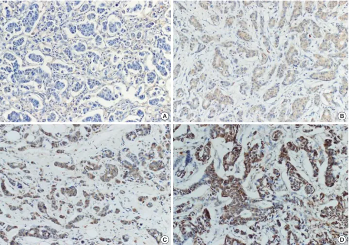
DUSP4 expression was evaluated semiquantitatively by two independent pathologists (H.K. and S.S.P.) who were blind to the patients’ clinical outcome. We categorized cytoplasmic DUSP4 expression in terms of both staining intensity and ex-tent, as described previously [11]. Staining intensity was graded as negative ( 0), weak ( 1), moderate ( 2), and strong ( 3), and staining extent was graded as 0% ( 0), 1%–25% ( 1), 26%–50% ( 2), 51%–75% ( 3), and 76%–100% ( 4). The product of intensity and extent grade was used as the final staining score. Thus, the maximum combined score was 12 and the minimum score was 0. Representative photomicrographs of DUSP4 immuno-staining in invasive ductal carcinoma are shown in Figure 1. For the purpose of statistical analysis, a cutoff value of 4 was adopt-ed according to the receiver operating characteristic curve. Therefore, the samples were finally classified as either negative (score 0–3) or positive (score 4–12) for DUSP4 expression. ER, PR, and c-erbB-2 expression status was interpreted according to the American Society of Clinical Oncology/College of American Pathologists guidelines for ER/PR/c-erbB-2 testing in breast cancer. When ≥1% of the tumor cell nuclei were stained, it was classified as ER or PR positive. Positive c-erbB-2 staining was determined by complete and intense membran-ous patterns in >10% of the tumor cells.
Statistical analysis
Statistical analysis was performed using the SPSS software version 19.0 (IBM Corp., Armonk, USA). The Mann-Whitney U test, chi-square test for linear trend, and chi-square test for independence were used to examine the association between DUSP4 expression and the clinicopathological parameters in-cluding age; histological grade; primary tumor (T) category; regional lymph nodes (N) category; AJCC stage; lymphatic in-vasion; perinodal tumor extension; expression of ER, PR, and c-erbB-2; and triple negativity. Spearman analysis was used to
obtain the correlation coefficient. We used the Kaplan-Meier method with the log-rank test to perform analyses of overall and disease-free survival. To identify the independent prog-nostic factors, the Cox proportional hazards regression model was used in both univariable and multivariable analyses. A p-value <0.05 was considered significant.
RESULTS
Patterns of DUSP4 expression in breast tissue
In this study, we evaluated DUSP4 expression in 16, 12, 47,
Figure 1. Representative microphotographs of dual-specificity protein phosphatase 4 (DUSP4) immunostaining in invasive ductal carcinoma (×200). (A) Negative, (B) weak, (C) moderate, and (D) strong. The tumor cells showed cytoplasmic DUSP4 staining.
A B
C D
Table 1. Dual-specificity protein phosphatase 4 expression in normal breast tissue/usual ductal hyperplasia, ductal carcinoma in situ, and in-vasive ductal carcinoma (n=341)
Tissue sample
DUSP4 expression Negative (n=247)
No. (%) Positive (n=94)No. (%) p-value* rs
NL/UDH 25 (89.3) 3 (10.7)
DCIS 36 (76.6) 11 (23.4) 0.024 0.113
IDC 186 (69.9) 80 (30.1)
DUSP4=dual-specificity protein phosphatase 4; rs=Spearman rank correla-tion coefficient; NL=normal breast tissue; UDH=usual ductal hyperplasia; DCIS=ductal carcinoma in situ; IDC=invasive ductal carcinoma.
*Chi-square test for linear trend.
4 3 2 1 0
NL/UDH DCIS IDC
Figure 2. Mean dual-specificity protein phosphatase 4 (DUSP4) expres-sion score. Mean DUSP4 expresexpres-sion score was significantly higher in malignant tumors than in benign lesions (Kruskal-Wallis test).
NL=normal breast tissue; UDH=usual ductal hyperplasia; DCIS=ductal carcinoma in situ; IDC=invasive ductal carcinoma.
p=0.019
0.86±2.16
and 266 cases of normal breast tissue, usual ductal hyperplasia, ductal carcinoma in situ, and invasive ductal carcinoma, re-spectively. Various grades of cytoplasmic DUSP4 expression were observed. DUSP4 expression was positive in 3 of 28 (10.7%) benign cases (normal breast tissue and usual ductal hyperplasia), 11 of 47 (23.4%) ductal carcinoma in situ cases, and 80 of 266 (30.1%) invasive ductal carcinoma cases (Table 1). DUSP4 was more frequently expressed in malignant than in benign cases (p=0.024). The mean DUSP4 expression score was 0.86±2.16 in normal breast tissue and usual ductal hyper-plasia, 2.19±2.98 in ductal carcinoma in situ, and 2.40±3.30 in invasive ductal carcinoma (Figure 2). It was significantly higher in malignant tumors than in benign lesions (p=0.019, Krus-kal-Wallis test).
Correlation between DUSP4 expression and clinicopathological parameters
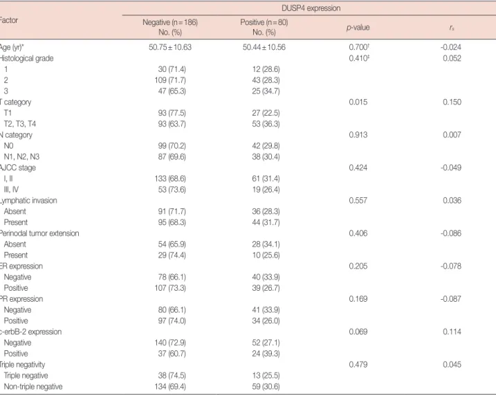
We evaluated the correlation between DUSP4 expression and the clinicopathological parameters to assess the significance of its expression in invasive ductal carcinoma. DUSP4 expression was more frequent in the higher T-stage categories (T2 to T4) than in the lower one (T1) (Table 2), and significantly correlat-ed with a larger tumor size (>2 cm, p=0.015). However, there was no correlation with histological grade, AJCC stage, lym-phatic invasion, perinodal tumor extension, hormone receptor status, c-erbB-2 expression, or triple negativity.
Correlation between DUSP4 expression, and overall and disease-free survival
The impact of DUSP4 expression on survival in 266 patients
Table 2. Correlation between dual-specificity protein phosphatase 4 expression and clinicopathological factors in invasive ductal carcinoma Factor
DUSP4 expression Negative (n=186)
No. (%) Positive (n=80)No. (%) p-value rs
Age (yr)* 50.75±10.63 50.44±10.56 0.700† -0.024 Histological grade 0.410‡ 0.052 1 30 (71.4) 12 (28.6) 2 109 (71.7) 43 (28.3) 3 47 (65.3) 25 (34.7) T category 0.015 0.150 T1 93 (77.5) 27 (22.5) T2, T3, T4 93 (63.7) 53 (36.3) N category 0.913 0.007 N0 99 (70.2) 42 (29.8) N1, N2, N3 87 (69.6) 38 (30.4) AJCC stage 0.424 -0.049 I, II 133 (68.6) 61 (31.4) III, IV 53 (73.6) 19 (26.4) Lymphatic invasion 0.557 0.036 Absent 91 (71.7) 36 (28.3) Present 95 (68.3) 44 (31.7)
Perinodal tumor extension 0.406 -0.086
Absent 54 (65.9) 28 (34.1) Present 29 (74.4) 10 (25.6) ER expression 0.205 -0.078 Negative 78 (66.1) 40 (33.9) Positive 107 (73.3) 39 (26.7) PR expression 0.169 -0.087 Negative 80 (66.1) 41 (33.9) Positive 97 (74.0) 34 (26.0) c-erbB-2 expression 0.069 0.114 Negative 140 (72.9) 52 (27.1) Positive 37 (60.7) 24 (39.3) Triple negativity 0.479 0.045 Triple negative 38 (74.5) 13 (25.5) Non-triple negative 134 (69.4) 59 (30.6)
DUSP4=dual-specificity protein phosphatase 4; rs=Spearman rank correlation coefficient; AJCC=American Joint Committee on Cancer; ER=estrogen receptor; PR=progesterone receptor.
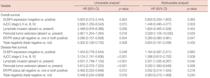
with invasive ductal carcinoma was evaluated. We observed that AJCC stage, lymphatic invasion, perinodal tumor exten-sion, ER/PR status, and triple negativity showed a significant effect on overall and disease-free survival in univariable and/or
multivariable analyses (Table 3). However, there was no signifi-cant correlation between overall survival or disease-free sur-vival and DUSP4 expression (p=0.924 and p=0.246, respec-tively; log-rank test) according to the Kaplan-Meier survival
100 50 0 100 50 0 100 50 0 100 50 0 0 50 100 150 0 50 100 150 0 50 100 150 0 50 100 150
Follow-up period (mo)
Follow-up period (mo)
Follow-up period (mo)
Follow-up period (mo)
p=0.924 p=0.034 p=0.246 p=0.045 Overall survival (%) Overall survival (%) Disease-fr ee survival (%) Disease-fr ee survival (%)
Figure 3. Cumulative overall and disease-free survival curves according to dual-specificity protein phosphatase 4 (DUSP4) expression. There was no significant difference of overall and disease-free survival in all 266 patients with invasive ductal carcinoma (A, B). However, there was significant differ-ence of overall and disease-free survival in 120 patients with the T1-stage tumor (C, D) (Kaplan-Meier method with log-rank test).
DUSP4-negative DUSP4-negative DUSP4-negative DUSP4-negative DUSP4-positive DUSP4-positive DUSP4-positive DUSP4-positive A C B D Table 3. Variables associated with the risks of death and recurrence in invasive ductal carcinoma (n=266)
Variable Univariable analysis* Multivariable analysis*
HR (95% CI) p-value HR (95% CI) p-value
Overall survival
DUSP4 expression (negative vs. positive) 0.955 (0.373–2.444) 0.924 0.600 (0.200–1.803) 0.363 AJCC stage (I, II vs. III, IV) 2.836 (1.230–6.543) 0.015 1.448 (0.490–4.277) 0.503 Lymphatic invasion (absent vs. present) 2.499 (0.978–6.386) 0.056 1.604 (0.465–5.528) 0.454 Perinodal tumor extension (absent vs. present) 2.957 (1.204–7.264) 0.018 3.329 (1.105–10.030) 0.033 ER/PR status (all negative vs. one or both positive) 0.266 (0.107–0.658) 0.004 0.280 (0.080–0.981) 0.047 Triple negativity (triple negative vs. not) 0.305 (0.126–0.735) 0.008 0.624 (0.191–2.039) 0.435 Disease-free survival
DUSP4 expression (negative vs. positive) 1.434 (0.778–2.644) 0.248 1.164 (0.587–2.311) 0.663 AJCC stage (I, II vs. III, IV) 3.124 (1.741–5.606) <0.001 1.286 (0.612–2.702) 0.508 Lymphatic invasion (absent vs. present) 3.531 (1.748–7.133) <0.001 2.551 (1.035–6.287) 0.042 Perinodal tumor extension (absent vs. present) 3.813 (2.070–7.023) <0.001 3.030 (1.380–6.649) 0.006 ER/PR status (all negative vs. one or both positive) 0.462 (0.252–0.846) 0.012 0.552 (0.214–1.420) 0.218 Triple negativity (triple negative vs. not) 0.448 (0.234–0.859) 0.016 0.563 (0.215–1.469) 0.240 HR=hazard ratio; CI=confidence interval; DUSP4=dual-specificity protein phosphatase 4; AJCC=American Joint Committee on Cancer; ER=estrogen receptor; PR=progesterone receptor.
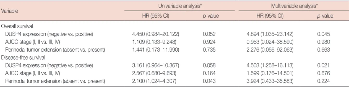
curves for all 266 patients with invasive ductal carcinoma (Fig-ure 3A, B). We evaluated the impact of DUSP4 expression on survival in 120 patients with T1-stage tumors (Table 4). We found that DUSP4 expression had a significant effect on over-all and disease-free survival in multivariable analysis. Kaplan-Meier survival curves revealed a significant effect of DUSP4 expression on overall survival (p=0.034, log-rank test) and disease-free survival (p=0.045, log-rank test) in early T-stage breast cancer (Figure 3C, D).
DISCUSSION
In the present study, we investigated DUSP4 expression in 16, 12, 47, and 266 cases of normal breast tissue, usual ductal hyperplasia, ductal carcinoma in situ, and invasive ductal carci-noma, respectively. We evaluated the correlation between DUSP4 expression, and the clinicopathological parameters and survival of patients with invasive ductal carcinoma. DUSP4 was more frequently expressed in malignant (ductal carcinoma in situ and invasive ductal carcinoma) than in be-nign cases (normal breast tissue and usual ductal hyperplasia). The mean DUSP4 expression score was also significantly high-er in malignant cases and DUSP4 expression was significantly correlated with a larger tumor size (>2 cm). There was a sig-nificant association between DUSP4 expression and overall and disease-free survival in patients with T1-stage tumors.
DUSPs are a heterogeneous group of protein phosphatases that can dephosphorylate both phosphotyrosine and phospho-serine/phosphothreonine residues [7]. They regulate members of the MAP-kinase superfamily. MKPs play an important role in regulating the tumor relevant MAP-kinase pathways. These pathways are associated with cellular proliferation, differentia-tion, apoptosis, and inflammation [9]. DUSP4 is a member of the inducible nuclear MKP group and specifically dephos-phorylates the MAP kinases, ERK1/2, p38, and JNK [8].
DUSP4 is expressed in many different tumor types including colorectal and pancreatic cancer, malignant melanoma, ovari-an serous borderline tumors, lung covari-ancer, glioblastomas, ovari-and breast cancer [9]. The role of DUSP4 in cancer development and progression appears to vary with the type of malignancy.
Whether DUSP4 acts as a tumor promoter or tumor sup-pressor is still controversial and there is no consensus on the exact role of DUSP4 expression in human cancer. Recently, Saigusa et al. [8] suggested that DUSP4 might be involved in the suppression of tumor progression and metastasis in colorectal cancer. They showed that decreased DUSP4 expres-sion was associated with advanced T-stage categories, lym-phatic invasion, vascular invasion, advanced stage, and distant metastasis. Increased DUSP4 expression was associated with a better prognosis. Waha et al. [12] demonstrated that glioblas-toma cell growth was inhibited by exogenous DUSP4 over-expression. Armes et al. [13] showed that DUSP4 was expressed in primary tumors, but could be lost in early-onset and high-grade breast cancers. Recently, Baglia et al. [14] described that low DUSP4 expression levels, particularly of variant 1, were as-sociated with both increased recurrence/breast cancer mortali-ty and increased overall mortalimortali-ty.
Some authors have reported that DUSP4 may play a role in promoting cancer progression. It has been proposed that DUSP4 may not act as a tumor suppressor factor, because its expression was found to be upregulated in some malignancies including breast and rectal cancer, and pancreatic and melano-ma cell lines [15-18]. Liu et al. [9] demonstrated that over-expression of DUSP4 may play an important role in promoting the epithelial-mesenchymal transition in breast cancer, and suggested that it may be a marker of adverse prognosis. Gröschl et al. [6] found that DUSP4 was frequently over-expressed in colorectal cancer with high frequent microsatellite instability (MSI-H) compared to microsatellite-stable colo-rectal cancer. They posited that DUSP4 may act as an important
Table 4. Variables associated with the risks of death and recurrence in T1-ranked invasive ductal carcinoma by the American Joint Committee on Cancer breast cancer staging system (n=120)
Variable Univariable analysis* Multivariable analysis*
HR (95% CI) p-value HR (95% CI) p-value
Overall survival
DUSP4 expression (negative vs. positive) 4.450 (0.984–20.122) 0.052 4.894 (1.035–23.142) 0.045 AJCC stage (I, II vs. III, IV) 1.109 (0.133–9.248) 0.924 0.953 (0.024–38.590) 0.980 Perinodal tumor extension (absent vs. present) 1.441 (0.173–11.990) 0.735 2.276 (0.056–92.063) 0.663 Disease-free survival
DUSP4 expression (negative vs. positive) 3.161 (0.964–10.367) 0.058 4.503 (1.258–16.113) 0.021 AJCC stage (I, II vs. III, IV) 2.567 (0.680–9.693) 0.164 1.599 (0.176–14.501) 0.676 Perinodal tumor extension (absent vs. present) 2.100 (1.024–4.307) 0.043 3.924 (0.433–35.583) 0.224 HR=hazard ratio; CI=confidence interval; DUSP4=dual-specificity phosphatase 4; AJCC=American Joint Committee on Cancer.
regulator of cell growth within the MAPK pathway and may cause enhanced cell growth in MSI-H colorectal cancer. Wang et al. [15] reported that the expression of MKP1 and MKP2 displayed a significant increase in human breast cancer com-pared to normal breast tissue.
In our study, we found that DUSP4 was more frequently ex-pressed in malignant (ductal carcinoma in situ and invasive ductal carcinoma) than in benign cases (normal breast tissue and usual ductal hyperplasia). The mean DUSP4 expression score was also significantly higher in malignant cases. These results suggest that DUSP4 may be involved in the carcinogen-esis of breast cancer. DUSP4 expression was significantly cor-related with a larger tumor size (>2 cm), indicating that it may be involved in breast cancer tumor progression. In survival analyses, the Kaplan-Meier survival curves revealed a signifi-cant effect of DUSP4 expression on both overall and disease-free survival in T1-stage tumors. These results suggest that DUSP4 may be a marker of adverse prognosis, especially in patients with early-stage breast cancer. Our results imply that DUSP4 may play a role as a cancer promoter, not a tumor sup-pressor in invasive ductal carcinoma.
In conclusion, our findings show that DUSP4 is frequently upregulated in breast malignancy, and may play an important role in cancer development and progression. Furthermore, it may be a marker of adverse prognosis, especially in patients with early T1-stage cancer. The exact role of DUSP4 and its potential as a novel therapeutic target for breast cancer should be investigated in future studies.
CONFLICT OF INTEREST
The authors declare that they have no competing interests.
REFERENCES
1. DeSantis C, Siegel R, Bandi P, Jemal A. Breast cancer statistics, 2011. CA Cancer J Clin 2011;61:409-18.
2. Schmidt M, Voelker HU, Kapp M, Krockenberger M, Dietl J, Kammer-er U. Glycolytic phenotype in breast cancKammer-er: activation of Akt, up-regu-lation of GLUT1, TKTL1 and down-reguup-regu-lation of M2PK. J Cancer Res Clin Oncol 2010;136:219-25.
3. Malhotra GK, Zhao X, Band H, Band V. Histological, molecular and functional subtypes of breast cancers. Cancer Biol Ther 2010;10:955-60. 4. Jang SM, Han H, Jang KS, Jun YJ, Jang SH, Min KW, et al. The glycolytic phenotype is correlated with aggressiveness and poor prognosis in in-vasive ductal carcinomas. J Breast Cancer 2012;15:172-80.
5. Bertolo C, Guerrero D, Vicente F, Cordoba A, Esteller M, Ropero S, et al. Differences and molecular immunohistochemical parameters in the subtypes of infiltrating ductal breast cancer. Am J Clin Pathol 2008;130: 414-24.
6. Gröschl B, Bettstetter M, Giedl C, Woenckhaus M, Edmonston T, Hof-städter F, et al. Expression of the MAP kinase phosphatase DUSP4 is as-sociated with microsatellite instability in colorectal cancer (CRC) and causes increased cell proliferation. Int J Cancer 2013;132:1537-46. 7. De Vriendt V, De Roock W, Di Narzo AF, Tian S, Biesmans B, Jacobs B,
et al. DUSP 4 expression identifies a subset of colorectal cancer tumors that differ in MAPK activation, regardless of the genotype. Biomarkers 2013;18:516-24.
8. Saigusa S, Inoue Y, Tanaka K, Toiyama Y, Okugawa Y, Shimura T, et al. Decreased expression of DUSP4 is associated with liver and lung me-tastases in colorectal cancer. Med Oncol 2013;30:620.
9. Liu Y, Du F, Chen W, Yao M, Lv K, Fu P. Knockdown of dual specificity phosphatase 4 enhances the chemosensitivity of MCF-7 and MCF-7/ ADR breast cancer cells to doxorubicin. Exp Cell Res 2013;319:3140-9. 10. Jang SM, Sim J, Han H, Ahn HI, Kim H, Yi K, et al. Clinicopathological
significance of CADM4 expression in invasive ductal carcinoma of the breast. J Clin Pathol 2013;66:681-6.
11. Jang SM, Han H, Jun YJ, Jang SH, Min KW, Sim J, et al. Clinicopatho-logical significance of CADM4 expression, and its correlation with ex-pression of E-cadherin and Ki-67 in colorectal adenocarcinomas. J Clin Pathol 2012;65:902-6.
12. Waha A, Felsberg J, Hartmann W, von dem Knesebeck A, Mikeska T, Joos S, et al. Epigenetic downregulation of mitogen-activated protein kinase phosphatase MKP-2 relieves its growth suppressive activity in glioma cells. Cancer Res 2010;70:1689-99.
13. Armes JE, Hammet F, de Silva M, Ciciulla J, Ramus SJ, Soo WK, et al. Candidate tumor-suppressor genes on chromosome arm 8p in early-onset and high-grade breast cancers. Oncogene 2004;23:5697-702. 14. Baglia ML, Cai Q, Zheng Y, Wu J, Su Y, Ye F, et al. Dual specificity
phos-phatase 4 gene expression in association with triple-negative breast can-cer outcome. Breast Cancan-cer Res Treat 2014;148:211-20.
15. Wang HY, Cheng Z, Malbon CC. Overexpression of mitogen-activated protein kinase phosphatases MKP1, MKP2 in human breast cancer. Cancer Lett 2003;191:229-37.
16. Gaedcke J, Grade M, Jung K, Camps J, Jo P, Emons G, et al. Mutated KRAS results in overexpression of DUSP4, a MAP-kinase phosphatase, and SMYD3, a histone methyltransferase, in rectal carcinomas. Genes Chromosomes Cancer 2010;49:1024-34.
17. Yip-Schneider MT, Lin A, Marshall MS. Pancreatic tumor cells with mutant K-ras suppress ERK activity by MEK-dependent induction of MAP kinase phosphatase-2. Biochem Biophys Res Commun 2001;280: 992-7.
18. Teutschbein J, Haydn JM, Samans B, Krause M, Eilers M, Schartl M, et al. Gene expression analysis after receptor tyrosine kinase activation re-veals new potential melanoma proteins. BMC Cancer 2010;10:386.