INTRODUCTION
Cystic hypersecretory carcinoma (CHC) and cystic hyper- secretory hyperplasia (CHH) were first described in 1984 (1).
The characteristic features of these lesions are dilated ducts and cysts containing an eosinophilic secretory product resem- bling thyroid colloid. After the initial report of CHC and CHH, the entire spectrum of cystic hypersecretory lesions of the breast was described by Guerry et al. in 1988 (2). These lesions range from benign CHH to the intermediate CHH with atypia and the frankly malignant CHC.
There have been only seven cases of invasive CHC report- ed in the literature (1-5). We describe an additional case of invasive CHC in a 45-yr-old female.
CASE REPORT
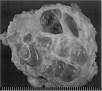
A 45-yr-old woman was admitted to the hospital for diag- nosis and treatment of a palpable mass in the lower quadrant of the left breast. The mass was soft and focally hard. She had no past or family history of a breast disease. The sonograph showed a cystic and lobulated mass. An excisional biopsy of the left breast was performed. Gross pathologic examination of the excisied specimen revealed an ill-defined, mucoid mass.
The cut surface of the mass, which was 4.7×3.7×3 cm, revealed multiple cystic spaces, and the cysts were filled with thick, gelatinous secretions. The individual cysts varied from
0.1 cm to 1.2 cm in dimension (Fig. 1). Intervening solid areas were also noted. Microscopically, many cystically dilated ducts contained thyroid colloid-like eosinophilic secretions.
The homogeneous secretions were retracted from the sur- rounding epithelia, producing scalloped margins (Fig. 2A).
The secretory material also showed linear cracking artifact.
The cyst-lining epithelium exhibited variable patterns. The Ji Shin Lee, Young Jik Lee*
Department of Pathology, Seonam Univerisy, College of Medicine, Namwon; Department of Pathology*, St. Carollo Hospital, Suncheon, Korea
Address for correspondence Ji Shin Lee, M.D.
Department of Pathology, Seonam University, College of Medicine, 720 Kwangchi-dong, Namwon 590-711, Korea
Tel: +82.63-620-0352, Fax: +82.63-620-0355 E-mail: [email protected]
149 J Korean Med Sci 2004; 19: 149-51
ISSN 1011-8934
Copyright � The Korean Academy of Medical Sciences
Invasive Cystic Hypersecretory Carcinoma of the Breast : A Case Report
Cystic hypersecretory lesions of the breast are rare. These breast lesions include cystic hypersecretory hyperplasia (CHH), atypical CHH, and cystic hypersecretory carcinoma (CHC). The characteristic features are dilated ducts and cysts filled with thyroid colloid-like eosinophilic secretion. Only seven cases of invasive CHC have been reported in the literature. Here, we report an additional case of invasive CHC.
The histologic features of the tumor showed both micropapillary intraductal carcino- ma and focal high-grade invasive carcinoma in a background of CHH. This case sug- gests that cystic hypersecretory breast lesions encompass a spectrum of patholog- ic lesions including CHH, atypical CHH, CHC, and invasive CHC.
Key Words : Breast; Neoplasms; Carcinoma; Cystic Hypersecretory Carcinoma; Cystic Hypersecreto- ry Hyperplasia
Received : 15 January 2003 Accepted : 7 March 2003
Fig. 1.The cut surface of the mass shows numerous cysts, mea- suring up to 1.2 cm in diameter, with a gelatinous secretion.
150 J.S. Lee, Y.J. Lee
lining of the cysts in most areas was of flat or cuboidal epithe- lium and devoid of cellular atypia (Fig. 2B). The epithelium of some cysts showed proliferative change ranging from atypi- cal hyperplasia to intraductal carcinoma, micropapillary type (Fig. 2C). The intraductal carcinoma component was accom- panied by an invasive component with small solid nests (Fig.
2D). The invasive component was a high-grade carcinoma lacking cystic and papillary traits. Histochemical staining of the secretory material in the cysts was positive with periodic acid-Schiff (PAS) and alcian blue. Immunohistochemically, the cystic contents were positive for carcinoembryonic antigen
(CEA) (Zymed, San Franciscoa, CA, U.S.A., predilute), but negative for thyroglobulin (Zymed, predilute). Immunostains for estrogen and progesterone receptors (DAKO, Glostrup, Denmark, predilute) and p53 protein (DAKO, dilution 1:100) were negative in the neoplastic epithelial cells.
The diagnosis was invasive CHC. Modified radical mastec- tomy with axillary lymph node dissection was performed.
Axillary lymph nodes were free of tumor metastasis. Subse- quent radiotherapy was performed. The seven-month follow- up period was uneventful.
Fig. 2.Microscopic findings. The lesion is composed of multiple cysts and ducts containing eosinophilic secretion (A, H&E, ×20). Most of the cysts are lined by flat epithelium. The secretion retracts from the surrounding epithelium (B, H&E, ×200). The epithelium of some cysts grows as micropapillary intraductal carcinoma (C, H&E, ×200). An invasive component (arrow) is found adjacent to the intraductal carcinoma component (D, H&E, ×100).
A B
C D
Invasive Cystic Hypersecretory Carcinoma of the Breast 151
DISCUSSION
Cystic hypersecretory lesions of the breast have a spectrum of morphologic features ranging from the clearly benign (CHH), a combination of benign and atypical epithelium (CHH with atypia), to cases that combine benign, atypical, and frankly malignant epithelium (CHC) (2). The characteris- tic gross features are the formation of dilated ducts and cysts filled with a colloid-like secretion. Although cystic hypersecre- tory lesions have a distinctive gross appearance, it is usually not possible to distinguish CHC from CHH grossly. CHC is differentiated from CHH by a micropapillary cyst lining with cytologic atypia. If no cytologic atypia is present and the epithelium is flat or cuboidal, the lesion is characterized as CHH. Invasion is heralded by solid nests of invasive carci- noma and is usually poorly differentiated with no secretory characteristics. As a consequence, total excisional biopsy is required for definitive diagnosis of cystic hypersecretory lesions of the breast.
About 50 cases of cystic hypersecretory breast lesions have been reported (1-7). Most cases of CHC have been intraduc- tal and only seven cases of invasive CHC have been reported (1-5). Most invasive carcinomas have been poorly differentiated duct carcinomas with a solid growth pattern. Invasive CHC tends to have an aggressive behavior. Four cases were diagnosed with lymph node metastases (2, 5). Metastatic foci in the axil- lary lymph nodes had cystic foci that contained eosinophilic secretion. One patient developed invasive lobular carcinoma of the contralateral breast 10 yr after ipsilateral mastectomy for invasive CHC (3). The patient described herein had neg- ative lymph nodes.
The present case is the eighth case of invasive CHC. In this case all features of CHH were identified. In addition, micro- papillary intraductal CHC and focal high-grade invasive car- cinoma were also observed. Our case supports the concept that cystic hypersecretory breast lesions encompass a spectrum of pathologic lesions including CHH, atypical CHH, CHC, and invasive CHC. The progression of these lesions from CHH, through intraductal CHC, to invasive CHC may be possible.
The differential diagnosis of invasive CHC includes secreto- ry carcinoma, mucinous carcinoma, malignant mucocele-like
tumor, and metastatic thyroid carcinoma. Secretory carcino- ma contains vacuolated cytoplasm and more bubbly secre- tions, which are not typical features of CHC (8). Mucinous carcinoma and malignant mucocele-like tumor also show cystically dilated ducts (9). However, the secretions in these lesions are rather pale and basophilic and do not show linear cracking artifacts. Metastatic follicular thyroid carcinoma of the breast may mimic CHC. Histologic differentiation of these two conditions may require immunohistochemical stain for thyroglobulin. Negative reaction for thyroglobulin was observed in the cyst contents of our case.
REFERENCES
1. Rosen PP, Scott M. Cystic hypersecretory duct carcinoma of the breast.
Am J Surg Pathol 1984; 8: 31-41.
2. Guerry P, Erlandson RA, Rosen PP. Cystic hypersecretory hyperpla- sia and cystic hypersecretory duct carcinoma of the breast: patholo- gy, therapy, and follow-up of 39 patients. Cancer 1988; 61: 1611-20.
3. Kim MK, Kwon GY, Gong GY. Fine needle aspiration cytology of cystic hypersecretory carcinoma of the breast: a case report. Acta Cytol 1997; 41: 892-6.
4. Herrmann ME, McClatchey KD, Siziopikou KP. Invasive cystic hyper- secretory ductal carcinoma of breast: a case report and review of the literature. Arch Pathol Lab Med 1999; 123: 1108-10.
5. Lee WY, Cheng L, Chang TW. Diagnosing invasive cystic hypersecre- tory duct carcinoma of the breast with fine needle aspiration cytology:
a case report. Acta Cytol 1999; 43: 273-6.
6. Shah AK, Banerjee SN, Sehonanda AS, Girishkumar H, Gerst PH.
Cystic hypersecretory duct carcinoma of the breast. Breast J 2000;
6: 269-72.
7. Park JM, Seo MR. Cystic hypersecretory duct carcinoma of the breast:
report of two cases. Clin Radiol 2002; 57: 312-5.
8. Lamovec J, Bracko M. Secretory carcinoma of the breast: light micro- scopical, immunohistochemical and flow cytometric study. Mod Pathol 1994; 7: 475-9.
9. Lee JS, Kim HS, Jung JJ, Lee MC. Mucocele-like tumor of the breast associated with ductal carcinoma in situ and mucinous carcinoma: a case report. J Korean Med Sci 2001; 16: 516-8.