JKSMRM 14:21-30(2010)
1Department of Radiology, College of Medicine, The Catholic University of Korea
2Department of Pathology, College of Medicine, The Catholic University of Korea Received; January 20, 2010, revised; April 7, 2010, accepted; April 27, 2010
Corresponding author : Sung Hun Kim, M.D., Department of Radiology, Seoul St. Mary’ Hospital, College of Medicine, The Catholic University of Korea, 505 Banpo-dong, Seocho-gu, Seoul 137-701, Korea.
Tel. 82-2-2258-6250 Fax. 82-2-599-6771 E-mail: [email protected]
Imaging Features of Mucinous Breast Carcinoma
Hye Jung Han1, Sung Hun Kim1, Eun Suk Cha1, Hyun Sook Kim1, Bong Joo Kang1, Jae Jung Choi1, Jee Hye Lee1, Ah Won Lee2
Purpose : To examine the imaging findings of mucinous breast carcinoma and to evaluate the difference in these findings based on the histopathologic grade.
Materials and Methods : We retrospectively analyzed the imaging features according to BI-RADS in 29 patients with surgically proven mucinous carcinoma. The histopathologic grade was classified as well-differentiated, moderately-differentiated and poorly-differentiated. Based on these criteria, the differences in imaging findings were statistically analyzed.
Results : Mammography was available in 20 cases, which contained 17 mass lesions (85%) and 3 cases of normal findings. On ultrasonography (27 cases), mucinous carcinoma was observed as a mass with an oval shape (59.3%), a microlobulated margin (55.6%) or an inhomogeneous isoechogenicity (74.1%). On MRI (21 cases), mucinous carcinoma was commonly observed to have a lobular shape (76%), smooth margin (86%) or heterogeneous contrast-enhancement (61.9%). On the kinetic curve, there was a delayed wash-out pattern (52.3%). There were no significant differences in the imaging findings for each histopathologic grade except that a well- differentiated tumor had an abrupt interface.
Conclusion : A well-differentiated mucinous carcinoma tended to have an abrupt interface on ultrasonography, as compared with the moderately-differentiated one.
Mucinous carcinoma showed a heterogeneous enhancement and a delayed washout kinetic curve pattern on dynamic MRI.
Index words :Adenocarcinoma, mucinous Breast neoplasms
Mammography Ultrasonography
Magnetic resonance imaging (MRI)
Introduction
Mucinous carcinoma is a relatively rare cancer that accounts for approximately 1-7% of total cases of primary breast cancer (1). On mammography, mucinous carcinoma is often misdiagnosed as a benign tumor that leads to a delayed diagnosis (1, 2). On ultrasonography, mucinous carcinoma has an isoechogenicity similar to that of adjacent adipose tissue, which often limits detection (2).
To date, several studies have reported the mammographic (1-14), ultrasonographic (2, 12-16) and MRI findings (17-20) of mucinous carcinoma.
Mammography showed that mucinous carcinoma was an oval or a lobular shaped mass with a circumscribed margin and no calcification (1-14). And most of mucinous carcinoma showed oval-shaped and microlobulated mass with isoechogenecity on ultrasonography (2, 13, 15). MRI showed mucinous carcinoma was a mass with a lobular shape, a smooth margin and a very high signal intensity on a T2- weighted image (17, 18, 20). And most of them showed heterogeneous (18, 20) or rim enhancement (17, 18, 20).
To our knowledge, there have been no studies that
describe these imaging findings simultaneously. We evaluated the mammographic, ultrasonographic and MRI findings of mucinous carcinoma and evaluated the difference of these findings based on the histopathologic grade of the carcinoma.
Materials and Methods
Patient Group
The current study was conducted in 29 female patients who were surgically diagnosed with mucinous carcinoma between 2000 and 2008. The patients’ mean age was 49.6 years (range, 34-82 years). Twenty-three patients underwent wide excision, five underwent modified radical mastectomy and one underwent quadrantectomy. Tumor size, histopathologic grade and immunohistochemical staining were referenced to the histopathologic results on medical records. Tumor size was defined as the length of the long diameter, measured on surgical specimens. The Bloom- Richardson grade was used as the histologic grade in this study, which is scored on the degree of tubule formation, the nuclear pleomorphism and the mitotic activity. Then, this grade comprises three classifications: well-differentiated, moderately-
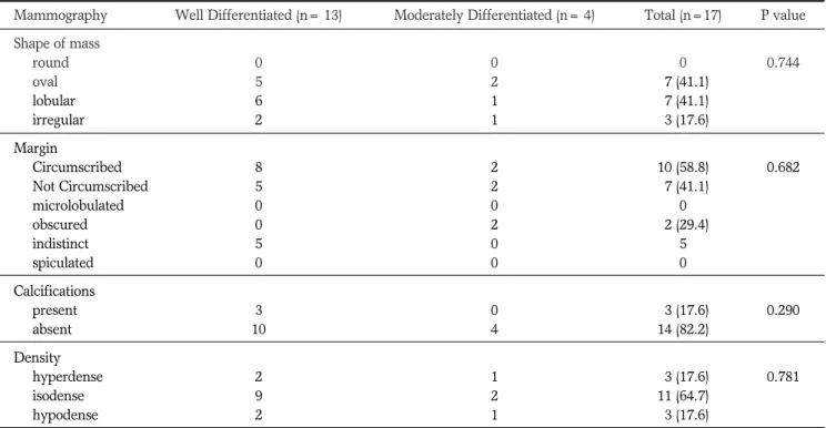
Table 1. Mammographic Features of Mucinous Breast Cancer
Mammography Well Differentiated (n= 13) Moderately Differentiated (n= 4) Total (n=17) P value Shape of mass
round 0 0 0 0.744
oval 5 2 07 (41.1)
lobular 6 1 07 (41.1)
irregular 2 1 03 (17.6)
Margin
Circumscribed 8 2 10 (58.8) 0.682
Not Circumscribed 5 2 07 (41.1)
microlobulated 0 0 0
obscured 0 2 02 (29.4)
indistinct 5 0 5
spiculated 0 0 0
Calcifications
present 3 0 03 (17.6) 0.290
absent 10 4 14 (82.2)
Density
hyperdense 2 1 03 (17.6) 0.781
isodense 9 2 11 (64.7)
hypodense 2 1 03 (17.6)
Note- Percentages are in parentheses
differentiated and poorly-differentiated. We made comparisons between the imaging and histopathologic findings.
Radiological Acquisition
Of the 29 patients, mammography was available in 20 cases, ultrasonography was available in 27 cases and MRI was available in 21 cases. Two radiologists who had 4- and 7-years of experience in breast imaging, evaluated the imaging with consensus interpretation according to the lexicon of Breast Imaging Reporting and Data System (BI-RADS), retrospectively.
Mammography was performed for the craniocaudal and the mediolateral oblique views using a Mammomat Nova 3000 (Siemens Medical Solutions, Solna, Sweden) and a Hologic system (Lorad Selenia;
Danbury, U.S.A.). Images were analyzed for the presence of a mass, the shape of a mass (round, oval, lobular or irregular), the margin of a mass (circumscribed, microlobulated, obscured, indistinct or spiculated), the presence of calcification and the density of a mass (hyperdense, isodense or hypodense).
Ultrasound was performed with a linear transducer using a frequency of 5-12 MHz (HDI 3000 and HDI 5000, Advanced Technology Laboratories; Bothell, WA, USA). Tumors were analyzed based on several features: the shape of a mass (oval, round or irregular), orientation (parallel or non-parallel), margin
(circumscribed, indistinct, angular, microlobulated or spiculated), lesion boundary (abrupt interface or echogenic halo), echogenicity (anechoic, hyperechoic, complex echo, hypoechoic or isoechoic), posterior echo features (no posterior acoustic features, posterior acoustic enhancement, posterior shadowing or combined pattern), calcification and vascularity (not present, present in lesion, immediately adjacent or diffusely increased vascularity in surrounding tissue).
Tumor echogenicity was also evaluated as being either homogeneous or inhomogeneous.
MR images were acquired with a 1.5T scanner (Signa; GE Medical Systems, Milwaukee, WI, U.S.A.
and Achieva; Philips Medical system, Best, the Netherlands) using a breast coil. MRI with the Signa scanner was performed using the following sequences;
sagittal, fat-suppressed, fast spin-echo T2-weighted imaging, and axial or sagittal, fat-suppressed, fat-spoiled gradient-echo T1-weighted imaging (TR/TE=6.2/3.1, flip angle of 10 o, 2.6 mm section thickness, and an acquisition time of 1 min 31 minutes) obtained before and 91, 182, 273, 364 and 455 sec after the rapid bolus injection of 0.2 mmol/kg body weight of Gd-DPTA (Magnevist, Schering, Berlin, Germany). MRI with the Achieva scanner was performed using the following sequences; sagittal, fat-suppressed, fast spin-echo T2- weighted imaging and axial fat-suppressed, fat-spoiled gradient-echo T1-weighted imaging obtained before
a b
Fig. 1. Left mucinous carcinoma in a 54-year-old woman.
(a) Mammogram showed a lobular, circumscribed, isodense mass without calcifications.
(b) Ultrasonography showed an oval, microlobulated, heterogeneously isoechoic mass with posterior enhancement and abrupt interface.
and 0, 91, 182, 273, 364 and 455 sec after the rapid bolus injection of the same contrast agent. We analyzed the kinetic curve pattern (persistent, plateau or washout) on delayed images. Kinetic curve was obtained automatically after the manual placement of
region of interest (ROI) within the tumor. The ROI was manually placed on the area demonstrating the highest enhancement within each tumor.
MRI images were analyzed for several tumor features: the shape of a mass (round, oval, lobular or
a b
c d
e
Fig. 2. Right mucinous carcinoma in a 41-year-old woman.
(a) Ultrasonography showed an oval, microlobulated, complex echoic mass with posterior enhancement and abrupt interface.
(b) Axial T2-weighted image showed a hyperintense, lobular mass with a smooth border.
(c) Axial contrast-enhanced T1-weighted image showed rim enhancement.
(d) Kinetic curve showed the delayed washout pattern.
(e) Mucinous carcinoma consisted of hypocellular areas with large lakes filled with mucin and sparse clusters of tumor cells (black stars), and hypercellular areas with crowded clusters of tumor cells in the mucin (black arrowheads) (Hematoxylin-eosin; magnification ×40).
irregular), margin (smooth, irregular or spiculated), mass enhancement (homogeneous, heterogeneous or rim enhancement) and signal intensity of the mass on T2-weighted images.
Results
The average size of mass was 2.3 cm (range: 0.8-7 cm). The histopathologic stage was identified in 93% of cases (27 cases) as either T1 or T2. Lymph node metastasis was surgically confirmed in 7% (2 cases). A well-differentiated grade was observed in 79% (23
cases), and a moderately-differentiated grade was observed in 21% (six cases). No poorly-differentiated grades were observed. On immunohistochemical staining, there was a positive response to estrogen receptor in 97% of cases (28 cases), a positive response to progesterone receptor in 90% of cases (26 cases) and a positive response to C-erbB2 in 14% of cases (4 cases).
Imaging Findings
The results of the mammographic findings are summarized in Table 1.
3 of 20 cases showed normal findings. The mucinous
c d
Fig. 3. Left mucinous carcinoma in a 52-year-old woman.
(a) Ultrasonography showed an irregular, microlobulated and heterogeneously isoechoic mass with posterior- enhancement and an echogenic halo (white arrowheads).
(b, c) Axial and sagittal contrast-enhanced T1-weighted image showed an oval, heterogeneously enhancing mass with irregular margin.
(d) Mucin pools of variable sizes were separated by fibrous septae (black stars) and clusters of tumor cells floating in the mucin pools (Hematoxylin-eosin; magnification ×40).
a b
carinomas commonly presented as a isodense mass (64%) with oval (41%) or lobular shape (41%) and circumscribed margin (59%) (Fig. 1).
The results of the ultrasonographic findings are summarized in Table 2. All of mucinous carcinoma showed a detectable mass. The most commonultra- sonographic appearance of mucinous carcinomas was inhomogenous isoechoic mass (74%) with oval (59%) or irregular (41%) shape, microlobulated margin (56%) and posterior acoustic enhancement (89%) (Figs. 1-3).
Most of them (93%) showed long axis of mass, parallel to the skin axis, abrupt interface(67%) (Figs. 1, 2) and the remaining 33% had an echogenic halo (Fig. 3).
Vascularity was noted within (38%) or adjacent to the mass (24%).
The results of MRI findings are summarized in Table 3. On MRI, the mucinous carcinomas commonly showed lobular shape (76%), smooth margin (86%) and high signal intensity on T2-weighted images (100%)
(Fig. 2). After contrast enhancement, there were 13 cases (61.9%) of heterogeneous enhancement (Fig. 3) and 6 cases (29%) of rim enhancement (Fig. 2). A kinetic curve was obtained for 17 of 21 cases. These curves revealed that there were 11 cases (52.3%) with a delayed wash-out pattern (Fig. 2).
Statistical Analysis
In accordance with the histopathologic grade based on the Bloom-Richardson grade, all cases of mucinous carcinoma had a well-differentiated or a moderately- differentiated grade. In cases in which a well- differentiated grade was observed (23 cases, 79%), the mean age of the patients was 48.1 years. In cases in which a moderately-differentiated grade was observed (6 cases, 21%), the mean age of the patient was 55.8 years.
A mass was palpated in 17 of 29 patients (58.6%). In 12 cases (41.1%), the mass was detected at screening.
Table 2. Ultrasonographic Features of Mucinous Carcinoma
Ultrasonography Well differentiated (n=21 ) Moderately differentiated (n=6 ) Total (n=27) P value Shape of mass
oval 12 4 16 (59.2) 0.675
round 00 0 0
irregular 09 2 11 (40.7)
Orientation
parallel 20 5 25 (92.5) 0.326
nonparallel 01 1 2 (7.4)
Margin
Circumscribed 05 0 05 (18.5) 0.185
Not Circumscribed 16 6 22 (81.4)
indistinct 05 2 07 (25.9)
angular/spiculated 00 0 0
microlobulated 11 4 15 (55.5)
Lesion boundary
abrupt interface 16 2 18 (66.6) *0.050*
echogenic halo 05 4 09 (33.3)
Echo pattern
anechoic/ Hypo 00 0 0 0.695
hyperechoic 02 0 2 (7.4)
complex echo 02 1 03 (11.1)
isoechoic (hetero) 15 5 20 (74.0)
isoechoic (homo) 02 0 2 (7.4)
Posterior acoustic
no 03 0 03 (11.1) 0.326
enhancement 18 6 24 (88.8)
shadowing/combined 00 0 0
Note. - percentages are in parentheses.
All 12 of these cases had a well-differentiated histologic grade. Of 17 cases with a palpable mass, 11 had a well- differentiated histologic grade, and the remaining 6 had a moderately-differentiated grade. There was a significant correlation between the presence of symptoms and the histopathologic grade (p < 0.05).
There were no significant differences in the mammographic, ultrasonographic and MRI findings according to the histopathologic grade. On breast ultrasound, however, the lesion boundary of a mass with an abrupt interface was observed more prevalently in cases with a well-differentiated grade than those with a moderately-differentiated grade (p=0.05).
Discussion
Mucinous carcinoma has a 10-year survival rate of approximately 70-90%, with a better prognosis than other carcinomas. It slowly grows and rarely shows lymph node metastasis (1, 4).
The prevalence of mucinous carcinoma has a higher correlation with age, and it commonly occurs in elderly women (3). In the current study, the mean age of the patients was 49.7 years (range 34-82 years), and the proportion of women aged from 30 to 49 was 66% (19 cases). This population is younger than those in
previous reports, likely due to the dermographic differences. Mucinous carcinoma is detected in approximately 50% of cases with a palpable mass and approximately 50% in asymptomatic cases (15). Also, in the current study, 59% of patients had the chief complaint of a palpable mass. In the remaining 41% of patients, a lesion was incidentally identified during a routine screening.
Histopathologically, mucinous carcinoma is a well- differentiated or moderately-differentiated tumor (2, 21). The current study reports similar results. Most of the previous studies have reported that mucinous carcinoma could be classified into one of two histologic types, pure or mixed type, based on the differences in each types radiologic findings (1, 3-5, 7, 13, 14, 18, 20).
However, to our knowledge, there have been no studies that have examined the differences between the clinical characteristics and the radiological findings depending on the histologic grade, as shown in our study. Clinical findings depending on the histologic grade showed a significant difference in the present study. That is, it was noted that a well-differentiation was associated with screen-detected mucinous carcinoma, and a moderate differentiation was associated with mucinous carcinoma with a palpable mass (p<0.05). Imaging findings depending on the histopathologic grade did not show a significant Table 3.MRI Features of Mucinous Carcinoma
MRI Well differentiated (n=16) Moderately differentiated (n=5) Total (n=21) P value Shape
round/oval 04 1 05 (23.8) 0.849
lobular 12 4 16 (76.2)
irregular 00 0 0 (0)0.
Margin
smooth 14 4 18 (85.7) 0.676
irregular 02 1 03 (14.3)
spiculated 00 0 0 (0)0.
Enhancement
homogenous 02 0 2 (9.5) 0.133
heterogenous 08 5 13 (61.9)
rim 06 0 06 (28.6)
Kinetic curve
not available 03 1 04 (19.0) 0.539
persistent 01 0 1 (4.7)
pleteau 03 2 05 (23.8)
washout 09 2 11 (52.3)
Note. - Percentages are in parentheses.
difference. On the ultrasound, however, a well- differentiated mucinous carcinoma tended to have an abrupt interface, as compared with the moderately- differentiated one (p=0.05).
Mammography showed that mucinous carcinoma was an oval or a lobular shaped mass with a circumscribed margin and no calcification (1-14). In our study, mammography showed that mucinous carcinoma had almost no calcification (82.4%, 14 cases), and was oval or lobular shaped (82.2%, 14 cases) with a circumscribed margin (58.8%, 10 cases) and isodensity (64.7%, 11 cases). These findings are in agreement with the previous reports of mammography of mucinous carcinoma.
On ultrasonography, most of the cases showed an oval-shaped mass (59.2%, 16 cases) with a microlobu- lated margin (56%, 15 cases). Lam et al. (13) reported that mucinous carcinoma is observed to be an oval- shaped and microlobulated mass (56.8%). This is in agreement with results of the present study. In our study, the echogenicity showed an inhomogenous isoechogenicity (74%, 20 cases), a complex echogenicity (11.1%, 3 cases), a homogeneous isoechogenicity (7.4%, 2 cases) and a hyperechogenicity (7.4%, 2 cases).
According to Dhillon et al. (2) 39% of lesions visible by mammography were not identified on ultrasonography. These authors noted that the mass had an isoechogenicity, so it could not be differentiated from the adjacent adipose tissue. Stavros et al. (15) also reported that mucinous carcinoma was observed to be a mass with isoechogenicity. In particular, according to these authors, a small mucinous carcinoma of < 1.5 cm is observed to have an echogenicity that is equivalent to that of adjacent adipose tissue. These authors noted however, that the echogenicity was slightly coarse and heterogeneous as compared with the adjacent adipose tissue, which could be compared to the “salt and pepper”. These results were in agreement with the inhomogenous isoechogenicity that was seen in our study. As compared with histopathologic findings, the solid component with an isoechogenicity corresponded to the malignant cells floating on the mucous pool with a variable size. A hyperechogenicity with a point or a linear shape corresponded to the fibrous septa. Lam et al. (13) concluded that the echogenicity of mucinous carcinoma was observed to have a heteroechogenecity
(43.7%), a complex echogenicity (37.5%), a homogeneous echogenicity (12.5%) and a homogeneous hypoechogenicity (6.5%). Compared to our study, the proportion of a complex echogenicity was relatively higher and that of a homogeneous isoechogenicity was similar. Because a heteroechoic feature was not listed in the lexicon of BI-RADS however, it could not be compared with an inhomogenously isoechoic feature seen in our study. According to Dihillon R et al. (2), there was no complex echogenicity on ultrasonography in 26 patients (28 cases) with mucinous carcinoma. The proportion of the mucinous carcinoma with complex echogenicity is also small in our study. When correlated with histopathologic findings, the central anechoic portion corresponded to a mucinous pool with a relatively greater size. There were two cases of hyperechoic mass. Presumably, this might be because mixed cancer cells and mucin were scattered through the ultrasonography (16).
In our study, the posterior acoustic enhancement was present in 89% (24 cases) of cases of mucinous carcinoma. This originated from the high water content and the increased transmission of ultrasonography due to the mucinous components. This concurrent presence is easily observed in cases in which the mucinous components are abundant in the tumor, and has been reported to be the typical ultrasonographic findings of mucinous carcinoma (13).
There have been few studies of MRI findings of mucinous carcinoma (17-20). The common MRI features of mucinous carcinoma are a mass with a lobular shape, a smooth margin and a very high signal intensity, on a T2-weighted image (17, 18, 20).
Mucinous carcinomas have previously showed heterogeneous (18, 20) or rim enhancement (17, 18, 20).
Our study showed similar results, with lobular shape, (76%, 16 cases), smooth margin (86%, 18 cases) and a high signal intensity on T2-weighted images (100%).
Additionally, in our study, mucinous carcinoma showed heterogeneous enhancement (61.9%, 13 cases), and there were many cases in which a rim enhancement was observed (28.6%, 6 cases). A heterogeneous or rim enhancement is indicative of the presence of malignant tumor, which has been reported to be one of the typical radiological findings of mucinous carcinoma (18, 20). It is widely known that this heterogeneous enhancement originates from a
mixture of two different histologic components, the mucin pool and the tumor cell nest (18, 20). It is known that the thick fibrous septum dividing the mucin pool was not enhanced on MRI (20). Also, in our study, the point or linear hyperechogenicity on ultrasonography corresponded to a fibrous septum on histopathologic findings, which was not well-enhanced (unlike the tumor cells on MRI). This contributed to the heterogeneous enhancement pattern. A rim enhancement has been shown in cases with a viable tumor cell nest in the margin and a necrotic or fibrotic scar in the tumor center (22). However, in our study, central non-enhancing areas corresponded to the large mucin pool, which was shown as anechoic area on ultrasonography.
In general, malignant masses show a delayed wash- out pattern on the kinetic curves, but mucinous carcinoma showed the delayed persistent enhancement, which was thought to be typical findings of mucinous carcinoma (17, 18, 20). In our study, there were 11 cases (65%) of a washout pattern, 5 cases (29%) of a plateau pattern and 1 case of a persistent pattern. These results were not in agreement with previous reports.
We selected the pure mucinous carcinoma and manually placed region of interest (ROI)s on the area demonstrating the highest enhancement within each tumor. These might result in different results from previsou studies. So, these results may be another radiologic finding of mucinous carcinoma and suggest of further studies with larger case numbers are required.
Our study has some limitations. First, this was a retrospective study which enrolled a small number of patients. Second, some of the radiologic examinations (mammography, ultrasonography and MRI) were not available on the review. Third, the imaging findings of each modality were not blindly interpreted by the two radiologists.
In conclusion, only significant different imaging finding depending on the histopathologic grade is that a well-differentiated mucinous carcinoma tended to have an abrupt interface on ultrasonography, as compared with the moderately-differentiated one. Mucinous carcinoma showed a heterogeneous enhancement and a delayed washout kinetic curve pattern on dynamic MRI.
References
1.Conant EF, Dillon RL, Palazzo J, Ehrlich SM, Feig SA.
Imaging findings in mucin-containing carcinomas of the breast: correlation with pathologic features. AJR Am J Roentgenol 1994;163:821-824
2.Dhillon R, Depree P, Metcalf C, Wylie E. Screen-detected mucinous carcinoma: potential for delayed diagnosis. Clin Radiol 2006;61:423-430
3.Cardenosa G, Doudna C, Eklund GW. Mucinous (colloid) breast cancer: clinical and mammographic findings in 10 patients. AJR Am J Roentgenol 1994;162:1077-1079
4.Wilson TE, Helvie MA, Oberman HA, Joynt LK. Pure and mixed mucinous carcinoma of the breast: pathologic basis for differences in mammographic appearance. AJR Am J Roentgenol 1995;165:285-289
5.Matsuda M, Yoshimoto M, Iwase T, et al. Mammographic and clinicopathological features of mucinous carcinoma of the breast. Breast Cancer 2000;7:65-70
6.Kushwara AC, Whitman GJ, Williamson JD. Mucinous carcinoma of the breast. AJR Am J Roentgenol 1999;173:290 7.Goodman DN, Boutross-Tadross O, Jong RA. Mammographic
features of puremucinous carcinoma of the breast with pathological correlation. Can Assoc Radiol J 1995;46:296-301 8.Svane G, Potchen EJ, Sierra A, Azavedo E. Screening
mammography. St. Louis, MO: Mosby 1993;296-299, 308-311 9.Kopans DB. Breast imaging. Philadelphia: Lippincott 1989;
298
10.Parsons CA. Diagnosis of breast disease: imaging, clinical features and pathology. Baltimore: University Park Press 1983;142-162
11.Pina Insausti LJ, Soga Garcia E. Mucinous breast carcinoma showing as a cluster of suspicious microcalcifications on mammography. Eur Radiol 1998;8:1666-1668
12.Memis A, Ozdemir N, Parildar M, Ustunn EE, Erhan Y.
Mucinous breast cancer: mammographic and US features with histologic correlation. Eur J Radiol 2000;35:39-43 13.Lam WW, Chu WC, Tse GM, Ma TK. Sonographic
appearance of mucinous carcinoma of the breast. AJR Am J Roentgenol 2004;182:1069-1074
14.Chopra S, Evans AJ, Pinder SE, et al. Pure mucinous breast cancer: mammographic and ultrasound findings. Clin Radiol 1996;51:421-424
15.Stavros AT. Malignant solid breast nodules: specific types. In:
Stavros AT, editor. Breast ultrasound. Philadelphia: Lipincott Williams & Wilkins 2003;645-648
16.Kamitani Kumiko, Ono Minoru, Toyoshima Satoshi, et al.
Isoechoic axillary lymph node metastases of mucinous carcinoma of the breast: a case report. Breast cancer 2006;
13:382-385
17.Kawashima M, Tamaki Y, Nonaka T, et al. MR imaging of mucinous carcinoma of the breast. AJR Am J Roentgenol 2002; 179:179-183
18.Takashi Okafuji, Hidetake Yabuuchi, Shuji Sakai. MR imaging
features of pure mucinous carcinoma of the breast. Eur J Radiol 2006;60:405-413
19.Yuen S, Uematsu T, Kasami M, et al. Breast carcinomas with strong high-signal intensity on T2-weighted MR images:
pathological characteristics and differential diagnosis. J Magn Reson Imaging 2007;25:502-510
20.Shuichi Monzawa, Masaki Yokokawa, et al. Mucinous Carcinoma of the Breast: MRI Features of Pure and Mixed Forms with Histopathologic Correlation. AJR Am J Roentgenol
2009;192:125-131
21.Clayton F. Pure mucinous carcinoma of the breast:
morphologic features and prognostic correlates. Hum Pathol 1986;17:34-38
22.Wedegartner U, Bick U, Wortler K, Rummery E, Bongartz G.
Differentiation between benign and malignant findings on MR-mammography: usefulness of morphological criteria. Eur Radiol 2001;11:1645-1650
통신저자 : 김성헌, (137-701) 서울시 서초구 반포동 505, 가톨릭 의과대학 성모병원 영상의학과
Tel. 82-2-2258-6250 Fax. 82-2-599-6771 E-mail: [email protected]
점액암의 영상소견
1가톨릭대학교 의과대학 영상의학과
2가톨릭대학교 의과대학 병리학과
한혜정1∙김성헌1∙차은숙1∙김현숙1∙강봉주1∙최재정1∙이지혜1∙이아원2
목적: 점액암의 영상소견에 대해서 알아 보고, 조직학적 분화도에 따른 영상 소견의 차이를 알아보고자 하였다.
대상과 방법: 수술을 통해 점액암으로 확진된 29명의 환자를 대상으로 유방촬영술과 초음파 검사, 그리고 자기 공명 영상 소견을 BI-RADS에 따라서 후향적으로 분석하였다. 또한 점액암의 조직학적 분화도를 고분화, 중증도분화, 미 분화 등급으로 나눠서 이에 따른 영상소견의 차이를 통계학적으로 분석하였다.
결과: 총 20예의 유방 촬영에서 17예 (85%)가 종괴로 보였고 3예는 정상소견이었다. 총 27예의 초음파 검사에서 점액암은 난원형 (59.2%)의 미세소엽성 변연 (55.5%)을 가지거나 불균일한 등에코 (74%)의 종괴로 보였다. 총 21예의 자기공명영상에서 점액암은 소엽상의 변연(76.2%), 매끄러운 변연(86%)의 종괴로 불균일한 조영증강 (61.9%)을 보이는 경우가 흔하였다. 역동성 조영 증가 그래프에서 지연시 유실형 양상 (52.3%)을 보였다. 고분화 점액암이 갑자기 끝나는 종괴의 경계를 보이는 것 외에는 점액암의 분화도에 따른 영상소견의 차이는 없었다.
결론: 점액암의 분화도에 따른 차이는 중등도 분화 점액암과 비교하여 고분화 점액암이 갑자기 끝나는 종괴의 경계를 보이는 것이었다. 대부분의 점액암은 자기공명영상에서 불균일한 조영증강과 지연시 유실형 양상을 보였다.
대한자기공명의과학회지 14:21-30(2010)