475
Print ISSN 1738-5520 / On-line ISSN 1738-5555 Copyright © 2010 The Korean Society of Cardiology CASE REPORT
DOI 10.4070 / kcj.2010.40.9.475
Open Access
A Case of Guide Wire Fracture With Remnant Filaments in the Left Anterior Descending Coronary Artery and Aorta
Young-Min Hong, MD and Sang-Rok Lee, MD
Department of Internal Medicine, Chonbuk National University Medical School, Jeonju, Korea ABSTRACT
Guide wire fractures during percutaneous coronary intervention (PCI) are very rare, but when they do occur they may lead to life-threatening complications, such as embolization, thrombus formation and perforation. In cases when percutaneous retrieval has failed, surgical extraction of the remnant fragments is recommended. We present a case of remnant guide wire filaments that remained in place without complications, over a one-year clinical follow up period. (Korean Circ J 2010;
40:475-477)
KEY WORDS: Percataneous transluminal coronary angioplasty; Coronary artery disease.
Received: January 27, 2010 Accepted: February 19, 2010
Correspondence: Sang-Rok Lee, MD, Division of Cardiology, Depart- ment of Internal Medicine, Chonbuk National University Medical School, 42 Wonjam 5-gil, Deokjin-gu, Jeonju 561-712, Korea
Tel: 82-63-250-2418, Fax: 82-63-250-1680 E-mail: [email protected]
cc This is an Open Access article distributed under the terms of the Cre- ative Commons Attribution Non-Commercial License (http://creativecom- mons.org/licenses/by-nc/3.0) which permits unrestricted non-commer- cial use, distribution, and reproduction in any medium, provided the origi- nal work is properly cited.
Introduction
The increasing incidence of complex percutaneous coro- nary intervention (PCI) is accompanied by risks of device fracture or dislodgement. Guide wire fractures during PCI are very rare, but in such cases, life-threatening complications such as embolization, thrombus formation and perforation may occur. Although, in most cases, percutaneous retrieval te- chniques of fractured guide wires are recommended, there have been several reports of fragments being left in place with- out complications.1-4)
We present the case of a 78-year-old female patient who was diagnosed with non-ST segment elevation myocardial in- farction (NSTEMI) and treated with PCI. The patient had re- mnant guide wire filaments in the left anterior descending artery (LAD) and aorta but did not experience any serious complications during a one-year follow up period.
Case
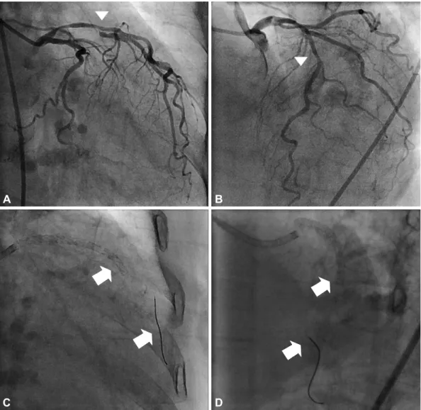
A 78-year-old woman presented with NSTEMI. Echocar- diography showed decreased left ventricular systolic function (ejection fraction 45%). A diagnostic coronary angiogram showed diffuse significant stenosis from the proximal to mid- dle LAD and first diagonal branch (D1) (Fig. 1). There was to- tal thrombotic obstruction at the middle right coronary artery.
We initially treated the patient with a single paclitaxel-eluting stent (2.75×32 mm Taxus®, Boston Scientific, Natick, MA, USA).
Four days later, two coronary angioplasty 0.014 guide wires were inserted {one hi-torque Balance Middleweight (BMW) univer- sal coronary guide wire (Abott Vascular, Santa Clara, CA, USA) into the LAD, and one high-torque Whisper coronary guide wire (Abott Vascular) into the D1}. An intravascular ultrasound after predilatation with a 2.5×20 mm Voyager® balloon (Abott Vascular) revealed a large plaque burden at both the LAD and the D1. We deployed two overlapped sirolimus-eluting stents at the proximal and middle LAD: a 2.75×33 mm Cypher® at the middle LAD and a 2.75× 18 mm Cypher® at the proximal LAD (Cordis Corporation, Miami Lakes, FL, USA). When we exchanged the guide wires to perform kissing balloon angio- plasty, a fracture occurred at the distal tip of the BMW guide wire (Fig. 1). Another guide wire was inserted to perform a beaded wire rotation, and distal balloon inflation retrieval was attempt- ed, in order to remove the fractured guide wires. This attempt, however, was ineffective. Finally, we used a goose neck loop- snare (Microvena Corporation, St. Paul, MN, USA) to remove the fractured guide wires. Multiple forward and backward
476 Guide Wire Fracture
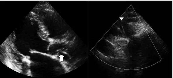
movements of the snare, combined with distal balloon infla- tion retrieval, successfully removed most of the fractured guide wires (Fig. 2) but we later observed retained filaments during echocardiography (Figs. 3 and 4). The patient declined surgical
intervention for removal of these stray filaments and was dis- charged from our hospital, with triple anti-platelet medication and no complications. The patient did not experience any th- rombotic or embolic events and did not suffer from any sub- jective symptoms over the one year of clinical follow up.
Discussion
Guide wire fractures during PCI are very rare, occurring in approximately 0.1-0.2% of cases.5) Guide wire remnants could lead to life threatening complications such as thrombosis, em- boli, and perforation. Therefore, in the event of failed percu- taneous retrieval and persistent signs of ischemia, patients should be urgently referred for surgical intervention. There are several methods recommended for the management of frac- tured guide wires, including emergent surgery, loop snare re- moval, two- or three-wire rotation, stenting over the retained wire, and conservative treatment.6-8) Surgical extraction is st- rongly recommended in cases of protrusion of the guide wire A
C
B
D
Fig. 1. A diagnostic coronary angiogram showed diffuse and significant stenosis from the proximal to middle LAD (arrow head) and first di- agonal branch (arrow head) (A and B). After deployment of two sirolimus-eluting stents over the LAD, a fracture occurred at the distal tip of the BMW guide wire (arrow) (C and D). LAD: left anterior descending coronary artery, BMW: Balance Middleweight.
Fig. 2. A fracture occurred at the distal tip of the BMW guide wire (white arrow). In addition, un-coiled filaments of the BMW guide wire were left in situ (arrow head). BMW: Balance Middleweight.
Young-Min Hong, et al. 477
into the ascending aorta.9-11) However, guide wire segments retained within the coronary circulation may remain benign for a long time, particularly if they are entrapped within a dis- tal part of the vessel and do not have accompanying total cor- onary occlusions.3) Vascular endothelial cell covering over the guide wire fragments may render them immobile and non- thrombogenic.
Hi-torque BMW guide wires consist of a distal core and a stainless proximal shaft, facilitating treatment of multiple le- sions and tortuous vessels. However, guide wire fractures may occur if the distal core and stainless proximal shaft are sepa- rated by either the trapping of the distal tip or by vascular resistance. Because the fracture in the LAD developed after stent deployment at the main branch, we could not determine the mechanism of fracture in this particular case. However, tr- apping of the distal tip of the BMW wire, or stent deployment over a severely angulated guide wire are two possible expla-
nations.
In conclusion, even though the most ideal management option for remnant guide wires is their removal, conservative treatment with the fragments left in situ may be successful in cases in which patients remain asymptomatic and hemo- dynamically stable. However, life-long administration of in- tensive anti-platelet medications and close observation are recommended for these patients.
REFERENCES
1) Cafri C, Rosenstein G, Ilia R. Fracture of a coronary guidewire during graft thrombectomy with the X-sizer device. J Invasive Cardiol 2004;
16:263-5.
2) Doorey AJ, Stillabower M. Fractured and retained guide-wire frag- ment during coronary angioplasty: unforeseen late sequelae. Cathet Cardiovasc Diagn 1990;20:238-40.
3) van Gaal WJ, Porto I, Banning AP. Guide wire fracture with retained filament in the LAD and aorta. Int J Cardiol 2006;112:e9-11.
4) Lee HJ, Son MS, Ju KT, et al. An unusual case of guide wire fracture during coronary artery stenting for bifurcation lesion. Korean Circ J 2001;31:1200-2.
5) Khonsari S, Livermore J, Mahrer P, Magnusson P. Fracture and dis- lodgment of floppy guidewire during percutaneous transluminal cor- onary angioplasty. Am J Cardiol 1986;58:855-6.
6) Woodfield SL, Lopez A, Heuser RR. Fracture of coronary guidewire during rotational atherectomy with coronary perforation and tam- ponade. Cathet Cardiovasc Diagn 1998;44:220-3.
7) Collins N, Horlick E, Dzavik V. Triple wire technique for removal of fractured angioplasty guidewire. J Invasive Cardiol 2007;19:E230-4.
8) Chang CP, Lin JJ, Hung JS, Pai PY, Hsu CH. Retrieval of dislodged coronary intravascular ultrasound catheter with embolic protection de- vice. Int Heart J 2009;50:121-5.
9) Alexiou K, Kappert U, Knaut M, Matschke K, Tugtekin SM. En- trapped coronary catheter remnants and stents: must they be surgical- ly removed? Tex Heart Inst J 2006;33:139-42.
10) Hartzler GO, Rutherford BD, McConahay DR. Retained percutane- ous transluminal coronary angioplasty equipment components and th- eir management. Am J Cardiol 1987;60:1260- 4.
11) Kilic H, Akdemir R, Bicer A. Rupture of guide wire during percuta- neous transluminal coronary angioplasty, a case report. Int J Cardiol 2008;128:e113-4.
Fig. 3. Two-dimensional echocardiography showed string-like echogenicity from the ascending aorta (arrow) to the aortic arch (arrow head).
RCA LAD
LM
Stent
Fig. 4. Remnant guide wire filaments were located from the coro- nary artery to the aortic arch. LAD: left anterior descending coro- nary artery, LCX: left circumflex coronary artery, LM: left main cor- onary artery, RCA: right coronary artery.
Aorta Remnant guide wire
LCX