723
Role of Anti-Endothelial Cell Antibody in the Development of Coronary Arterial Lesions in Kawasaki Disease
Shun Ji Liang, MD, Hae II Cheong, MD, Chung II Noh, MD and Yong Soo Yun, MD Department of Pediatrics, Seoul National University Children’s Hospital, Seoul, Korea
ABSTRACT
Background and Objectives:Anti-endothelial cell antibodies (AECA) are found in the sera of many patients with Kawasaki disease (KD). In this study, the pathogenic role of AECA in the development of coronary arterial lesions of KD was investigated. Subjects and Methods:Serum IgM-AECA concentrations were measured in 22 KD patients. Cultured human coronary artery endothelial cells (HCAEC) were incubated with either acute or convalescent phase sera, and their expressions of intercellular adhesion molecule-1 (ICAM-1) assessed. IgM fractions of the sera were purified, and their ability to induce ICAM-1 mRNA and protein expressions evaluated.
To address the signal transduction pathways involved in IgM-AECA-induced ICAM-1 expression, the blocking effect of four protein kinase inhibitors, PD98059, SB203580, dimethylaminopurine (DMAP) and parthenolide were measured. Results:IgM-AECA was present in 14 out of 22 (64%) acute KD sera. ICAM-1 expression of HCAEC incubated with acute KD sera (117.1±46.7) and AECA-positive acute KD sera (143.3±37.5) were significantly higher than those of the convalescent KD sera (88.9±14.4, p<0.05) or AECA-negative acute KD sera (71.2±11.8, p<0.05), respectively. IgM-AECA from KD patients significantly induced ICAM-1 protein and mRNA expression. The upregulation of ICAM-1 expression was significantly inhibited by SB203580, DMAP and parthenolide, but not by PD98059. Conclusion:IgM-AECA was detected in the sera of about 2/3 of acute KD patients, which activated endothelial cells by upregulation of ICAM-1 expression, possibly via p38, JNK MAPK and NF-κB signal transduction pathways. Thus, IgM-AECA may play a pathogenic role in the develop- ment of coronary arterial lesions in KD patients. (Korean Circulation J 2006;36:723-731)
KEY WORDS:Kawasaki disease;Coronary vessel anomalies;Anti-endothelial cell antibody;Intercellular ad- hesion molecule-1.
Introduction
Kawasaki disease(KD) is an acute childhood illness characterized by prolonged fever, diffuse mucosal infla- mmation, conjunctival injection, edema of the hands and feet, skin rash and non-suppurative lymphadeno- pathy.1) Without appropriate treatment, many patients may develop carditis,2) coronary abnormalities3) and even acute myocardial infarction as late cardiac sequelae.4) However, the pathogenesis of KD is still not completely understood.
The recruitment and adhesion of circulating poly- morphonuclear cells(PMNs) to the vascular endothe-
lium play a critical role in the inflammation response.
This event is mediated through the expression of ad- hesive molecules onto the cell surface of endothelial cells(EC) and PMNs.5) Although the molecular mech- anism of adherence is not completely understood, in vitro studies have identified three molecules that con- tribute to cell adhesion: intercellular adhesion mole- cule-1(ICAM-1), vascular cell adhesion molecule-1 and E-selectin. These adhesion molecules can be induced on the endothelial cell surface following stimulation.
ICAM-1 is an inducible cell surface glycoprotein found on several cell types, which plays an important role in a number of inflammatory and immune responses. Up- regulation of the expression of ICAM-1 on the surface of vascular EC enhances the targeted transmigration of PMNs into the extravascular space of inflammation.6)
Several studies have demonstrated the presence of anti-endothelial cell antibodies(AECA) in the sera of the KD patients.7) Although both IgG- and IgM-AECA can induce cytolysis, IgM-AECA is more effective.8) Ka-
Received:August 9, 2006 Accepted:September 26, 2006
Correspondence:Chung II Noh, MD,Department of Pediatrics, Seoul Na- tional University Children’s Hospital, 28 Yeongeon-dong, Jongno-gu, Seoul 110-744, Korea
Tel: 82-2-2072-3632, Fax: 82-2-743-3455 E-mail: [email protected]
neko et al.9) demonstrated that significant cytolysis of cultured human umbilical vein endothelial cells (HUV EC) was induced by the IgM-rich fractions of the sera of KD patients, but not by the IgM-poor fractions. This finding suggested the cytotoxicity of the sera of KD pa- tients was mainly caused by IgM AECA. The correlation between the levels of AECA and the disease activity, as well as the decline in AECA titres following treatment, suggest that AECA may be important in the develop- ment of autoimmune and vasculitic diseases, as well as in KD.10) Despite some conflicting data concerning the ability of AECA to affect resting vs. pre-stimulated cells, several in vitro studies have revealed that KD sera induced activation or damage of EC.8)9)11)
The expressions of ICAM-1 and other genes appear to be highly regulated by a number of mitogen-activated protein kinases(MAPKs) as well as nuclear factor-κ B(NF-κB). In mammalian cells, three MAPK modu- les have been well characterized; ERK1/2, which prefer- entially regulates cell growth and differentiation, c-Jun N-terminal kinase(JNK), and the p38 cascades, which function mainly in response to stress, such as inflamm- ation and apoptosis.12) Depending on the cell type, these MAPKs can be independently and simultaneously acti- vated by extracellular agonists. Following activation, MAPKs can induce nuclear transcription factors, which regulate gene expression and may promote the expre- ssion of adhesion molecules.13)
In this study, the levels of IgM-AECA in the sera of KD patients were measured, and whether sera contain- ing IgM-AECA could induce the expression of ICAM-1 in cultured human coronary artery endothelial cells (HCAEC) determined. Then, in order to directly assess the role of AECA, the ICAM-1 expressions of HCAEC, both before and after treatment with IgM-AECA iso- lated from the KD, were compared. In addition, the in- volvement of MAPK was addressed using specific pro- tein kinase inhibitors.
Subjects and Methods Patients
Twenty two patients meeting the diagnostic criteria for KD1) were enrolled. All patients received intravenous gamma-globulin and aspirin after the diagnosis of KD.
Thirteen age-matched consecutive patients with trivial congenital heart diseases were recruited as controls. No patient had signs of infection or congestive heart failure and no previous history of KD, and none were being treated with any drug.
Blood samples
Paired blood samples were collected from all KD pati- ents. Acute and convalescent phase samples, between the third and eighth day of illness before any treatment,
and more than 4 weeks later, when the C-reactive pro- tein concentration had declined to below 0.3 mg/dL, respectively, were taken. Sera and plasmas were separat- ed from the blood samples by centrifugation, and stored at -70℃ prior to use.
Concentrations of plasma tumor necrosis factor-α (TNF-α) measured by ELISA
The plasma TNF-α concentration was determined with a Human TNF-α/TNFSF1A immunoassay kit (Quantikine HS, R&D System Inc, Minneapolis, MN, USA), according to the manufacture’s instructions.
Briefly, 50 μL Assay Diluent HD1-11 was added to each well, and 200 μL of standard or samples then in- cubated in duplicate wells for 3 hours at room temper- ature. After six washes with wash buffer, 200 μL TNF- α HS conjugate was added to each well and incubated for 2 hours at room temperature. After six washes, 50 μL of substrate solution was added to each well, incu- bated for a further 1 hour at room temperature, and 50 μL amplifier solution then added and incubated for 30 minutes at room temperature. The chromogenic react- ion was stopped by the addition of 50 μL stop solution within 30 minutes. The absorbance was read spectroph- otometrically at 490 nm using a microplate reader (VE RSA max, Molecular Devices, CA, USA). Wavelength correction was performed at either 650 or 690 nm.
Concentration of IgM-AECA in the sera measured by ELISA
The concentration of AECA in the sera was measur- ed using a cyto-ELISA method employing unfixed HCAEC. The cells were seeded in 96-well microtitre plates, and allowed to grow to confluence over a 48 hour period, at a concentration of 4×10⁴ cells per well.
Cells were washed twice with phosphate-buffered saline (PBS). A total of 100 μL of serum samples, diluted 1:50 in Hanks balanced salt solution(HBSS) with divalent cations(Irvine Scientific, Santa Ana, CA, USA) and 1% bovine serum albumin(SIGMA, St. Louis, MO, USA), were added to each well and incubated for 60 minutes at 37℃. Cells were washed a further twice and incubated for 60 minutes at 37℃ with the secondary antibody, peroxidase-conjugated goat anti-human IgM (SIGMA, St. Louis, MO, USA), diluted 1:1000 with HBSS containing divalent cations and 5% new-born calf serum(Gibco, Gaithersburg, MD, USA). After wa- shing, the binding of the antibody was quantified colo- rimetrically, using tetramethylbenzidine(TMB, SIGMA, St. Louis, MO, USA). A 1 mg/mL stock solution of TMB in acetone was added to 10 mL of distilled water. 1 μL of 30% H2O2 was immediately added prior to use. The chromogenic reaction was stopped by the addition of 25 μL 4M H2SO4. The absorbance was read spectropho- tometrically at 450 nm on an ELISA reader(VERSA max,
Molecular Devices, CA, USA). Serum was taken from a positive control patient with systemic lupus erythemato- sus, as well as from a negative control patient. The res- ults were expressed as an ELISA ratio(ER), which was calculated as ER=100×(S-A)/(C-A); where S is the absorbance of the sample, and A and C the absorbance of the standard negative and positive controls. Values are the means of duplicate determinations.
Culture of HCAEC
HCAEC were purchased from Cambrex Bio Science Walkersville, Inc(Walkersville, MD, USA). Cells were cultured in EGM-2(endothelial basal media-2 supple- mented with 5% FBS, hEGF, hydrocortisone, GA-1000, VEGF, hFGF-B, R3-IGF-1, ascorbic acid;Cambrex Bio Science Walkersville, Inc, Walkersville, MD, USA) at 37℃ in a humidified 5% CO2 atmosphere. When the cultures reach confluence(5 days), the cells were detach- ed from the culture flasks using 0.025% trypsin/ 0.01%
EDTA, neutralized with trypsin neutralizing solution, washed and then resuspended in complete medium.
The culture medium was changed after 24 hours, and every other day thereafter. Cells were starved in serum- free EGM-2 for 24 hours prior to treatment with sera or purified IgM fractions. All experiments were performed with the cells kept in culture for between four and seven passages.
Flow cytometric analysis of ICAM-1 expression by HCAEC treated with sera
The sera were diluted 25% with culture medium, and sterilized using a 0.2 μm pore sized filter(PALL, MI, USA). Diluted sera were added to confluent monola- yers of HCAEC and incubated for 24 hours. The nega- tive controls used in the experiments included culture medium alone and control subjects. After incubation, the HCAEC monolayers were washed once with 0.5%
BSA/PBS. The cells were then stained with fluorescein- conjugated antibody directed against ICAM-1(CD54, R&D system Inc, Minneapolis, MN, USA) for 45 mi- nutes at 4℃. After three washes with 0.5% BSA/PBS, the cells were resuspended in 500 μL of 0.5% BSA/
PBS, and then enumerated in a flow cytometer(Becton Dickinson, Franklin, NJ, USA). The appropriate settings of the forward and side scatter gates were used to examine 20,000 cells per experiment. The number of fluorescent molecules per cell was indirectly measured as the mean fluorescence intensity of the cells analyzed in each test.
Immunoglobulin purification
The IgM fractions of the sera were purified by gel filtration using Ultrogel AcA 34 columns(SIGMA, St.
Louis, MO, USA), and stored at 4℃ while in use, or at -20℃ when stored for long periods. The IgM fractions were sterilized using a 0.2 μm pore sized filter(PALL,
MI, USA), with the protein concentration determined spectrophotometrically immediately prior to use.
The ICAM-1 expression of HCAEC measured by ELISA
The cells were seeded in 96-well microtitre plates, and allowed to grow to confluence over 48 hours, at a con- centration of 4×10⁴ cells per well. Cells were incubated with the purified IgM fractions(200 μg/mL at the final volume of 100 μL) of AECA-positive patients(AECA- positive IgM) or AECA-negative patients (AECA-nega- tive IgM) for 24 hours. A positive control was stimu- lated by 20 ng/mL TNF-α(SIGMA, St, Louis, MO, USA), and two negative controls were incubated with medium alone and with IgM fraction from control pa- tients, respectively. Parallel experiments were performed with the inhibitors added prior to the application of the AECA-positive IgM. After incubation, the cells were washed twice with PBS, and incubated for 60 minutes at 37℃ with 100 μL/well monoclonal mouse anti-ICAM- 1 antibody(R&D system Inc, Minneapolis, MN, USA), to a final dilution of 1:50. After two further washes, the cells were incubated with 100 μL peroxidase-con- jugated goat anti-mouse IgG, diluted 1:200, for a fu- rther 60 minutes at 37℃. After two washes with PBS, the binding of the antibody was quantified colorime- trically by the addition of 100 μL TMB. The chromo- genic reaction was stopped by the addition of 25 μL 4 M H2SO4, and the plates read spectrophotometrically at 450 nm on an ELISA reader(VERSA max, Molecular Devices, CA, USA).
Signal transduction inhibitors
Four different signal transduction inhibitors were purchased from SIGMA(St, Louis, MO, USA): PD 98059 [2-(2'-amino-3'-methoxyphenyl) oxanaphthalen- 4-one] for specifically blocking MEK1, SB203580 [4-(4- fluorophenyl)-2-(4-ethylsulfinyl)-5-(4-pyridyl) imida- zole] for blocking p38, DMAP(dimethylaminopurine) for blocking JNK and parthenolide for blocking NF-κ B. Stock solutions of PD98059(50 mM), SB203580 (10 mM) and parthenolide(10 mM) in dimethyl sul- foxide(DMSO) and DMAP(40 mM) in H2O were st- ored at -20℃. All the inhibitors were diluted in EGM-2 prior to their addition to the cells. HCAEC were starv- ed in serum-free EGM-2 for 24 hours. The inhibitors were added prior to the application of AECA-positive IgM [PD98059(50 μM for 1 hour), SB203580(10 μM for 1 hour), DMAP(1 mM for 15 minutes) and parth- enolide(10 μM for 1 hour)].
Total RNA extraction and reverse transcriptionpo- lymerase chain reaction(RT-PCR)
After 4 hours of incubation with purified IgM fra- ctions, HCAEC grown in 10-cm culture dishes were ha-
rvested. Total RNA was isolated from the cells using TRIZOL reagent(Invitrogen, Carlsbad, CA, USA), acc- ording to the protocol of the manufacturer. The RNA concentration and purity were determined spectropho- tometrically by measuring the absorbance at 260 and 280 nm.
First strand cDNA synthesis was performed using an Advantage RT-for-PCR Kit(Carpentaria, Palo Alto, CA, USA), in a final volume of 20 μL. 1 μg(12.5 μL) of total RNA and 1 μL of oligo-dT primer were heated at 70℃ for 2 minutes, and then quickly chilled on ice.
The following components were then added: 4 μL 5×
reaction buffer [final concentrations were 50 mM, Tris- HCl(pH 8.3), 75 mM KCl, 3 mM MgCl2], 1 μL dNTP mix(10 mM, each final concentration 0.5 mM), 0.5 μL RNase inhibitor and 1 μL MMLV reverse transcriptase (200 units/μg RNA). The reaction was carried out at 42℃ for 60 minutes, and terminated by incubating at 94℃ for 5 minutes.
For the PCR reaction, 2 μL of the diluted cDNA, 2 μL 10×PCR buffer, 1 μL dNTP mix(10 mM each), 1.2 μL MgCl2, 1 μL of ICAM-1 and GAPDH(glyce- raldehyde-3-phosphate dehydrogenase) primer and 1 Taq Bead(1.25 u/bead) hot start polymerase(Promega, Madison, USA) were added, to a final volume of 20 μL.
The oligonucleotide primers for ICAM-1 and GAP DH were as follows:
ICAM-1:5'-CGACTGGACGAGAGGGATTGT-3' (sense)
5'-ATTATGACTGCGGCTGCTACC-3'(anti-sense) GAPDH:5'-TCACCAGGGCTGCTTTTAACTC-3' (sense)
5'-GGTGAAGACGCCAGTGGACTC-3' (anti-sense)
The amplification profile included; 1 cycle of initial denaturation at 94℃ for 5 minutes, 35 cycles of dena- turation at 94℃ for 30 seconds, primer annealing at 58℃ for 30 seconds and extension at 72℃ for 30 se- conds, followed by 1 cycle of final extension at 72℃ for 5 minutes. The sizes of the PCR products were 290 and 257 bp for ICAM-1 and GAPDH, respectively. The ex- pression of GAPDH was used as an internal control for the assay of a constitutively expressed gene.
Statistics
The results are expressed as the mean±SD. Stati- stical analysis was performed using the non-parametric Mann-Whitney U test for inter-group comparison, co- rrected using the Bonferroni method. Values of p<0.05 were considered statistically significant.
Results Patients
Of the twenty-two patients 14 were male and 8 were
female, with a mean age of 2.4±1.7 years(0.5-6 years) and duration of fever of 7.4±0.8 days. Conjunctivial injection was noted in 18(82%), oral mucosal changes in 17(77%), a rash in 21(95%), extremity changes in 15(68%) and cervical enlargement in 14(64%). Two pa- tients developed coronary aneurysms. The initial white blood cells(WBC), C-reactive protein(CRP), erythro- cyte sedimentation rate(ESR) and TNF-α in the pla- sma of acute stage were 14.6±8.2×10³/mm3, 8.9±6.9 mg/dL, 71.5±40.3 mm/1 hour and 4.4±1.6 pg/mL, respectively. Among the thirteen children in the control group, 7 and 6 were male and female, respectively, with a mean age of 2.8±1.8 years, admitted for the evalu- ation of noncyanotic congenital heart disease (5 with a small ventricular septal defect, 4 with a small atrial sepal defect and 4 with patent ductus arteriosus).
Plasma TNF-α concentrations
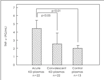
The concentration of TNF-α in the plasmas of the acute stage KD patients(4.4±1.6 pg/mL) was signifi- cantly higher than in those of the convalescent stage (2.5±1.4 pg/mL, p<0.05) or in the control subjects (1.9±0.5 pg/mL, p<0.01)(Fig. 1). However, the level was lower than the rhTNF-α concentration needed to up-regulate ICAM-1(10 pg/mL).14)
Serum IgM-AECA concentrations
The IgM-AECA titers were significantly higher in the sera of KD patients in the acute stage(0.85±0.64) than those in the convalescent stage(0.36±0.27, p<
0.01) or in the sera of the control subjects(0.35±0.14, p<0.01). Using a cut-off point of 0.63, the mean+2SD in the sera of the control subjects, 14 of the 22(64%) KD patients in the acute stage and 2(9%) in the conva- lescent stage were positive for IgM-AECA. None of the sera of the control subjects were positive for IgM-AECA
7
6
5
4
3
2
1
0
TNF-α(PG/mL)
p<0.05 p<0.01
Acute KD plasmas
n=22
Convalescent KD plasmas
n=22
Control plasmas n=13
Fig. 1. Comparison of plasma TNF-α measured by ELISA. The con- centration of TNF-α in the acute KD plasmas was significantly hi- gher than those in the convalescent KD (p<0.05) or control plasmas (p<0.01). TNF-α: tumor necrosis factor-α, KD: Kawasaki disease.
(Fig. 2). The titers in the sera of those in the acute stage declined in the convalescent stage in all KD pa- tients. Two KD patients with highest IgM-AECA levels developed coronary aneurysms, and their sera in the convalescent stage remained IgM-AECA positive.
The clinical and laboratory findings were compared between the sera of KD patients with positive IgM-AECA in the acute stage(n=14) and those with negative IgM- AECA(n=8)(Table 1). The initial serum CRP and ESR levels in the former group were significantly higher than those in the latter(p<0.01 and p<0.05, respectively).
Effects of the KD sera on the ICAM-1 expression of cultured HCAEC
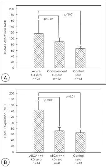
The ICAM-1 expression of HCAEC treated with the sera of KD patients in the acute stage was significantly higher than that of HCAEC treated with the sera of those in the convalescent stage(117.1±46.7 vs. 88.9±
14.4, p<0.05) or the sera of the control subjects(117.1
±46.7 vs. 65.2±9.5, p<0.01)(Fig. 3A). The IgM-AE CA-positive KD sera of the acute stage significantly in- duced the ICAM-1 expression of HCAEC compared to the IgM-AECA-negative KD sera of the acute stage (143.3±37.5 vs. 71.2±11.8, p<0.01) or the sera of the control subjects(143.3±37.5 vs. 65.2±9.5, p<0.01).
There was no significant difference between the effect of
Table 1. Comparison of the clinical and laboratory findings of the KD patients according to the positivity of IgM-AECA in the acute sera
AECA (+) KD patients
AECA (-) KD patients p
No. of patients 14 8
Age (years) 02.6±01.6 02.2±01.2 NS
Duration of fever (days) 07.8±00.7 06.7±00.5 NS Conjunctivial injection 11 (79%) 7 (88%)
Oral mucosal changes 11 (79%) 6 (75%)
Rash 13 (93%) 8 (100%)
Extremity changes 10 (71%) 5 (63%) Cervical enlargement 10 (71%) 4 (50%) Coronary aneurysm 02 (14%) 0 (0%)
Initial WBC (×103/mm3) 13.7±04.2 16.2±04.8 Initial CRP (mg/dL) 10.8±06.0 05.5±01.4 <0.01 Initial ESR (mm/1 hr) 78.4±22.3 59.5±19.7 <0.05
TNF-α* (pg/mL) 04.5±01.6 04.1±01.3 NS
IgM-AECA 01.1±00.6 00.3±00.1 <0.01
*: in the plasma of the acute stage. KD: Kawasaki disease, AECA:
anti-endothelial cell antibody, WBC: white blood cell, CRP: C-re- active protein, ESR: erythrocyte sedimentation rate, TNF-α: tumor necrosis factor-α, NS: no significance
Fig. 3. Effects of the KD sera on the ICAM-1 expression of HCAEC.
HCAEC were incubated for 24 hours with 1:4 diluted sera from KD patients, with/without ACEA or control subjects, and were st- ained with a fluorescent monoclonal antibody against human ICAM- 1 prior to flow cytometry. Expression was indicated as the mean fluo- rescence intensity (MFI). A: the ICAM-1 expression of HCAEC tre- ated with the acute KD sera was significantly higher than those for HCAEC treated with the convalescent KD or control subjects. B: the IgM-AECA-positive acute KD sera significantly induced the ICAM-1 expression of HCAEC higher than the IgM-AECA-negative acute KD or control sera. KD: Kawasaki disease, ICAM-1: intercellular ad- hesion molecule-1, HCAEC: human coronary artery endothelial cells, AECA: anti-endothelial cell antibody.
200 180 160 140 120 100 80 60 40 20 0
ICAM-1 expression (MFI) p<0.05
p<0.01
Acute KD sera
n=22
Convalescent KD sera
n=22
Control sera n=13
A
200 180 160 140 120 100 80 60 40 20 0
ICAM-1 expression (MFI)
p<0.01
p<0.01
AECA (+) KD sera
n=14
AECA (-) KD sera
n=8
Control sera n=13
B
Fig. 2. Serum IgM-AECA levels measured by ELISA. The IgM- AECA titers were significantly higher in the acute KD sera than those in the convalescent KD or control sera (p<0.01, respectively). Using a cut-off point of 0.63, the mean±2SD in control sera, 14 of the 22 (64%) acute KD sera and 2 (9%) of the convalescent KD sera were positive for IgM-AECA. The lower solid line indicates the mean level of the control subjects, and the upper dotted line indicates the mean
±2SD of the control subjects. AECA: anti-endothelial cell antibody, KD: Kawasaki disease, CA: coronary aneurysm.
3.0
2.5
2.0
1.5
1.0
0.5
0
IgM-AECA ELISA ratio
○:Case with CA p<0.01 p<0.01
Acute n=22
Convalescent n=22
Control n=13
AECA-negative sera of the acute stage and that of the control subjects(Fig. 3B).
Effects of IgM-AECA and signal transduction inhi- bitors on the ICAM-1 protein expression of HCAEC The ICAM-1 expression of cultured HCAEC, incub- ated for 24 hours with 200 μg/mL of the AECA-po- sitive IgM, was significantly higher than that of HC AEC incubated with AECA-negative IgM(0.54±0.07 vs. 0.27±0.05, p<0.05) or the control IgM(0.54±0.07 vs. 0.29±0.05, p<0.05)(Fig. 4).
Pretreatment with SB203580(a P38 inhibitor) prior to exposure to AECA-positive IgM inhibited the ICAM-1 expression of HCAEC by 93%(0.27±0.02, p<0.05).
Pretreatment of DMAP(a JNK inhibitor) decreased the ICAM-1 expression by 83%(0.30±0.03, p<0.05), and that of parthenolide(an NF-κB inhibitor) reduced the ICAM-1 expression by 41%(0.41±0.04, p<0.05). In contrast, pretreatment with PD98059(an ERK1/2 in- hibitor) did not reduce the IgM-AECA-induced ICAM- 1 expression of HCAEC(0.61±0.05, p>0.05)(Fig. 5).
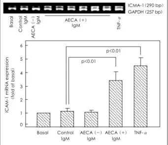
Effects of IgM-AECA and signal transduction inhi- bitors on the ICAM-1 mRNA expression of HCAEC The ICAM-1 mRNA expression of HCAEC incub- ated for 4 hours, with 200 μg/mL of the AECA-posi- tive IgM, was significantly higher than those of HCAEC incubated with the AECA-negative IgM(p<0.01) or the control IgM(p<0.01)(Fig. 6).
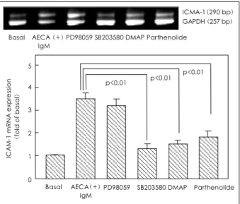
Pretreatment of the cells with SB203580, DMAP or parthenolide prior to exposure to AECA-positive IgM significantly inhibited the ICAM-1 mRNA expression (p<0.01 in all three comparisons). However, pretreat-
ment with PD98059 had no effect on the IgM-AECA- induced ICAM-1 mRNA expression of HCAEC(Fig. 7).
These results suggested that IgM-AECA induces the ICAM-1 expression of HCAEC via p38 and JNK MAPK pathways and activation of NF-κB.
0.8 0.7 0.6 0.5 0.4 0.3 0.2 0.1 0.0
ICAM-1 expression (O.D. at 450 nm)
p<0.05
p<0.05
Basal AECA (+)
IgM
Fig. 4. Effects of the IgM-AECA on the ICAM-1 expression of HCA EC. HCAEC were incubated for 24 hours with 200 μg/mL of IgM fractions purified from KD sera, with/without AECA or control sera.
The ICAM-1 expression was measured by ELISA, with the results ob- tained from eight independent experiments. Basal, unstimulated cells;
TNF-α, a positive control treated with 20 ng/mL of TNF-α. AECA:
anti-endothelial cell antibody, ICAM-1: intercellular adhesion mole- cule-1, HCAEC: human coronary artery endothelial cells, KD: Kaw- asaki disease, TNF-α: tumor necrosis factor-α.
Control IgM
TNF-α AECA (-)
IgM
0.8 0.7 0.6 0.5 0.4 0.3 0.2 0.1 0.0
ICAM-1 expression (O.D. at 450 nm)
p<0.05
Basal
Fig. 5. Effects of four different signal transduction inhibitors on the IgM-AECA-induced ICAM-1 protein expression of HCAEC. Cells were pretreated with PD98059 (50 μM for 1 hour), SB203580 (10 μM for 1 hour), DMAP dimethylaminopurine (DMAP, 1 mM for 15 minutes) or parthenolide (10 μM for 1 hour) prior to incubation with AECA-positive IgM (200 μg/mL) for 24 hours. The ICAM-1 expression was measured by ELISA, with the results obtained from eight independent experiments. Basal, unstimulated cells. AECA: anti- endothelial cell antibody, ICAM-1: intercellular adhesion molecule- 1, HCAEC: human coronary artery endothelial cells.
AECA (+) IgM
PD98059 SB203580 DMAP Parthenolide p<0.05 p<0.05
ICMA-1(290 bp) GAPDH (257 bp)
Basal Control IgM AECA (-) IgM TNF-αAECA (+) IgM
6
5
4
3
2
1 0 ICAM-1 mRNA expression (fold of basal)
p<0.01
p<0.01
Basal AECA (+)
IgM Control
IgM
TNF-α AECA (-)
IgM
Fig. 6. IgM-AECA-induced ICAM-1 mRNA expression of HCAEC.
Cells were incubated with 200 μg/mL of AECA-positive IgM, AE CA-negative IgM or control IgM. The isolated RNA samples were an- alyzed by reverse transcription-polymerase chain reaction (RT-PCR), using primers specific for ICAM-1 and GAPDH. Densitometric eva- luation of ethidium bromide stained gels was performed. The density of the ICAM-1 PCR products were corrected by that of the corres- ponding GAPDH PCR products, and then expressed as the fold va- lues of unstimulated cells. Data represent the mean±SD of six ex- periments. Basal, unstimulated cells; TNF-α, a positive control tre- ated with 20 ng/mL of TNF-α. AECA: anti-endothelial cell antibo- dy, ICAM-1: intercellular adhesion molecule-1, HCAEC: human coronary artery endothelial cells, TNF-α: tumor necrosis factor-α.
Discussion
Although the pathogenesis of KD is still not comple- tely understood, several pieces of clinical and laboratory evidence suggest a central role of EC dysfunction in the development of the disease and its cardiac complicat- ions. The cause of the EC dysfunction is unclear, but either a profound disturbances of immunoregulation associated with abnormal apoptosis of neutrophils or mononuclear cells, endothelial tissue infiltration by in- flammatory cells, or circulating immune complexes and diverse autoantibody have been proposed as possible immunologic causes. AECA is one of the pathogenic autoantibody candidates.
AECA were originally identified in the sera of patients with rheumatic diseases, due to their ability to react with rodent EC.15) The largest and most consistent groups of diseases in which AECA have been demonstrated are the systemic autoimmune and vasculitic diseases.16) The majority of these conditions share some degree of vas- cular injury, which initially led to the suspicion that AECA are merely epi-phenomena to the disruption of the vessel wall integrity.17) However, the correlation bet- ween AECA serum levels and disease activity, as well as the decline in their titers after treatment that led to a halt in disease progression, are indirect evidence for the significance of AECA.10)
Several studies have demonstrated the presence of
increased AECA titers in 26 to 72% of the patients with KD.7) The correlation between the levels of AECA and the disease activity, as well as the decline in AECA titers after treatment, are also evident in KD.18) Indeed, se- veral in vitro studies have reported the sera of patients with KD induced activation or damage of EC. However, the actual role of AECA in the development of KD re- mains debatable, as conflicting data exist concerning the ability of AECA to activate or damage EC, or even the frequency of AECA in KD patients.
In this study, about 2/3 of the KD patients had ab- normally high levels of IgM-AECA in their acute sera, and the serum levels of IgM-AECA declined in the con- valescent phase in all patients. The two patients with the highest AECA developed coronary aneurysms. These fi- ndings suggest the serum IgM-AECA levels may be rela- ted to the disease activity, and high serum levels may pre- dispose patients to the development of coronary lesions.
This study has also documented that acute KD sera induced the activation of HCAEC, which was assessed by the degree of ICAM-1 expression in the cells. IgM- AECA may be the very substance associated with this action of the sera. Previous studies have shown that ICAM-1 is involved in the pathogenesis of coronary artery lesions associated with KD. During the acute phase, KD patients with coronary artery lesions had a higher soluble ICAM-1 concentration in the sera than KD patients without coronary artery lesions.19) An im- munohistological study revealed that the EC of coro- nary arteries and skin of KD patients expressed ICAM- 1 as well as E-selectin.20) The pathogenetic consequences of an increased expression of ICAM-1 on the surface of EC in KD may be deduced as follows. ICAM-1 is con- stitutively present on the surface of EC, and its expre- ssion is augmented by several stimulating factors. ICAM- 1 has an important role in the migration of leukocytes to sites of inflammation, enabling firm adhesion and diapedesis of leukocytes via the interaction with αmβ 2 and αLβ2.21) The expression of ICAM-1 induced by sera from KD patients is likely to promote adhesion and transendothelial migration of leukocytes, resulting in the progression of vasculitis in KD. Indeed, the his- topathological findings in KD include; EC injury and the infiltration of neutrophils, monocytes and lympho- cytes into the walls of small and medium-sized blood vessels.22)
In this study, the plasma concentrations of TNF-α in the KD patients were measured to ascertain whether this cytokine induces activation or damage of EC. The mean concentration of TNF-α in the plasma of acute KD patients was 4.4 pg/mL, which was far less than the concentration of rhTNF-α needed to induce upregu- lation of ICAM-1, i.e. 10 pg/mL.14) Thus, it is suggested that the induction of ICAM-1 expression of HCAEC is mainly caused by IgM-AECA, but not by TNF-α, al-
ICMA-1(290 bp) GAPDH (257 bp) Basal AECA (+) PD98059 SB203580 DMAP Parthenolide
IgM
5
4
3
2
1
0 ICAM-1 mRNA expression (fold of basal)
p<0.01 p<0.01
Basal AECA(+) IgM
Fig. 7. Effects of four different signal transduction inhibitors on the IgM-AECA-induced ICAM-1 mRNA expression of HCAEC. Cells were pretreated with PD98059 (50 μM for 1 hour), SB203580 (10 μM for 1 hour), dimethylaminopurine (DMAP, 1 mM for 15 min- utes) or parthenolide (10 μM for 1 hour) prior to incubation with 200 μg/mL of AECA-positive IgM for 4 hours. The isolated RNA samples were analyzed by RT-PCR, using primers specific for ICAM- 1 and GAPDH, with the amount of ICAM-1 mRNA determined as described in Fig. 6. Basal, unstimulated cells. AECA: anti-endothelial cell antibody, ICAM-1: intercellular adhesion molecule-1, HCAEC:
human coronary artery endothelial cells.
PD98059 SB203580 DMAP Parthenolide p<0.01
though the plasma TNF-α levels are increased in KD patients.
Conflicting data have been presented on the necessi- ty of EC preactivation in other reports. Namely, AECA lyses cytokine activated cells only in some studies,8) while other studies have demonstrated that AECA from KD patients also influences resting cells.9)11) In this study, IgM-AECA was able to induce ICAM-1 expression of HCAEC in the resting state, and co-stimulation with IgM-AECA and 10 pg/mL of TNF-α resulted in no further increase of the ICAM-1 expression of HCAEC (data not shown).
In this study, the signal transduction pathways involv- ed in the regulation of ICAM-1 gene expression indu- ced by IgM-AECA were addressed using several specific inhibitors. SB203580 is a specific inhibitor of p38,23) and the kinase directly upstream to p38 MAPK. SB20 3580 attenuated IgM-AECA-induced ICAM-1 mRNA and the protein expression of HCAEC almost comple- tely, indicating the p38 MAPK signal transduction pa- thway is important to the upregulation of ICAM-1 expression in vascular EC.24) Recently, DMAP, a protein kinase inhibitor, has been identified as an useful reag- ent, which inhibits JNK MAPK by increasing the ex- pression of mitogen-activated protein phosphatase 2.25) Pretreatment of HCAECs with DMAP decreased the IgM-AECA-induced ICAM-1 mRNA in addition to the protein expression, suggesting the involvement of JNK kinase as well as p38 MAPK. It has been well establish- ed that inflammatory responses following exposure to various stimuli are highly dependent on the activation of NF-κB transcription factor, which plays an important role in the regulation of several gene expressions.26)27) The sequestration of NF-κB in the cytoplasm, and IκB phosphorylation leading to proteasomal degradation of IκB-α, result in activation and translocation of NF-κB into the nucleus, which is essential in the expression of several genes, such as ICAM-1.27) In this study, the ex- pression of ICAM-1 induced by IgM-AECA was parti- ally abolished by the specific NF-κB inhibitor, parthe- nolide, indicating that activation of NF-κB, at least pa- rtially, is involved in the AECA-induced expression of ICAM-1. This result is consistent with the report that AECA reacts with endothelial membrane antigen and induces a pro-inflammatory endothelial phenotype th- rough the activation of NF-κB.26) Thus, the activations of p38 MAPK and JNK, as well as NF-κB, appear to be involved in the IgM-AECA-induced expression of ICAM- 1 of HCAEC. However, it remains unclear how the ac- tivations of p38 MAPK and JNK are associated with ICAM-1 gene expression. In EC, p38 and JNK MAPK can be activated and rapidly translocated to the nucleus, where they phosphorylate and activate the transcription factors AP-1, Ets, C/EBP and NF-κB, etc. The bindings sites of these factors exist in the ICAM-1 promoter,
which suggests that the upregulation of ICAM-1 expre- ssion of EC may be mediated by p38 and JNK MAPK activating these transcription factors.28)29)
In summary, about 2/3 of patients with KD had IgM-AECA in their acute sera, and the titers of IgM- AECA decreased in the convalescent phase. Patients with higher AECA titers were prone to develop coronary aneurysms. Acute KD sera and purified IgM-AECA were able to induce upregulation of the ICAM-1 expre- ssion of HCAEC in vitro. JNK, p38 MAPK and NF-κ B signaling pathways may be involved in the upregula- tion of the ICAM-1 expression of HCAEC induced by IgM-AECA. Thus, the IgM-AECA in KD patients may play an important role in the development of coronary artery lesions. Further studies will be required to inve- stigate the signal transduction pathways involved in the regulation of ICAM-1 gene expression induced by IgM- AECA.
■ Acknoewledgments
This study was supported by a grant from the Korean Society of Cir- culation(Industrial-educational cooperation 2003) and a grant (04- 2003-016-0) from the Seoul National University Hospital Research Fund.
REFERENCES
1) Kawasaki T, Kosaki F, Okawa S, Shugematsu I, Yanagawa H. A new infantile acute febrile mucocutaneous lymph node syndrome (MLNS) prevailing in Japan. Pediatrics 1974;54:271-6.
2) Cho HJ, Yu JJ, Kim HS. The detection of acute phase Kawasaki myocardites via echocardiographic functional studies. Korean Circ J 2001;31:1318-23.
3) Kim BK, Lee BK, Choi DH, et al. Coronary stenting in 15 year- old boy with artery stenosis seconary to Kawasaki disease.
Korean Circ J 2000;30:1300-6.
4) Lee SY, Gwon HC, Park SW, et al. Acute myocardial infarction in young patient probably due to Kawasaki disease. Korean Circ J 2001;31:119-24.
5) Lo SK, van Seventer GA, Levin SM, Wright SD. Two leukocyte receptors (CD11a/CD18 and CD11b/CD18) mediate transient adhesion to endothelium by binding to different ligands. J Imm- unol 1989;143:3325-9.
6) Dustin ML, Singer KH, Tuck DT, Springer TA. Adhesion of T lymphoblasts to epidermal keratinocytes is regulated by inter- feron gamma and is mediated by intercellular adhesion molecule 1 (ICAM-1). J Exp Med 1988;167:1323-40.
7) Grunebaum E, Blank M, Cohen S, et al. The role of anti-endoth- elial cell antibodies in Kawasaki disease: in-vitro and in-vivo st- udies. Clin Exp Immunol 2002;130:233-40.
8) Leung DY, Collins T, Lapierre LA, Geha RS, Pober JS. Im- munoglobulin M antibodies present in the acute phase of Kawa- saki syndrome lyse cultured vascular endothelial cells stimulated with gamma interferon. J Clin Invest 1986;77:1428-35.
9) Kaneko K, Savage CO, Pottinger BE, Shah V, Pearson JD, Ki- llon MJ. Antiendothelial cell antibodies can be cytotoxic to en- dothelial cells without cytokine pre-stimulation and correlate with ELISA antibody measurement in Kawasaki disease. Clin Exp Immunol 1994;98:264-9.
10) Chan TM, Frampton G, Jayne DR, Perry GJ, Lockwood CM, Cameron JS. Clinical significance of anti-endothelial cell anti-
bodies in systemic vasculitis: a longitudinal study comparing anti-endothelial cell antibodies and anti-neutrophil cytoplasm antibodies. Am J Kidney Dis 1993;22:387-92.
11) Fujieda M, Oishi N, Kurashige T. Antibodies to endothelial cells in Kawasaki disease lyse endothelial cells without cytokine pre- treatment. Clin Exp Immunol 1997;107:120-6.
12) Chang L, Karin M. Mammalian MAP kinase signalling cascades.
Nature 2001;410:37-40.
13) Cohen P. The search for physiological substrates of MAP and SAP kinases in mammalian cells. Trends Cell Biol 1997;7:353-61.
14) Inoue Y, Kimura H, Kato M, Okada Y, Morikawa A. Sera from patients with Kawasaki disease induce intercellular adhesion molecule-1 but not Fas in human endothelial cells. Int Arch All- ergy Immunol 2001;125:250-5.
15) Lindquist KJ, Osterland CK. Human antibodies to vascular en- dothelium. Clin Exp Immunol 1971;9:753-60.
16) Meroni PL, Youinou P. Endothelial cell antibodies. In: Peter J, Shoenfeld Y, editors. Textbook of Autoantibodies. Amsterdam:
Elsevier;1996. p.245-8.
17) Meroni PL, D’Cruz D, Khamashta MA, Youinou P, Hughes GR.
Anti-endothelial cell antibodies: only for scientist or for clini- cians too? Clin Exp Immunol 1996;104:199-202.
18) Tizard EJ, Baguley E, Hughes GR, Dillon MJ. Antiendothelial cell antibodies detected by a cellular based ELISA in Kawasaki disease. Arch Dis Child 1991;66:189-92.
19) Furukawa S, Imai K, Matsubara T, et al. Increased levels of cir- culating intercellular adhesion molecule 1 in Kawasaki disease.
Arthritis Rheum 1992;35:672-7.
20) Leung DY, Contran RS, Kurt-Jones E, Burns JC, Newburger JW, Pober JS. Endothelial cell activation and high interleukin-1 se- cretion in the pathogenesis of acute Kawasaki disease. Lancet 1989;2:1298-302.
21) Butcher EC. Leukocyte-endothelial cell recognition: three (or
more) steps to specificity and diversity. Cell 1991;67:1033-6.
22) Fujiwara H, Hamashima T. Pathology of the heart in Kawasaki’s disease. Pediatrics 1978;61:100-7.
23) Beyaert R, Cuenda A, Vanden Berghe W, et al. The p38/RK mi- togen-activated protein kinase pathway regulates interleukin-6 synthesis response to tumor necrosis factor. EMBO J 1996;15:
1914-23.
24) Yan W, Zhao K, Jiang Y, et al. Role of p38 MAPK in ICAM-1 expression of vascular endothelial cells induced by lipopolysa- ccharide. Shock 2002;17:433-8.
25) de Cesaris P, Starace D, Starace G, Filippini A, Stefanini M, Ziparo E. Activation of Jun N-terminal kinase/stress-activated protein kinase pathway by tumor necrosis factor α leads to in- tercellular adhesion molecule-1 expression. J Biol Chem 1999;
274:28978-82.
26) Yazici ZA, Raschi E, Patel A, et al. Human monoclonal anti- endothelial cell IgG-derived from a systemic lupus erythemato- sus patient binds and activates human endothelium in vitro. Int Immunol 2001;13:349-57.
27) Chen CC, Chen JJ, Chou CY. Protein kinase C but not p44/42 mitogen- activated protein kinase, p38, or c-Jun NH(2)-terminal kinase is required for intercellular adhesion molecule-1 expre- ssion mediated by interleukin-1: Involvement of sequential acti- vation of tyrosine kinase, nuclear factor-kappaB-inducing kinase, and IkappaB kinase 2. Mol Pharmacol 2000;58:1479-89.
28) Arbabi S, Garcia I, Bauer GJ, Maier RV. Alcohol (ethanol) in- hibits IL-8 and TNF: role of the p38 pathway. J Immunol 1999;
162:7441-5.
29) Yoshizumi M, Fujita Y, Izawa Y, et al. Ebselen inhibits tumor ne- crosis factor-induced c-Jun N-terminal kinase activation and adhesion molecule expression in endothelial cell. Exp Cell Res 2004;292:1-10.