INTRODUCTION
Cleft lip and palate are most frequent congenital mal- formation in oral and maxillofacial region
1). In general, the palatogenesis can be divided by the exogenous fac- tors as well as the intrinsic factors. The exogenous factors are usually related to the growth of tongue, nasal carti- lage and primary palate, Meckel’s cartilage and cranial base. The intrinsic factors are usually related to cell migra-
tion, epithelial-mesenchymal transformation, synthesis and accumulation of extracellular matrix, and cell divi- sion
2). Palatal mesenchymes originated from neural crest cells are gradually located in the inner side of left and right maxillary process, and rapidly grow transversely to fuse each other to form palatal processes and shelves.
However, the merging processes of palate and lip should undergo rapid tissue degradation and regeneration in a short time. Thus, cleft formation is not directly resulted from fusion failure in the merging of palatal process, mesial and lateral nasal process which begins in the 6th week of gestation, but also the cleft can be resulted from the fusion defects in the tri-dimensional organization of palatal shelves and lip in the conjunction with the growth of adjacent structure including the secondary
* Corresponding author Suk-Keun Lee, D.D.S., Ph.D.
Department. of Oral Pathology, College of Dentistry Kangnung National University, Kangnung Daehangno 120 Gangneung, Gangwon, 210-702, Korea
Tel: 82-33-640-2228 FAX: 82-33-642-6410
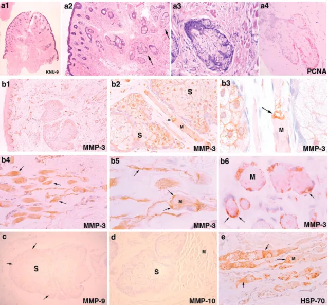
Over-expression of MMP-3 in the fissured tissue of cleft lip and palate
Young-Wook Park
1, Bong-Gi Min
1, Ji-Hyuck Kim
1, Soung-Min Kim
1, Young-Joon Lee
2, Sang-Shin Lee
2, Suk-Keun Lee
2, Huck-Soo Moon
3, Je-Geun Chi
41
Department of Oral and Maxillofacial Surgery,
2Department of Oral Pathology, College of Dentistry, Kangnung National University,
3
Department of Preventive Dentistry, College of Dentistry, Seoul National University,
4