https://doi.org/10.22283/qbs.2018.37.1.57
pISSN 2288-1344 eISSN 2508-7185 Original Article
─57─
Introduction
Obesity is a high risk factor for developing many human ill-
nesses, including type 2 diabetes, hyperlipidemia, and cancer [1]. It can be induced by many causatives, including imbal- anced energy homeostasis, nutritional and environmental fac- tors, and genetic and endocrine abnormalities[2,3]. Accumu- lating evidence indicates that adipocytes are highly special- ized cells that are involved in energy homeostasis by regulat- ing energy storage and release in response to changing nutri-
Anti-adipogenic Effect and Mechanism in 3T3-L1 Preadipocytes by Cyclosporin A, an Immunosuppressant
Tae-Yun Lee
1, Yu-Kyoung Park
2, Byeong-Churl Jang
2,*
1Department of Microbiology, College of Medicine, Yeungnam University, 170 Hyeonchung-ro, Nam-gu, Daegu 42415, Korea
2Department of Molecular Medicine, College of Medicine, Keimyung University, 1095 Dalgubeoldaero, Dalseo-gu, Daegu 42601, Korea
(Received May 13, 2018; Revised May 23, 2018; Accepted May 25, 2018)
AbstrAct
Adipogenesis, also called the differentiation of preadipocytes, leads to the phenotype of mature adipocytes. However, excessive adipogenesis is closely linked to the development of obesity. Cyclosporin A, a neutral lipophilic cyclic undecapeptide isolated from the fungus Hypocladium inflatum gams, is known for several biological activities, including immunosuppression and anti-inflammation. Little is known about the relationship between cyclosporin A and obesity. In this study, we investigated the effect of cyclosporin A on adipogenesis in 3T3-L1 murine preadipocytes. 3T3-L1 preadipocyte differentiation was evaluated by Oil Red O staining and AdipoRed assay. The expression and phosphorylation levels of adipogenesis-related proteins in differentiating 3T3-L1 cells were evaluated by Western blotting. The mRNA expression levels of adipogenesis-related proteins in differentiating 3T3-L1 cells were measured by RT-PCR. Cyclosporin A considerably inhibited lipid accumulation and reduced triglyceride(TG) contents during the differentiation of 3T3-L1 preadipocytes into adipocytes in a concentration-dependent manner, suggesting its anti-adipogenic effect. Treatment with cyclosporin A at the concentrations tested was not cytotoxic to 3T3-L1 cells. Mechanistically, at a 10μM concentration, cyclosporin A strongly down-regulated the protein and mRNA expressions of two adipogenic transcription factors, CCAAT/enhancer-binding protein-α(C/EBP-α) and peroxisome proliferator-activated receptor-γ(PPAR-γ) in differentiating 3T3-L1 cells. Furthermore, at a 10μM concentration, cyclosporin A reduced the phosphorylation levels of signal transducers and activator of transcription-3(STAT-3), other adipogenic transcription factor, and suppressed the protein and mRNA expressions of perilipin A, a protein that binds to and stabilizes lipid droplets, in differentiating 3T3- L1 cells. However, treatment with 10μM of cyclosporin A did not modulate the protein and mRNA expressions of fatty acid synthase during 3T3-L1 preadipocyte differentiation. Collectively, these results show that cyclosporin A inhibits adipogenesis in differentiating 3T3-L1 cells, and that this inhibition is mediated through the reduced expression and phosphorylation levels of C/EBP-α, PPAR-γ, perilipin A, and STAT-3.
Key words : Cyclosporin A, Adipogenesis, C/EBP-α, PPAR-γ, 3T3-L1
* Correspondence should be addressed to Byeong-Churl Jang, Professor, Department of Molecular Medicine, College of Medicine, Keimyung University, 1095 Dalgubeol-daero, Dalseo-Gu, Daegu 42601, Republic of Korea. Tel: +82-10-2743-4377, Fax: +82-53-580-3792, E-mail: jangbc123@
gw.kmu.ac.kr
Available Online at https://qbs.kmu.ac.kr:442/
tional needs[4]. It also has been recently demonstrated that adipose tissue plays a critical role in the control of energy meta- bolism by secreting adipocytokines[5,6]. However, excessive expansion of the adipose tissue, mainly due to abnormal in- crease in preadipocyte differentiation, also called adipogene- sis, leads to the development of obesity and obesity related diseases[7,8]. Thus, any substance that blocks excessive adi- pogenesis could be a preventive and/or therapeutic option against obesity.
Adipogenesis is the process during which fibroblast-like preadipocytes develop into mature adipocytes[9]. This pro- cess is largely regulated by the action of many adipogenic transcription factors, including CCAAT/enhancer-binding proteins(C/EBPs), peroxisome proliferator-activated recep- tors(PPARs), and Janus-activated protein kinases(JAKs)/sig- nal transducer and activator of transcriptions(STATs) signal- ing complexes[10-12]. In addition, there are many studies showing that the expression and activity of lipogenic enzymes, such as fatty acid synthase(FAS) and lipid droplet(LD) asso- ciated proteins like perilipin A, are crucial for adipocyte dif- ferentiation[13,14]. Participation of a number of signaling protein kinases and factors, including extracellular signal-reg- ulated protein kinase-1/2(ERK-1/2), protein kinase A(PKA), and adenosine 3′,5′-cyclic monophosphate(cAMP) in the con- trol adipocyte differentiation also has been previously pro- posed[15,16].
Cyclosporin A(CsA) is a neutral lipophilic cyclic undeca- peptide isolated from the fungus Hypocladium inflatum gams and is known for immunosuppressive, anti-nephrotic, and anti-skin inflammatory activities[17-19]. As of now, neither the anti-adipogenic effect nor the action mechanism of cyclo- sporin A is reported. In this study, we investigated whether cyclosporin A inhibits adipogenesis in 3T3-L1 murine preadi- pocytes. Our study shows that cyclosporin A inhibits lipid accumulation in differentiating 3T3-L1 adipocytes, and that this inhibition is in part due to the reduced expression and phosphorylation levels of C/EBP-α, PPAR-γ, perilipin A, and STAT-3.
Materials and Methods
1. Materials
Cyclosporin A was purchased from Selleckchem(Houston, TX, USA). Anti-C/EBP-α, anti-PPAR-γ, anti-phospho(p)-
STAT-3(Y705), and anti-STAT-3 antibodies were purchased from Santa Cruz Biotechnology(Delaware, CA, USA). Anti- perilipin A antibody was purchased from Bio Vision.(Milpi- tas, CA, USA). Anti-FAS antibody was purchased from BD Bioscience(San Jose, CA, USA). Enhanced chemilumines- cence(ECL) reagent was bought from Advansta(Menlo Park, CA, USA). Anti-β-actin antibody, 3-isobutyl-1-methylxan- thine(IBMX), dexamethasone, and insulin were purchased from Sigma(St. Louis, MO, USA).
2. 3T3-L1 cell culture and differentiation
3T3-L1 preadipocytes(ATCC, Manassas, VA, USA) were grown up to the contact inhibition stage in DMEM supple- mented with 10% heat-inactivated fetal calf serum(FBS) (Gibco, Grand Island, NY, USA) and penicillin/streptomycin (Welgene, Daegu, Korea). 3T3-L1 preadipocytes were in- duced to differentiate with DMEM supplemented with 10%
FBS plus a cocktail of hormones(MDI), containing 0.5mM IBMX(M), 0.5μM dexamethasone(D), and 5μg/mL insulin (I) in the absence(vehicle control, 0.1% DMSO) or presence of cyclosporin A at the indicated concentrations for 2 days.
The differentiation media was removed from cells, and cells were grown with fresh DMEM supplemented with 10% FBS and 5μg/mL insulin in the absence or presence of cyclosporin A at the indicated concentrations for additional 3 days. The conditioned cells were fed with DMEM containing 10% FBS in the absence or presence of cyclosporin A for additional 3 days.
3. Oil Red O staining
On day 8 of differentiation, the control or cyclosporin A- treated 3T3-L1 cells were washed with PBS, fixed with 10%
formaldehyde for 2h at room temperature(RT) washed with 60% isopropanol and dried. The fixed cells were stained with Oil Red O working solution(Sigma, St. Louis, MO, USA) for 1h at RT and washed with distilled water. Lipid droplets were observed using a light microscopy(Nikon, TS100, Japan).
4. Cell count analysis
On day 8 of differentiation, the survival of control or cyclo- sporine A-treated 3T3-L1 cells was assessed by trypan blue exclusion assay. The number of cells survived was counted using phase contrast microscopy in triplicate. Data are mean±
standard deviation(SD) of three independent experiments.
Survival was expressed as a percentage of vehicle control.
5. Measurement of cellular triglyceride(TG) contents by AdipoRed assay
On day 8 of differentiation, cellular lipid contents were measured with AdipoRed Assay Reagent kit in consonance with the manufacturer’s instructions(Lonza, Basel, Switzer- land). Fluorescence was measured after a 10min incubation on Victor3 (Perkin Elmer, Waltham, MA, USA) with excita- tion at 485nm and emission at 572nm. The AdipoRed assay was done in triplicates.
6. Preparation of cellular proteins
3T3-L1 cells were washed with PBS and lysed in a modi- fied RIPA buffer at a designated time point. The cell lysates were collected and centrifuged at 12,000rpm for 15min at 4°C. The supernatant containing cellular proteins was saved, and protein concentrations were determined with Pierce BCA Protein Assay Kit(Thermo scientific, Rockford, IL, USA).
7. Western blot analysis
Forty micrograms of proteins were separated by SDS-PAGE and transferred onto nitrocellulose membranes(Millipore, Bedford, MA, USA). The membranes were washed with Tris- buffered saline(10mM Tris-Cl, 150mM NaCl, pH 7.5) sup- plemented with 0.05%(v/v) Tween 20(TBST) followed by blocking with TBST containing 5%(w/v) non-fat dried milk.
The membranes were incubated overnight with specific pri- mary antibodies at 4°C. The membranes were exposed to sec- ondary antibodies conjugated to horseradish peroxidase for 2 h at RT and treated with ECL reagent.
8. Reverse transcription-polymerase chain reaction (RT-PCR)
Total cellular RNA in the control or cyclosporin A-treated 3T3-L1 cells was isolated with the RNAiso Plus(TaKaRa, Kusatsu, Shiga, Japan). Three micrograms of total RNA were reverse transcribed using a random hexadeoxynucleotide primer and reverse transcriptase. Single stranded cDNA was amplified by PCR with the following primers: C/EBP-α sense 5′-TTACAACAGGCCAGGTTTCC-3′; antisense 5′-CTCTG GGATGGATCGATTGT-3′; PPAR-γ sense 5′-GGTGAAACT CTGGGAGATTC-3′; antisense 5′-CAACCATTGGGTCAGC TCTC-3′; FAS sense 5′-TTGCTGGCACTACAGAATGC-3′;
antisense 5′-AACAGCCTCAGAGCGACAAT-3′; perilipin A sense 5′-CTTTCTCGACACACCATGGAAACC-3′; antisense 5′-CCACGTTATCCGTAACACCCTTCA-3′; actin sense 5′- GGTGAAGGTCGGTGTGAACG-3′; antisense 5′-GGTAGG AACACGGAAGGCCA-3′. Expression levels of actin mRNA expression were used as an internal control as well as loading control.
9. Statistical analysis
The AdipoRed assay was done in triplicates and repeated three times. Data were expressed as mean±standard devia- tion(SD). The significance of difference was determined by One-Way ANOVA(Laerd Statistics, Chicago, IL, USA). All significance testing was based upon a p value of <0.05.
Results
1. Cyclosporin A inhibits lipid accumulation in differentiating 3T3-L1 cells
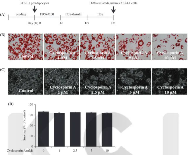
To see the cyclosporin A’s anti-adipogenic effect, we first investigated whether cyclosporin A inhibits lipid accumulation during the differentiation of 3T3-L1 preadipocytes into adipo- cytes by an Oil Red O staining. The experimental scheme of 3T3-L1 preadipocyte differentiation is shown in Fig. 1A. In the absence of cyclosporin A, there were many lipid droplets (LDs) in differentiating 3T3-L1 cells treated with vehicle con- trol(DMSO) on day 8(Fig. 1B). Remarkably, treatment with cyclosporin A for 8 days led to decrease in the amount of LDs in differentiating 3T3-L1 cells in a concentration-dependent manner. Light microscopy further revealed the cyclosporin A’s lipid-reducing effect on differentiating 3T3-L1 cells in a concentration-dependent manner(Fig. 1C). and no inhibitory effect of this drug at the concentrations tested on cell growth during the differentiation of 3T3-L1 preadipocytes into adipo- cytes(Fig. 1C). We next determined whether cyclosporine A treatment was cytotoxic to 3T3-L1 cells using cell count assay.
Cyclosporin A at the doses tested for 8 days had no inhibitory effect on growth(survival) of 3T3-L1 cells(Fig. 1D).
2. Cyclosporin A reduces TG contents in differentiating 3T3-L1 cells
We next sought to explore whether cyclosporin A reduces cellular triglyceride(TG) contents by an AdipoRed assay. Evi- dently, treatment with cyclosporin A for 8 days resulted in
decrease in the cellular levels of TG in differentiating 3T3-L1 cells in a concentration-dependent manner(Fig. 2). Similarly, 10μM cyclosporin A showed the most strong reductive effect on TG contents. Because of the most strong repressive effects on lipid accumulation and TG contents with no cytotoxicity, the concentration of 10μM of cyclosporin A was selected for further studies.
3. Cyclosporin A lowers expression and/or
phosphorylation levels of C/EBP-α, PPAR-γ, and STAT-3 in differentiating 3T3-L1 cells
In order to understand the mode of action of cyclosporin A-mediated anti-adipogenic effect, we next probed whether cyclosporin A affects cellular expression and phosphorylation
Fig. 1. Effect of cyclosporine A on lipid accumulation and growth in differentiating 3T3-L1 cells. (A) The experimental timescale of 3T3-L1 preadipocyte differentiation. (B-D) 3T3-L1 preadipocytes were induced to differentiate with induction medium in the absence(vehicle control;
0.1% DMSO) or presence of cyclosporine A at the indicated concentrations for 8 days. On day 8, cellular lipid contents were assessed by Oil Red O staining(B). On day 8, phase-contrast images of the cells were taken after the treatment(C). On day 8, the number of surviving cells in vehicle control or cyclosporine A-treated 3T3-L1 preadipocytes was measured by trypan blue dye exclusion(D). Data are mean±SD of three indepen- dent experiments, each done in triplicate. *p<0.05 vs. control.
(A)
(B)
(C)
(D)
3T3-L1 preadipocytes
Cyclosporin A(μM)
Survival(% of control)
120 90 60 30 0
Seeding FBS+MDI FBS+Insulin FBS
Day(D) 0 D2 D5 D8
Differentiated(mature) 3T3-L1 cells
0 1 2.5 5 10
Fig. 2. Effect of cyclosporine A on triglyceride(TG) contents in dif- ferentiating 3T3-L1 cells. 3T3-L1 preadipocytes were induced to differentiate with induction medium in the absence(vehicle control, 0.1% DMSO) or presence of cyclosporine A at the indicated concen- trations for 8 days. On day 8, cellular TG contents were quantified by AdipoRed assay. Values are mean±standard deviation(SD) of data from three independent experiments with three replicates. *p<0.05 vs. control.
Cyclosporin A(μM)
TG content(%)
120 90 60 30
0 0 1 2.5 5 10
* *
*
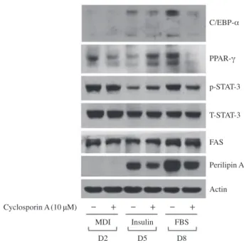
levels of adipogenesis-related proteins and enzymes during 3T3-L1 preadipocyte differentiation using Western blot analy- sis. As shown in Fig. 3A, cyclosporin A largely down-regulat- ed cellular protein levels of C/EBP-α and PPAR-γ in differen- tiating 3T3-L1 cells especially on day 8. Cyclosporin A also reduced STAT-3 phosphorylation levels in differentiating 3T3- L1 cells particularly on day 8. Total protein levels of STAT-3 were not affected by cyclosporin A treatment. Notably, as shown in Fig. 3B, cyclosporin A did not affect cellular protein levels of FAS but it considerably reduced cellular protein lev- els of perilipin A in differentiating 3T3-L1 cells on days 5 and 8. Control actin protein expression levels remained unchanged under these experimental conditions.
4. Cyclosporin A reduces mRNA levels of C/EBP-α and PPAR-γ in differentiating 3T3-L1 cells We next performed RT-PCR analysis to see whether down- regulation of C/EBP-α and PPAR-γ proteins by cyclosporin A is due to decrease in their transcripts. As shown in Fig. 4, cyclosporin A strongly reduced cellular mRNA levels of C/
EBP-α and PPAR-γ in differentiating 3T3-L1 cells on days 5
and 8. Control actin mRNA levels remained constant under these experimental conditions.
Discussion
Cyclosporin A is known for immunosuppressive, anti-neph- rotic, and anti-skin inflammatory activities[17-19]. In order to reposition cyclosporin A as an anti-obesity medicine, we here- in investigated the drug’s anti-adipogenic effects on 3T3-L1 adipocytes. Remarkably, our study shows that cyclosporin A inhibits adipogenesis in 3T3-L1 cells through control of the expression and/or phosphorylation levels of C/EBP-α, PPAR- γ, perilipin A, and STAT-3.
In initial experiments, we demonstrated that treatment with cyclosporin A at 10μM strongly inhibits lipid accumulation and reduces TG contents in differentiating 3T3-L1 cells, advocating the drug’s anti-adipogenic effect. It is well documented that the process of adipocyte differentiation is largely regulated by the expressions and activities of the family of C/EBPs and PPARs [20,21]. The present study clearly showed that cyclosporin A- treated 3T3-L1 cells have less expression levels of C/EBP-α and PPAR-γ, compared with the control cells. These data sug- gest that cyclosporin A may exerts its anti-adipogenic/lipid- lowering effect through down-regulation of these adipogenic transcription factors. On mechanistic levels, the cyclosporin A-induced protein down-regulation of C/EBP-α and PPAR-γ in differentiating 3T3-L1 cells especially on day 8 is associated with the drug’s ability to repress transcription(process) of C/
EBP-α and PPAR-γ, given that cyclosporin A reduces cellular mRNA levels of these transcription factors in the cells.
Fig. 3. Effect of cyclosporine A on expression and/or phosphoryla- tion levels of C/EBP-α, PPAR-γ, and STAT-3 in differentiating 3T3- L1 cells. 3T3-L1 preadipocytes were induced to differentiate with induction medium in the absence(vehicle control, 0.1% DMSO) or presence of cyclosporine A(10μM), and harvested at day 2, 5, and 8, respectively. At each time point, cellular proteins were extracted and analyzed by Western blot analysis. p-STAT-3, phosphorylated STAT- 3; T-STAT-3, total STAT-3.
Cyclosporin A(10μM)
Perilipin A Actin C/EBP-α
PPAR-γ p-STAT-3 T-STAT-3 FAS
MDI Insulin FBS D2 D5 D8 - + - + - +
Fig. 4. Effect of cyclosporine A on mRNA expression levels of FAS and perilipin A in differentiating 3T3-L1 cells. 3T3-L1 preadipocytes were induced to differentiate with induction medium in the absence (vehicle control, 0.1% DMSO) or presence of cyclosporine A(10μM), and harvested at day 2, 5, and 8, respectively. At each time point, cel- lular RNAs were extracted and analyzed by RT-PCR analysis.
Cyclosporin A(10μM)
PPAR-γ Actin C/EBP-α
MDI Insulin FBS D2 D5 D8 - + - + - +
Large body of evidence also indicate that increased phos- phorylation(activation) of the family of STATs, another adi- pogenesis-related transcription factor, is crucial for adipocyte differentiation[12,22]. In this study, we found that the cyclo- sporin A-treated 3T3-L1 cells have less phosphorylation levels of STAT-3 protein, compared with the control cells. These re- sults support the notion that cyclosporin A’s anti-adipogenic/
lipid-lowering effect herein is attributable to inhibition(dephos- phorylation) of STAT-3. Further assuming that cyclosporin A reduces levels of STAT-3 phosphorylation without affecting total STAT-3 protein levels in differentiating 3T3-L1 cells par- ticularly on day 8, the cyclosporin A-mediated STAT-3 dephos- phorylation in this study is likely to be not through suppression of de novo synthesis of STAT-3 protein but via inhibition of phosphorylation of the pre-existed STAT-3 in the cells. At this moment, how cyclosporin A inhibits STAT-3 phosphorylation in differentiating 3T3-L1 cells remains unclear. In general, cellular protein phosphorylation or dephosphorylation is regulated by protein kinase or phosphatase. Given that cyclosporin A is an inhibitor of calcineurin, a calcium-dependent serine/threonine protein phosphatase, it is hypothesized that cyclosporin A in- hibits STAT-3 phosphorylation by inhibiting calcineurin in differentiating 3T3-L1 cells. Future studies are warranted to see whether calcineurin expression and/or activity is altered in differentiating 3T3-L1 cells on day 8 and cyclosporin A inhib- its the expression and/or activity of this phosphatase. It has been previously shown that calcineurin mediates the calcium- dependent inhibition of 3T3-L1 adipocyte differentiation and inactivation of calcineurin in 3T3-L1 cells by its inhibitor cy- closporin A or FK506 enhances the efficiency of adipogenesis [4]. However, as of now, the role of calcineurin in 3T3-L1 ad- ipocyte differentiation remains un certain. It will be thus impor- tant to investigate, in the future, whether gene silencing of en- dogenous calcineurin will alter STAT-3 phosphorylation in dif- ferentiating 3T3-L1 cells and the adipocyte differentiation in the absence or presence of cyclosporin A, which will directly unravel the drug’s STAT-3 dephosphorylation through the cal- cineurin-dependent or independent mechanism as well as the positive or negative role of calcineurin in 3T3-L1 adipocyte differentiation.
A notable finding in this study is cyclosporine A-mediated differential regulation of perilipin A and FAS expressions in differentiating 3T3-L1 cells. FAS is a lipogenic enzyme invol- ved in the synthesis of fatty acid[13]. Perilipin A binds to and stabilizes the newly formed LDs during adipocyte differentia- tion[14]. In this study, cyclosporin A decreases cellular levels
of perilipin A, but not FAS, in differentiating 3T3-L1 cells.
These results point out that the cyclosporin A’s anti-adipogenic/
lipid-lowering effect herein is further attributable to inhibition of perilipin A expression at the protein level. It will be neces- sary to see, in future, whether cyclosporin A inhibits mRNA expression of perilipin A in differentiating 3T3-L1 cells and what factors or signaling pathways(components) are respon- sible for the drug-mediated perilipin A protein(and mRNA) down-regulation.
Increasing evidence indicates that additional signaling pro- teins, including cAMP-activated protein kinase(AMPK), par- ticipate in adipogenesis. Indeed, multiple studies have demon- strated that AMPK activation leads to suppression of adipo- genesis[23,24]. Interestingly, there is a recent study that CSA induces AMPK activation, which leads to improvement of cardiac function at an early stage of sepsis in rats[25]. Until now, little is known about CSA regulation of AMPK. It will be interesting to investigate, in the future, whether CSA acti- vates AMPK in differentiating 3T3-L1 cells, which will be a part of the drug’s anti-adipogenic effect.
In differentiating or mature adipocytes, lipid is stored and accumulated in the form of triglyceride(TG), consisting of one glycerol and three fatty acid molecules. Considering that cyclosporine A inhibits lipid accumulation and reduces TG contents in differentiating 3T3-L1 cells without affecting FAS protein expression level, it is speculative that cyclosporine A may inhibit the enzymatic activity of FAS in the cells. Future experiments to analyze the FAS enzyme activity level in dif- ferentiating 3T3-L1 cells treated without or with cyclosporine A would be necessary to prove this speculation. Moreover, assuming the CSA’s inhibitory effect on lipid accumulation in differentiating 3T3-L1 cells, one may raise a possibility that the drug-mediated decrease in lipid accumulation may be due to the degradation of lipid by the drug’s lipolytic activity in differentiating 3T3-L1 cells. It will be thus important to clari- fy whether CSA has a lipolytic activity in differentiating as well as differentiated 3T3-L1 cells, which will provide better understanding of the drug’s lipid lowering effect on fat cells.
In summary, this is the first study reporting that cyclosporin A has anti-adipogenic effect on differentiating 3T3-L1 cells through the reduced expression and/or phosphorylation levels of PPAR-γ, C/EBP-α, perilipin A, and STAT-3. Although important questions such as anti-adipogenic effect of cyclo- sporin A on obese animal models remain to be resolved, the findings presented herein advocate cyclosporin A as a poten- tial therapeutics for the treatment of obesity.
Acknowledgements
This work was supported by Yeungnam University Research Grant of 2016.
References
1. Kopelman PG. Obesity as a medical problem. Nature 2000;
404:635-643.
2. Ahima RS, Flier JS. Adipose tissue as an endocrine organ.
Trends Endocrinol Metab 2000;11:327-332.
3. Havel PJ. Control of energy homeostasis and insulin action by adipocyte hormones: leptin acylation stimulating protein, and adiponectin. Curr Opin Lipidol 2002;13:51-59.
4. Neal JW, Clipstone NA. Calcineurin mediates the Calcium- dependent inhibition of adipocyte differentiation in 3T3-L1 cells. J Biol Chem 2002;277:49776-49781.
5. Spiegelman BM, Flier JS. Obesity and the regulation of energy balance. Cell 2001;104:531-543.
6. Ouchi N, Parke JL, Lugus JJ, Walsh K. Adipokines in inflam- mation and metabolic disease. Nat Rev Immunol 2011;11:85- 97.
7. Melinikova I, Wages D. Anti-obesity therapies. Nat Rev Drug Discov 2006;5:369-370.
8. Bays HE. ‘‘Sick fat,’’ metabolic disease, and atherosclerosis.
Am J Med 2009;122:S26-S37.
9. Ali AT, Hochfeld WE, Myburgh R, Pepper MS. Adipocyte and adipogenesis. Eur J Cell Biol 2013;92:229-236.
10. Cao Z, Umek RM, McKnight SL. Regulated expression of three C/EBP isoforms during adipose conversion of 3T3-L1 cells. Genes Dev 1991;5:1538-1552.
11. Farmer SR. Transcriptional control of adipocyte formation.
Cell Metab 2006;4:263-273.
12. Zhang K, Guo W, Yang Y, Wu J. JAK2/STAT3 pathway is involved in the early stage of adipogenesis through regulating C/EBPbeta transcription. J Cell Biochem 2011;112:488-497.
13. Lakshmanan MR, Nepokroeff CM, Porter JW. Control of the synthesis of fatty-acid synthetase in rat liver by insulin, gluca-
gon, and adenosine 3′:5′ cyclic monophosphate. Proc Natl Acad Sci USA 1972;69:3516-3519.
14. Kern PA, Di Gregorio G, Lu T, Rassouli N, Ranganathan G.
Perilipin expression in humanadipose tissue is elevated with obesity. J Clin Endocrinol Metab 2004;89:1352-1358.
15. Martini CN, Plaza MV, Vila MC. PKA-dependent and indepen- dent cAMP signaling in 3T3-L1 fibroblasts differentiation. Mol Cell Endocrinol 2009;298:42-47.
16. Prusty D, Park BH, Davis KE, Farmer SR. Activation of MEK/
ERKsignaling promotes adipogenesis by enhancing peroxi- some proliferator-activated receptor gamma(PPARgamma) and C/EBPalpha geneexpression during the differentiation of 3T3- L1 preadipocytes. J Biol Chem 2002;277:46226-46232.
17. Kahan BD, Koch SM. Current immunosuppressant regimens:
considerations for critical care. Curr Crit Care 2001;7:242-250.
18. Seikaly MG, Prashner H, Nolde-Hurlbert B, Browne R. Long- term clinical and pathological effects of cyclosporine in chil- dren with nephrosis. Pediatr Nephrol 2000;14:214-217.
19. Harper JI, Berth-Jones J, Camp RD, Dillon MJ, Finlay AY, Holden CA, et al. Cyclosporin for atopic dermatitis in children.
Dermatol 2001;203:3-6.
20. Lehrke M, Lazar MA. The many faces of PPARgamma. Cell 2005;123:993-999.
21. Rosen ED, MacDougald OA. Adipocyte differentiation from the inside out. Nat Rev Mol Cell Biol 2006;7:885-896.
22. Wang D, Zhou Y, Lei W, Zhang K, Shi J, Hu Y, et al. Signal transducer and activator oftranscription 3(STAT3) regulates adipocyte differentiation via peroxisome-proliferator-activated receptor gamma(PPARgamma). Biol Cell 2009;102:1-12.
23. Kang J, Park J, Kim HL, Jung Y, Youn DH, Lim S, et al.
Secoisolariciresinol diglucoside inhibits adipogenesis through the AMPK pathway. Eur J Pharmacol 2018;820:235-244.
24. Han MH, Jeong JS, Jeong JW, Choi SH, Kim SO, Hong SH, et al. Ethanol extracts of Aster yomena(Kitam.) Honda inhibit adipogenesis through the activation of the AMPK signaling pathway in 3T3-L1 preadipocytes. Drug Discov Ther 2017;11:
281-287.
25. Liu J, Chen D, Liu X, Liu Z. Cyclosporine A attenuates cardiac dysfunction induced by sepsis via inhibiting calcineurin and activating AMPK signaling. Mol Med Rep 2017;15:3739-3746.