Introduction
Obesity is a serious public health epidemic with about 2 billion adults overweight or obese. The association between obesity and adverse health consequences, including hyper-lipidemia, cardiovascular disease, type 2 diabetes, and can-cer, is well established1). As a result, obesity is now a mong
the leading factors for global morbidity and mortality2).
Increasing evidence also indicates that obesity is induced as a result of excessive adipocyte differentiation, also known as adipogenesis, in the adipose tissues and adipocytes3,4).
Any compound that inhibits excessive adipogenesis could therefore be a potential therapeutic option for obesity.
Adipogenesis is a biological process characterized by
https://doi.org/10.15429/jkomor.2020.20.2.109
JKOMOR
pISSN 1976-9334, eISSN 2288-1522
Original Article
Tanshinone I, an Active Ingredient of Salvia miltiorrhiza, Inhibits
Differentiation of 3T3-L1 Preadipocytes and Lipid
Accumulation in Zebrafish
Hyo-Shin Kwon, Byeong-Churl Jang
Department of Molecular Medicine, College of Medicine, Keimyung University
Received: Revised: Accepted: December 8, 2020 December 13, 2020 December 14, 2020
Objectives: Tanshinone I is a bioactive constituent in Salvia miltiorrhiza. At present, the anti-obesity effect and mechanism of tanshinone I are not fully understood. Here we in-vestigated the effect of tanshinone I on lipid accumulation in 3T3-L1 preadipocytes and zebrafish.
Methods: Lipid accumulation and triglyceride (TG) content in 3T3-L1 cells were de-termined by Oil Red O staining and AdipoRed assay, respectively. The expression and phosphorylation levels of adipogenic/lipogenic proteins in 3T3-L1 cells were evaluated by Western blotting. The messenger RNA (mRNA) expression levels of adipogenic/lipo-genic markers and leptin in 3T3-L1 cells were measured by reverse transcription poly-merase chain reaction (RT-PCR). Lipid accumulation in zebrafish was assessed by LipidGreen2 staining.
Results: Tanshinone I at 5 μM largely blocked lipid accumulation and reduced TG content in differentiating 3T3-L1 cells. Furthermore, tanshinone I decreased the expression of CCAAT/enhancer-binding protein-α (C/EBP-α), peroxisome proliferator-activated re-ceptor-γ (PPAR-γ), fatty acid synthase (FAS), acetyl CoA carboxylase (ACC), and peril-ipin A but also the phosphorylation of signal transducer and activator of transcription-3 (STAT-3) in differentiating 3T3-L1 cells. In addition, tanshinone I increased the phos-phorylation of adenosine 3’,5’-cyclic monophosphate (cAMP)-activated protein kinase (AMPK) while decreased the intracellular adenosine triphosphate (ATP) content with no change in the phosphorylation and expression of liver kinase-B1 in differentiating 3T3-L1 cells. Importantly, tanshinone I also reduced the extent of lipid deposit formation in de-veloping zebrafish.
Conclusions: These findings demonstrate that tanshinone I has strong anti-adipogenic effects on 3T3-L1 cells and reduces adiposity in zebrafish, and these anti-adipogenic ef-fect in 3T3-L1 cells are mediated through control of C/EBP-α, PPAR-γ, STAT-3, FAS, ACC, perilipin A, and AMPK.
Key Words: Tanshinone, Adipogenesis, AMP-activated protein kinases, 3T3-L1 cells, Zebrafish
Correspondence to: Byeong-Churl Jang Department of Molecular Medicine, College of Medicine, Keimyung University, 1095 Dalgubeoldaero, Dalseo-gu, Daegu 42601, Korea. Tel: +82-53-258-7404 Fax: +82-53-258-7403 E-mail: [email protected]
Copyright © 2020 by The Society of Korean Medicine for Obesity Research
morphological, cellular, and biochemical changes4). The
dif-ferentiation process requires fibroblast-like preadipocytes to develop into mature adipocytes3) under the control of
adipo-genesis-related transcription factors, including the family of CCAAT/enhancer-binding proteins (C/EBPs), peroxisome proliferator-activated receptors (PPARs), and signal trans-ducer and activator of transcription (STAT) proteins5-8),
pro-tein kinase A, extracellular signal-regulated propro-tein kin-ase-1/2 (ERK-1/2), adenosine 3’,5’-cyclic monophosphate (cAMP)9-11), lipogenic enzymes, such as fatty acid synthase
(FAS) and lipid droplet (LD) associated proteins like peril-ipin A12,13), and cAMP-activated protein kinase (AMPK) and
its downstream effector acetyl CoA carboxylase (ACC)14-17).
Central to the early stage of 3T3-L1 adipocyte differentiation is the expression and phosphorylation of STAT-3, which then induces C/EBP-β transcription6,18).
The potential application of natural products as tradi-tional herbal medicines for the treatment of obesity by re-ducing lipid accumulation, adipocyte cell size and/or stim-ulating lipid metabolism are well documented19). Tanshinone
I is a bioactive constituent among the tanshinones found in a traditional herbal medicine Red sage, also called Salvia miltiorrhiza. Recently, Red sage and its bioactive phy-tochemicals have attracted much attention for their pharma-cological effects, including anti-inflammatory, anti-diabetic, anti-cancer, and cardio-cerebrovascular protective activ-ities20,21). We have recently demonstrated that tanshinone
IIA, one of the tanshinones in Red sage, greatly inhibits adipogenesis in 3T3-L1 cells as well as in zebrafish22). This
research led us to hypothesize that tanshinone I might have similar or potentially superior anti-adipogenic effects.
The present study examined the ability of tanshinone I to regulate lipid accumulation in 3T3-L1 adipocytes and zebrafish. We report that tanshinone I strongly inhibits lipid accumulation in 3T3-L1 cells and zebrafish, and its anti-adi-pogenic/lipogenic effect on 3T3-L1 cells is associated with dramatic changes in the expression or activation status of central adipogenic program mediators C/EBP-α, PPAR-γ, FAS, ACC, perilipin A, STAT-3, and AMPK.
Materials and Methods
1. Materials
Polyclonal C/EBP-α antibody, monoclonal PPAR-γ anti-body, monoclonal STAT-3 antianti-body, monoclonal phos-pho-STAT-3 (p-STAT-3) antibody, polyclonal STAT-5 anti-body and polyclonal phospho-STAT-5 (p-STAT-5) antianti-body were from Santa Cruz Biotechnology (Delaware, CA, USA). Polyclonal p-AMPK antibody, monoclonal AMPK antibody, polyclonal p-ACC antibody, polyclonal ACC antibody, poly-clonal phospho-liver kinase-B1 (p-LKB-1) antibody, and polyclonal LKB-1 antibody were from Cell Signaling Technology (Danvers, MA, USA). Monoclonal FAS antibody was from BD Bioscience (San Jose, CA, USA). Tanshinone I was purchased from Selleckchem (Houston, TX, USA). Tanshinone I was prepared as a 10 mM stock solution in dimethyl sulfoxide (DMSO). Monoclonal β-actin antibody was from Sigma (St. Louis, MO, USA). Polyclonal perilipin A antibody was from Bio Vision (Milpitas, CA, USA). Unless stated all other reagents were from Sigma.
2. Differentiation of 3T3-L1 preadipocytes
3T3-L1 preadipocytes (ATCC, Manassas, VA, USA) were grown in Dulbecco's modified Eagle's medium (DMEM) supplemented with 10% fetal bovine serum (FBS; Gibco, Rockville, MD, USA) and penicillin-streptomycin (Welgene, Daegu, Korea). Cells were grown to confluence and then incubated for a further 2 days before differentiation was induced. Differentiation was induced by incubating 3T3-L1 preadipcoytes with DMEM supplemented with 10% FBS (Welgene) plus a cocktail of hormones (MDI): 0.5 mM IBMX (M), 0.5 μM dexa metha sone (D), a nd 5 μg/mL in-sulin (I) in the absence (vehicle control; DMSO, 0.1%) or presence of tanshinone I at the indicated concentrations. After 48 h MDI-induction, the differentiation medium was replaced with DMEM supplemented with 10% FBS and 5 μg/mL insulin in the presence or absence of tanshinone I at the indicated concentrations. The cells were then fed every other day with DMEM containing 10% FBS in the presence or absence of tanshinone I at the indicated concentrations
until day 8. On day 8, the preadipocytes became mature adipocytes that rounded-up and filled with many oil droplets.
3. Oil Red O staining
On day 8 of differentiation, vehicle control (DMSO) or tanshinone I-treated 3T3-L1 cells were washed twice with phosphate buffer saline (PBS), fixed with 10% form-aldehyde for 2 h at room temperature (RT), washed with 60% isopropanol and dried completely. The fixed cells were then stained with Oil red O working solution for 1 h at RT and then washed twice with distilled water. In addition, quantitation of Oil red O staining was determined by Image J (U.S. National Institutes of Health, Bethesda, MD, USA). Lipid droplets were also observed by light microscopy (Nikon, Tokyo, Japan).
4. Cell count analysis
Vehicle or tanshinone I-treated 3T3-L1 cell viability was assessed by trypan blue exclusion assay. Viable cells were counted using phase contrast microscopy in triplicate. Data are mean±standard error (SE) of three independent experiments. Survival was expressed as a percentage of vehicle control.
5. Quantification of cellular triglyceride (TG) content by AdipoRed assay
Intracellular lipid content of vehicle or tanshinone I-treat-ed 3T3-L1 cells were measurI-treat-ed using a commercially avail-able AdipoRed Assay Reagent kit according to the manu-facturer's instructions (Lonza, Walkersville, MD, USA). After a 10 min incubation, fluorescence was measured on a Victor3 (Perkin Elmer Inc., Waltham, MA, USA) with ex-citation at 485 nm and emission at 572 nm. TG content was expressed as a percentage of vehicle control.
6. Preparation of whole cell lysates
At the designated time points, vehicle or tanshinone I-treated 3T3-L1 cells were washed twice with PBS and proteins extracted using a modified RIPA buffer (50 mM Tris-Cl [pH 7.4], 150 mM NaCl, 0.1% sodium dodecyl sul-fate, 0.25% sodium deoxycholate, 1% Triton X-100, 1%
Nonidet P-40, 1 mM ethylenediaminetetraacetic acid (EDTA), 1 mM ethylene glycol tetraacetic acid (EGTA), proteinase inhibitor cocktail [1x]). The cell lysates were collected and centrifuged at 12,000 rpm for 20 min at 4°C. The super-natants were saved and protein concentrations determined by Bradford reagent (Bio-Rad, Hercules, CA, USA).
7. Western blot analysis
Proteins (50 μg) were separated by sodium dodecyl sul-fate-poiyacrylmide gel eletrophoresis (SDS-PAGE) (10%) and transferred onto nitrocellulose membranes (Millipore, Burlington, MA, USA). The membranes were washed with TBS (10 mM Tris, 150 mM Na Cl) with 0.05% (vol/vol) tween 20 (TBST) followed by blocking with TBST containg 5% (w/v) non-fat dried milk. The membranes were in-cubated overnight with antibodies specific for C/EBP-α (1:1,000), PPAR-γ (1:1,000), STAT-3 (1:2,000), p-STAT-3 (1:2,000), STAT-5 (1:2,000), p-STAT-5 (1:2,000), p-LKB-1 (1:1,000), LKB-1 (1:1,000), p-ACC (1:2,000), ACC (1:2,000), FAS (1:1,000), perilipin A (1:2,000) or β-actin (1:10,000) at 4°C. The membranes were then exposed to secondary antibodies coupled to horseradish peroxidase for 2 h at RT. The membranes were washed three times with TBST at RT. Immunoreactivities were detected by enhanced chemiluminescence (ECL) reagents. Equal protein loading was assessed by the expression levels of β-actin protein.
8. Reverse transcription polymerase chain reaction (RT-PCR) analysis
At the designated time point, total cellular RNA in ve-hicle or tanshinone I-treated 3T3-L1 cells was isolated with the RNA-zol-B (Tel-Test, Friendswood, TX, USA). Three micrograms of total RNA were reverse transcribed using a random hexadeoxynucleotide primer and reverse transcriptase. Single stranded cDNA was amplified by PCR with the fol-lowing primers. Primer sequences used are listed in Table 1. The PCR conditions applied were: C/EBP-α, 30 cycles of denaturation at 95°C for 30 s, annealing at 62°C for 30 s, and extension at 72°C for 30 s; PPAR-γ, 30 cycles of dena-turation at 95°C for 30 s, annealing at 53°C for 30 s, and
extension at 72°C for 30 s; FAS, 30 cycles of denaturation at 95°C for 15 s, annealing at 55°C for 40 s, and extension at 68°C for 45 s; perilipin A, 30 cycles of denaturation at 95°C for 1 min, annealing at 55°C for 1 min, and extension at 72°C for 1 min; leptin, 30 cycles of denaturation at 95°C for 1 min, annealing at 57°C for 1 min, and extension at 72°C for 1 min; ACC, 30 cycles of denaturation at 95°C for 1 min, annealing at 62°C for 1 min, and extension at 72°C for 1 min; β-actin, 25 cycles of denaturation at 95°C for 30 s, annealing at 57°C for 30 s, and extension at 72°C for 1 min. Expression levels of β-actin messenger RNA (mRNA) were used as an internal control to evaluate the relative mRNA expression of adipocyte-specific genes and leptin.
9. Measurement of intracellular adenosine triphos-phate (ATP) levels
3T3-L1 preadipocytes were seeded in 96-well plates and grown in the presence of differentiation media in the ab-sence or preab-sence of tanshinone I or 2-deoxyglucose (2-DG). On day 2, 5, and 8 of differentiation, intracellular ATP lev-els were measured by luciferase activity using a lumines-cence assay kit according to the manufacturer’s protocol (ATPLite-1step; Perkin Elmer Inc.). After 2 min incubation,
luminescence was measured on a Victor3 (Perkin Elmer Inc.).
10. Zebrafish experiment
A number of studies have demonstrated that zebrafish can be used to study lipid biology including lipid accumu-lation and metabolism22,23). We and others also have recently
shown that LipidGreen2 selectively stains lipid deposits in live zebrafish22,24,25). Zebrafish was maintained under
stand-ard conditions as previously described26). All experimental
protocols involving zebrafish were approved by the Animal Care and Committee of the Korea Research Institute of Chemical Technology. Two dpf (day post fertilization) lar-vae were arrayed in 24-well plates in which each well con-tains 10 larvae (n=10) and 1 mL egg water (0.6 g/L sea salt). The indicated concentrations of tanshinone I dissolved in DMSO were a dded to ea ch well a nd incuba ted in 37°C incubator. After 24 h, larvae were washed with egg water and stained with 5 μM LipidGreen2 solution24). After 10
min, larvae were washed 3 times with egg water. To acquire images, larvae were anesthetized in tricaine (Sigma) and mounted in 3% methylcellulose (Sigma). Images were ac-quired with a Leica MZ10 F stereomicroscope, Leica DFC425 camera, and Leica Application Suite software v4.5 (Leica
Gene Sequence
C/EBP-α Sense 5′-TTACAACAGGCCAGGTTTCC-3′
Antisense 5′-CTCTGGGATGGATCGATTGT-3′
PPAR-γ Sense 5′-GGTGAAACTCTGGGAGATTC-3′
Antisense 5′-CAACCATTGGGTCAGCTCTC-3′
FAS Sense 5′-TTGCTGGCACTACAGAATGC-3′
Antisense 5′-AACAGCCTCAGAGCGACAAT-3′
Perilipin A Sense 5′-CTTTCTCGACACACCATGGAAACC-3′
Antisense 5′-CCACGTTATCCGTAACACCCTTCA-3′
Leptin Sense 5′-CCAAAACCCTCATCAAGACC-3′
Antisense 5′-CTCAAAGCCACCACCTCTGT-3′
ACC Sense 5′-GTCAGCGGATGGGCGGAATG-3′
Antisense 5′-CGCCGGATGCCATGCTCAAC-3′
Actin Sense 5′-TCATGAAGTGTGACGTTGACATCCGT-3′
Antisense 5′-CCTAGAAGCATTTGCGGTGCACGATG-3′
RT-PCR: reverse transcription polymerase chain reaction, C/EBP-α: CCAAT/enhancer-binding protein-α, PPAR-γ: peroxisome proliferator-activated receptor-γ, FAS: fatty acid synthase, ACC: acetyl CoA carboxylase.
Microsystems, Wetzlar, Germany). Yolk size and fluorescence intensity were measured by ImageJ software27). Data are
mean±standard error of the mean.
11. Statistical analysis
Cell count analysis was done in triplicates and repeated three times. Data were expressed as mean±SE. The sig-nificance of difference was determined by one-way analysis of variance. All significance testing was based upon a P-value of <0.05. For assessment of the size and intensity of yolk in zebrafish, unpaired t-test was used to measure difference versus DMSO group (n=10). The statistical sig-nificance was based upon a P-value of ≤0.05 or ≤0.01.
Results
1. Tanshinone I concentration-dependently inhibits lipid accumulation and reduces TG content in differentiating 3T3-L1 cells
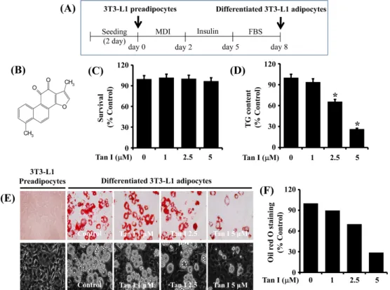
The protocol for 3T3-L1 preadipocyte differentiation is shown in Fig. 1A. Primarily, we investigated the treatment effect of various concentrations (1, 2.5, and 5 μM) of tan-shinone I (Fig. 1B) on cell growth (survival) during the dif-ferentiation of 3T3-L1 preadipocytes into adipocytes for 8 days using cell count analysis. Treatment with tanshinone I at the concentrations tested did not influence survival of 3T3-L1 cells (Fig. 1C). We next investigated the treatment
Fig. 1. Effects of tanshinone I on lipid accumulation, TG content, and cell survival during 3T3-L1 preadipocyte differentiation. (A) Protocol for 3T3-L1 preadipocyte differentiation. (B) is the chemical structure of tanshinone I. (C) 3T3-L1 preadipocytes were grown under the above-mentioned 3T3-L1 preadipocyte differentiation condition in Fig. 1A. On day 8, the number of surviving cells in vehicle control or tanshinone I-treated 3T3-L1 preadipocytes was measured by trypan blue dye exclusion. Data are mean±SE of three independent experiments, each done in triplicate. *P<0.05 vs. control (no chemical). (D) Quantification of the cellular TG content in vehicle control or tanshinone I-treated 3T3-L1 preadipocytes on day 8 by AdipoRed assay. Data are mean±SE of three independent experiments, each done in triplicate. *P<0.05 vs. control (no chemical). (E) Measurement of the cellular lipid (lipid droplets) in 3T3-L1 preadipocytes (undifferentiated) or differentiated adipocytes on day 8 of differentiation in the absence (vehicle control; DMSO, 0.1%) or presence of tanshinone I at the indicated concentrations by Oil red O staining (×400) (upper panels). Phase-contrast images were also taken after the treatment (lower panels). (F) Quantification of the Oil red O staining in vehicle control or tanshinone I-treated 3T3-L1 preadipocytes on day 8 by Image J. MDI: 0.5 mM IBMX, 0.5 μM dexamethasone, and 5 μ g/mL insulin, FBS: fetal bovine serum, Tan I: tanshinone I, TG: triglyceride, SE: standard error, DMSO: dimethyl sulfoxide.
effect of various concentrations of tanshinone I on intra-cellular TG content in 3T3-L1 cells on day 8 of differ-entiation using an AdipoRed assay. As shown in Fig. 1D, tanshinone I treatment concentration-dependently reduced cellular TG content during 3T3-L1 preadipocyte differentiation. We further investigated the treatment of tanshinone I at 1, 2.5, and 5 μM on lipid accumulation during the differ-entiation of 3T3-L1 preadipocytes into adipocytes using Oil red O staining. Treatment of the preadipocytes with tan-shinone I for 8 days markedly reduced the amounts of LDs in a concentration-dependent manner (Fig. 1E, upper pan-els). Evidently, treatment with 5 μM tanshinone I most strongly blocked the accumulation of LDs. In addition, the tanshinone I-mediated inhibition of LD accumulation in dif-ferentiating 3T3-L1 cells was observed by light microscopy (Fig. 1E, lower panels). Quantification of the Oil red O staining in vehicle control or tanshinone I-treated 3T3-L1
preadipocytes on day 8 was obtained by Image J (Fig. 1F). Since 5 μM of tanshinone I exhibited the most strong in-hibitory effect on lipid accumulation and TG content with no cytotoxicity, we chose this concentration for further studies.
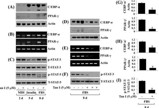
2. Tanshinone I decreases C/EBP-α, PPAR-γ, and phosphorylation levels of STAT-3 in differentiating 3T3-L1 cells
To gain a better understanding of the mechanisms asso-ciated with the anti-adipogenic effect of tanshinone I, we next measured the expression and phosphorylation levels (activity) of adipogenic transcription factors, C/EBP-α, PPAR-γ, and STAT-3/5, in differentiating 3T3-L1 cells treat-ed with vehicle control or tanshinone I (5 μM) using Western blotting and RT-PCR experiment. Tanshinone I considerably repressed protein (Fig. 2A) and mRNA (Fig. 2B) expressions of C/EBP-α and PPAR-γ, notably on days 5 and 8. Furthermore,
Fig. 2. Effect of tanshinone I on expression and/or phosphorylation of C/EBP-α, PPAR-γ, and STAT-3/5 in differentiating 3T3-L1 cells. (A-C) 3T3-L1 preadipocytes were induced to differentiate with induction medium in the absence (vehicle control, 0.1%) or presence of tanshinone I, and harvested at day 2, 5, and 8, respectively. Cellular protein and mRNA at the indicated time point were extracted and analyzed by Western blot (A, C) and RT-PCR (B) analysis, respectively. (D-F) Western blot (D,F) and RT-PCR (E) analysis in triplicate experiments on day 8, respectively. (G) and (H) are the densitometry data of (D) and (E), respectively. (I) is the densitometry result of (F). C/EBP-α: CCAAT/enhancer-binding protein-α, PPAR-γ: peroxisome proliferator-activated receptor-γ, STAT: signal transducer and activator of transcription, MDI: 0.5 mM IBMX, 0.5 μM dexamethasone, and 5 μg/mL insulin, FBS: fetal bovine serum, Tan I: tanshinone I, mRNA: messenger RNA, RT-PCR: reverse transcription polymerase chain reaction. *P<0.05 compared to the value of tanshinone I free control at the indicated day.
although tanshinone I did not inflect the phosphorylation or total expression levels of STAT-3 protein on days 2 and 5 of adipocyte differentiation, it considerably reduced STAT-3 phosphorylation levels without affecting the protein ex-pression levels on day 8 of adipocyte differentiation (Fig. 2C). Tanshinone I did not reduce phosphorylation and total expression levels of STAT-5 protein on days 2, 5, and 8 of adipocyte differentiation. Triplicate experiments confirmed the capability of tanshinone I to strongly reduce the protein and mRNA expressions of C/EBP-α and PPAR-γ, and the phosphorylation levels of STAT-3 on day 8 of differentiation (Fig. 2D-F). The densitometry data of Fig. 2D, 2E, and 2F a re shown in Fig. 2G, 2H, a nd 2I, respectively.
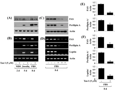
3. Tanshinone I reduces FAS, perilipin A, and leptin in differentiating 3T3-L1 cells
We next examined the effects of tanshinone I on ex-pressions of FAS (a lipogenic enzyme), perilipin A (an
LD-interacting protein), and leptin (an adipokine) during 3T3-L1 preadipocyte differentiation. Tanshinone I markedly reduced the protein (Fig. 3A) and mRNA (Fig. 3B) levels of FAS and perilipin A especially on days 5 and 8 of adipo-cyte differentiation. In addition, tanshinone I strongly re-duced cellular levels of the leptin mRNA transcripts on days 5 and 8. Triplicate experiments confirmed the inhibitory potential of tanshinone I on the protein expressions of FAS and perilipin A (Fig. 3C) and the mRNA expressions of FAS, perilipin A, and leptin (Fig. 3D) on day 8 of adipo-cyte differentiation. The densitometry data of Fig. 3C and 3D a re shown in Fig. 3E a nd 3F, respectively.
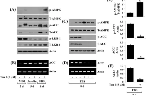
4. Tanshinone I differentially regulates the protein phosphorylation and total expression of AMPK, ACC, and LKB-1 in differentiating 3T3-L1 cells
AMPK is a major regulator of energy metabolism15,16),
and its activation (phosphorylation) leads to inhibition of
Fig. 3. Effect of tanshinone I on expression of FAS, perilipin A, and leptin in differentiating 3T3-L1 cells. (A, B) 3T3-L1 preadipocytes were induced to differentiate with induction medium in the absence (vehicle control, 0.1% DMSO) or presence of tanshinone I, and harvested at day 2, 5, and 8, respectively. Cellular protein and mRNA at the indicated time point were extracted and analyzed by Western blot (A) and RT-PCR (B) analysis, respectively. (C, D) Western blot (C) and RT-PCR (D) analysis in triplicate experiments on day 8, respectively. (E) and (F) are the densitometry data of (C) and (D), respectively. FAS: fatty acid synthase, MDI: 0.5 mM IBMX, 0.5 μM dexamethasone, and 5 μg/mL insulin, FBS: fetal bovine serum, Tan I: tanshinone I, DMSO: dimethyl sulfoxide, mRNA: messenger RNA, RT-PCR: reverse transcription polymerase chain reaction. *P<0.05 compared to the value of tanshinone I free control at the indicated day.
adipogenesis28). We thus probed whether tanshinone I affects
the expression and/or phosphorylation of AMPK during 3T3-L1 preadipocyte differentiation. In the absence of tan-shinone I, there was no or weak AMPK phosphorylation during 3T3-L1 preadipocyte differentiation (Fig. 4A). Tanshinone I slightly increased AMPK phosphorylation on day 2, while it caused a marked increase in AMPK phos-phorylation on day 8 of adipocyte differentiation. Tanshinone I did not affect total AMPK protein levels on days 2, 5, or 8 of adipocyte differentiation. ACC is a known downstream effector of AMPK and is involved in the biosynthesis of fatty acids14). We next measured the effect of tanshinone I
on the expression and/or phosphorylation levels of ACC during 3T3-L1 preadipocyte differentiation. There was a time-dependent increase in ACC phosphorylation during 3T3-L1 preadipocyte differentiation in the absence of tan-shinone I. However, total protein levels of ACC declined on days 2 and 8 of differentiating 3T3-L1 cells treated with tanshinone I, compared with the vehicle-treated cells. Given
that LKB-1 is a known upstream activator of AMPK phos-phorylation29), we next sought to explore whether LKB-1
protein is expressed and activated (phosphorylated) in dif-ferentiating 3T3-L1 cells and is regulated by tanshinone I. Notably, high levels of phosphorylated LKB-1 were ob-served on day 2 but greatly declined on days 5 and 8 of differentiating 3T3-L1 cells. Furthermore, there was a time-dependent increase in the total protein expression of LKB-1 during 3T3-L1 preadipocyte differentiation. Tanshinone I did not affect the phosphorylation or total protein levels of LKB-1 on days 2, 5, and 8 of adipocyte differentiation. In addition, although there was an enhancement of ACC mRNA on day 2 of differentiating 3T3-L1 cells treated with tanshinone I, there was a reduced expression of ACC mRNA on days 5 and 8 of differentiating 3T3-L1 cells treat-ed with tanshinone I, compartreat-ed with the mock-treattreat-ed cells (Fig. 4B). Triplicate experiments further revealed the ability of tanshinone I to largely increase levels of AMPK phos-phorylation but down-regulate those of ACC protein and
Fig. 4. Effect of tanshinone I on expression and phosphorylation of AMPK, ACC, and LKB-1 in differentiating 3T3-L1 cells. (A, B) 3T3-L1 preadipocytes were induced to differentiate with induction medium in the absence (vehicle control, 0.1% DMSO) or presence of tanshinone I, and harvested at day 2, 5, and 8, respectively. Cellular protein and mRNA at the indicated time point were extracted and analyzed by Western blot (A) and RT-PCR (B) analysis, respectively. (C, D) Western blot (C) and RT-PCR (D) analysis in triplicate experiments on day 8, respectively. (E) and (F) are the densitometry data of (C) and (D), respectively. AMPK: cAMP-activated protein kinase, ACC: acetyl CoA carboxylase, LKB-1: liver kinase-B1, MDI: 0.5 mM IBMX, 0.5 μM dexamethasone, and 5 μg/mL insulin, FBS: fetal bovine serum, Tan I: tanshinone I, DMSO: dimethyl sulfoxide, mRNA: messenger RNA, RT-PCR: reverse transcription polymerase chain reaction. *P<0.05 compared to the value of tanshinone I free control at the indicated day.
mRNA on day 8 of differentiation (Fig. 4C and 4D). The densitometry data of Fig. 4C and 4D are shown in Fig. 4E and 4F, respectively.
5. Tanshinone I reduces intracellular levels of ATP in differentiating 3T3-L1 cells
AMPK phosphorylation is induced by changes in cellular adenosine monophosphate (AMP)/ATP ratio30). We thus
ex-amined the effect of tanshinone I on intracellular ATP con-tents in differentiating 3T3-L cells. 2-DG, a glucose mim-etic that depletes intracellular ATP levels31), was included as
a control. Tanshinone I substantially reduced intracellular levels of ATP on days 2, 5, and 8 of adipocyte differ-entiation, which was comparable to the reductive effect by 2-DG (Fig. 5A-C).
6. Tanshinone I reduces lipid accumulation in the developing zebrafish
To test its in vivo efficacy, we next investigated the ef-fect of tanshinone I on lipid accumulation in zebrafish using LipidGreen2 staining. To this end, 2 dpf larvae were ex-posed to vehicle control (DMSO) or tanshinone I at the in-dicated concentrations (1, 10, or 100 μM) for 24 h. The conditioned larvae were washed, anesthetized, and stained with Lipid Green2 and visualized under bright field and fluorescence. In this study, the reduction of fluorescence was used as an indicator of inhibition of lipid accumulation. Notably, there was a significant decrease in the size of yolk in the group of larvae zebrafish treated with tanshinone I at the doses applied, compared with that in the control group (Fig. 6A). Moreover, there was a significant reduction in size (Fig. 6B and 6C) and intensity (Fig. 6B and 6D) of the fluorescence in the yolk in the group of larvae zebrafish
Fig. 5. Effect of tanshinone I on intracellular ATP levels in differentiating 3T3-L1 cells. (A-C) 3T3-L1 preadipocytes were induced to differentiate with induction medium in the absence (vehicle control, 0.1% DMSO) or presence of tanshinone I or 2-DG, a known ATP depleting agent, and harvested at day 2 (A), 5 (B), and 8 (C), respectively. The amounts of intracellular ATP at the indicated time point were measured by a ATP measurement kit. CPS: counts per second, 2-DG: 2-deoxyglucose, ATP: adenosine triphosphate, DMSO: dimethyl sulfoxide. *P<0.05 compared to the value of tanshinone I or 2-DG free control at the indicated day.
Fig. 6. Effect of tanshinone I on lipid accumulation in zebrafish. Two dpf larvae were exposed in the absence (DMSO) or presence of tanshinone I at the indicated concentrations. After 24 h, 3 dpf larvae were stained with LipidGreen2 and visualized under bright field (A) and fluorescence field (B). (C, D) Size and intensity of yolk (white dash in B) were quantified using the ImageJ software (U.S. National Institutes of Health, Bethesda, MD, USA), respectively. DMSO: dimethyl sulfoxide, Tan I: tanshinone I. Graph bars indicate mean±standard error of the mean. Unpaired t-test was used to analyze difference versus DMSO group, and statistical significance was set 0.05 and 0.01 (*P≤0.05, **P≤0.01). n=10.
treated with tanshinone I at the concentrations tested. Apparently, treatment with tanshinone I even at 1 μM was enough to significantly reduce the size and intensity of the fluorescence in the yolk in larvae zebrafish, supporting the potency of tanshinone I to inhibit lipid accumulation in de-veloping zebrafish.
Discussion
Obesity is associated with the development of type 2 dia-betes, insulin resistance, hyperlipidemia, and cancer1). There
is also a wealth of information that excessive preadipocyte differentiation (adipogenesis) and the resultant high fat ac-cumulation leads to the development of obesity2,3). Blockers
of excessive preadipocyte differentiation and adipogenesis may have therapeutic potential against obesity and related disorders. In this study, we demonstrated firstly that tan-shinone I has strong inhibitory effects on lipid accumulation in 3T3-L1 preadipocytes and zebrafish.
It is well documented that adipocyte differentiation is large-ly controlled by the expression and activity of adipogenic tran-scription factors C/EBP-α, PPAR-γ, and STAT-3/53-6,32). It has
been previously reported that other tanshinones such as tan-shinone IIA and cryptotantan-shinone inhibit adipogenesis in 3T3-L1 cells, and their anti-adipogenic effects are mediated through the reduced expression/activity/phosphorylation lev-els of C/EBP-α, PPAR-γ, and/or STAT-3/533,34). Of interest,
a recent study has demonstrated that tanshinone I has an-ti-obesity effects on a high fat diet fed mice and anti-adipo-genic effects on 3T3-L1 preadipocytes, which are mainly due to inhibition of early stage of differentiation through the attenuation of mitotic clonal expansion via cell cycle ar-rest, inactivation of p38 mitogen-activated protein kinase, ERK-1/2, and protein kinase B, and down-regulation of PPAR-γ35). However, a s of now, little is known a bout ta
n-shinone I regulation of C/EBP-α, PPAR-γ, and STAT-3/5 during 3T3-L1 preadipocyte differentiation. Here we found that tanshinone I reduced not only the protein and mRNA expressions of C/EBP-α and PPAR-γ but also the phosphor-ylation levels of STAT-3 during 3T3-L1 preadipocyte
differentiation. It is thus conceivable that the anti-adipo-genic effect of tanshinone I on 3T3-L1 cells is closely at-tributable to down-regulation of the expression and phos-phorylation levels of C/EBP-α, PPAR-γ, and STAT-3. It will be interesting to investigate, in the future, how tanshinone I regulates not only the transcription of C/EBP-α and PPAR-γ but also the phosphorylation of STAT-3 using phar-macological inhibition study and phosphoproteomics (kinome) approaches.
We have recently demonstrated that the cellular levels of FAS and perilipin A are increased during the differentiation of 3T3-L1 preadipocyte into adipocyte22,36). In a recent
study, it is also demonstrated that tanshinone I inhibits FAS expression in early stage of 3T3-L1 preadipocyte differ-entiation35). Until now, however, tanshinone I regulation of
FAS and perilipin A expression during 3T3-L1 preadipocyte differentiation process is unknown. Considering that FAS is a known lipogenic enzyme responsible for the synthesis of fatty acid37), and perilipin A interacts with and stabilizes the
newly formed LDs during 3T3-L1 preadipocyte differ-entiation12,38), the present findings that tanshinone I greatly
reduces the protein and mRNA levels of both FAS and per-ilipin A in differentiating 3T3-L1 cells strongly support the notion that down-regulation of FAS and perilipin A may further contribute to the tanshinone I’s anti-lipogenic and lipid-lowering effects. The adipose tissue, as an endocrine organ, synthesizes and secretes an array of adipocytokines, including leptin. The role of leptin in obesity and related disorders has been previously reported34). Tanshinone I
in-hibited leptin mRNA expression in differentiating 3T3-L1 cells, which further supports its potential utility for the pre-vention and/or treatment of obesity and related diseases in which excessive leptin production is problematic.
AMPK is a key regulator of energy metabolism and bal-ance15,30). It is a heterotrimeric protein kinase consisting of
a catalytic α subunit and regulatory β and γ subunits30).
There is accumulating evidence that AMPK activation leads to inhibition of adipogenesis28,39,40). AMPK activation
in-hibits ATP-consuming anabolic processes while activating ATP-producing catabolic processes16), in pa rt via the
phos-phorylation of metabolic enzymes, such as ACC41). It has
been previously reported that tanshinone IIA induces activa-tion of AMPK in cancer cells42) and increases AMPK
activ-ity in myoblasts and db/db mice43). Here we showed that
tanshinone I increases AMPK phosphorylation while de-creasing ACC phosphorylation/expression. It may therefore be possible that the anti-adipogenic effect of tanshinone I on 3T3-L1 cells is mediated through AMPK activation and ACC down-regulation, which would confer inhibition of ATP-consuming anabolic processes, such as fatty acid synthesis. It is known that activation of AMPK by phos-phorylation on T172 within the α subunit is affected by the expression/activity of LKB-1 and/or changes in the AMP/ATP ratio29,30). Tanshinone I did not alter the phosphorylation or
total expression levels of LKB-1, but it substantially re-duced intracellular ATP levels in differentiating 3T3-L1 cells. These results suggest that tanshinone I’s stimulating effect on AMPK phosphorylation is mediated by reducing intracellular ATP levels (or alteration of the AMP/ATP ra-tio) rather than the LKB-1 pathway.
It is evident that tanshinone I appears to have strong in-hibitory effects on differentiation of 3T3-L1 preadipocytes and lipid accumulation in zebrafish presented in the current study and anti-obesity effects on a high fat diet fed mouse model reported previously. However, it should be noted that 3T3-L1 cells, zebrafish, and mouse are the established in vi-tro and in vivo experimental model systems, and there is al-so a big difference of anti-adipogenic or lipid-lowering ef-fects by natural substance(s) between mouse preadipocytes and human ones, and among mouse, zebrafish, and human systems. Future studies are thus warranted to investigate if the treatment or administration of tanshinone I will truly in-hibit the differentiation of human preadipocytes and the adiposity of obese people or patients.
Conclusion
Our study shows that tanshinone I has strong anti-adipo-genic effects on 3T3-L1 adipocytes and zebrafish, and its lipid-lowering effect in 3T3-L1 cells is mediated through
control of the expression and/or phosphorylation levels of C/EBP-α, PPAR-γ, STAT-3, FAS, ACC, perilipin A, a nd AMPK. These findings advocate that this natural phytochem-ical tanshinone I may have therapeutic potential against obesity.
Acknowledgements
The authors sincerely thank Mr. Anil Yadav Kumar for the arrangement and reading of the manuscript. We also deeply thank Drs. Bae MA and Hwang KS (Korea Research Institute of Chemical Technology) for technical help and advice.
References
1. Kopelman PG. Obesity as a medical problem. Nature. 2000 ; 404(6778) : 635-43.
2. Abdelaal M, le Roux CW, Docherty, NG. Morbidity and mortality associated with obesity. Ann Transl Med. 2017 ; 5(7) : 161.
3. Ali AT, Hochfeld WE, Myburgh R, Pepper MS. Adipocyte and adipogenesis. Eur J Cell Biol. 2013 ; 92(6-7) : 229-36.
4. Ghaben AL, Scherer PE. Adipogenesis and metabolic health. Nat Rev Mol Cell Biol. 2019 ; 20(4) : 242-58. 5. Onal G, Kutlu O, Gozuacik D, Dokmeci Emre S. Lipid
droplets in health and disease. Lipids Health Dis. 2017 ; 16(1) : 128.
6. Cao Z, Umek RM, McKnight SL. Regulated expression of three C/EBP isoforms during adipose conversion of 3T3-L1 cells. Genes Dev. 1991 ; 5(9) : 1538-52. 7. Farmer SR. Transcriptional control of adipocyte formation.
Cell Metab. 2006 ; 4(4) : 263-73.
8. Lehrke M, Lazar MA. The many faces of PPARgamma. Cell. 2005 ; 123(6) : 993-9.
9. Lakshmanan MR, Nepokroeff CM, Porter JW. Control of the synthesis of fatty-acid synthetase in rat liver by insulin, glucagon, and adenosine 3′:5 cyclic monophosphate. Proc Natl Acad Sci USA. 1971 ; 69(12) : 3516-9.
syn-thesis and secretion: effects of concentration and type of fatty acids in adipocyte cell culture. J Lipid Res. 1993 ; 34(3) : 397-407.
11. Beller M, Bulankina AV, Hsiao HH, Urlaub H, Jäckle H, Kühnlein RP. PERILIPIN-dependent control of lipid droplet structure and fat storage in drosophila. Cell Meta b. 2010 ; 12(5) : 521-32.
12. Kern PA, Di Gregorio G, Lu T, Rassouli N, Ranganathan G. Perilipin expression in human adipose tissue is ele-vated with obesity. J Clin Endocrinol Metab. 2004 ; 89(3) : 1352-8.
13. Wolins NE, Brasaemle DL, Bickel PE. A proposed model of fat packaging by exchangeable lipid droplet proteins. FEBS Lett. 2006 ; 580(23) : 5484-91. 14. Duncan RE, Ahmadian M, Jaworski K, Sarkadi-Nagy
E, Sul HS. Regulation of lipolysis in adipocytes. Annu Rev Nutr. 2007 ; 27 : 79-101.
15. Greenberg AS, Shen WJ, Muliro K, Patel S, Souza SC, Roth RA, et al. Stimulation of lipolysis and hor-mone-sensitive lipase via the extracellular signal-regu-lated kinase pathway. J Biol Chem. 2001 ; 276(48) : 45456-61.
16. Tu Y. Artemisinin-A gift from traditional Chinese medi-cine to the world (nobel lecture). Angew Chem Int Ed Engl. 2016 ; 55(35) : 10210-26.
17. Sevene E, Banda CG, Mukaka M, Maculuve S, Macuacua S, Vala A, et al. Efficacy and safety of dihy-droartemisinin-piperaquine for treatment of Plasmodium falciparum uncomplicated malaria in adult patients on antiretroviral therapy in Malawi and Mozambique: an open label non-randomized interventional trial. Malar J. 2019 ; 18(1) : 277.
18. Qu C, Ma J, Liu X, Xue Y, Zheng J, Liu L, et al. Dihydroartemisinin exerts anti-tumor activity by induc-ing mitochondrion and endoplasmic reticulum apoptosis and autophagic cell death in human glioblastoma cells. Front Cell Neurosci. 2017 ; 11 : 310.
19. Liu X, Lu J, Lia o Y, Liu S, Chen Y, He R, et a l. Dihydroartemisinin attenuates lipopolysaccharide-in-duced acute kidney injury by inhibiting inflammation and oxidative stress. Biomed Pharmacother. 2019 ; 117 : 109070.
20. Lu P, Zhang FC, Qian SW, Li X, Cui ZM, Dang YJ,
et al. Artemisinin derivatives prevent obesity by induc-ing browninduc-ing of WAT and enhancinduc-ing BAT function. Cell Res. 2016 ; 26(10) : 1169-72.
21. Liu Y, Ga o S, Zhu J, Zheng Y, Zha ng H, Sun H. Dihydroartemisinin induces apoptosis and inhibits pro-liferation, migration, and invasion in epithelial ovarian cancer via inhibition of the hedgehog signaling pathway. Cancer Medicine. 2018 ; 7(11) : 5704-15.
22. Jang BC. Artesunate inhibits adipogeneis in 3T3-L1 preadipocytes by reducing the expression and/or phos-phorylation levels of C/EBP-α, PPAR-γ, FAS, peril-ipin A, and STAT-3. Biochem Biophys Res Commun. 2016 ; 474(1) : 220-5.
23. Zalatan F, Krause JA, Blask DE. Inhibition of iso-proterenol-induced lipolysis in rat inguinal adipocytes in vitro by physiological melatonin via a receptor-medi-ated mechanism. Endocrinology. 2001 ; 142(9) : 3783-90. 24. Rosen ED, Sarraf P, Troy AE, Bradwin G, Moore K,
Milstone DS, et al. PPARγ is required for the differ-entiation of adipose tissue in vivo and in vitro. Mol Cell. 1999 ; 4(4) : 611-7.
25. Linhart HG, Ishimura-Oka K, DeMayo F, Kibe T, Repka D, Poindexter B, et al. C/EBPα is required for differentiation of white, but not brown, adipose tissue. Proc Natl Acad Sci USA. 2001 ; 98(22) : 12532-7. 26. Stephens JM, Morrison RF, Pilch PF. The expression
and regulation of STATs during 3T3-L1 adipocyte differentiation. J Biol Chem. 1996 ; 271(18) : 10441-4. 27. Rosen ED, Hsu CH, Wang X, Sakai S, Freeman MW,
Gonzalez FJ, et al. C/EBPα induces adipogenesis through PPARγ: A unified pathway. Genes Dev. 2002 ; 16(1) : 22-6.
28. Kershaw EE, Flier JS. Adipose tissue as an endocrine organ. J Clin Endocrinol Metab. 2004 ; 89(6) : 2548-56. 29. Ahima RS. Adipose tissue as an endocrine organ.
Obesity (Silver Spring). 2006 ; 14 Suppl 5 : 242S-9S. 30. Hwang CS, Loftus TM, Mandrup S, Lane MD. Adipocyte
differentiation and leptin expression. Annu Rev Cell Dev Biol. 1997 ; 13 : 231-59.
31. Bastard JP, Maachi M, Lagathu C, Kim MJ, Caron M, Vidal H, et al. Recent advances in the relationship be-tween obesity, inflammation, and insulin resistance. Eur Cytokine Netw. 2006 ; 17(1) : 4-12.
32. Ceddia RB, Somwar R, Maida A, Fang X, Bikopoulos G, Sweeney G. Globular adiponectin increases GLUT4 translocation and glucose uptake but reduces glycogen synthesis in rat skeletal muscle cells. Diabetologia. 2005 ; 48(1) : 132-9.
33. Shehzad A, Iqbal W, Shehzad O, Lee YS. Adiponectin: regulation of its production and its role in human diseases. Hormones (Athens). 2012 ; 11(1) : 8-20. 34. Yadav A, Kataria MA, Saini V, Yadav A. Role of leptin
and adiponectin in insulin resistance. Clin Chim Acta Int J Clin Chem. 2013 ; 417 : 80-4.
35. Jung DY, Kim JH, Jung MH. Anti-obesity effects of tanshinone I from Salvia miltiorrhiza bunge in mice fed a high-fat diet through inhibition of early adipogenesis. Nutrients. 2020 ; 12(5) : 1242.
36. Friedman J. Fat in all the wrong places. Nature. 2002 ; 415(6869) : 268-9.
37. Miyoshi H, Souza SC, Zha ng HH, Strissel KJ, Christoffolete MA, Kovsan J, et al. Perilipin promotes hormone-sensitive lipase mediated adipocyte lipolysis via phosphorylation-dependent and –independent mechanisms. J Biol Chem. 2006 ; 281(23) : 15837-44.
38. Wolins NE, Brasaemle DL, Bickel PE, A proposed model of fat packaging by exchangeable lipid droplet
proteins. FEBS lett. 2006 ; 580 : 5484-91.
39. Vingtdeux V, Chandakkar P, Zhao H, Davies P, Marambaud P. Small-molecule activators of AMP-acti-vated protein kinase (AMPK), RSVA314 and RSVA405, inhibit adipogenesis. Mol Med. 2011 ; 17 : 1022-30. 40. Baek JH, Kim NJ, Song JK, Chun KH. Kahweol
in-hibits lipid accumulation and induces glucose-uptake through activation of AMP-activated protein kinase (AMPK). BMB Rep. 2017 ; 50 : 566-71.
41. Winder WW, Wilson HA, Hardie DG, Rasmussen BB, Hutber CA, Call GB, et al. Phosphorylation of rat mus-cle acetyl-CoA carboxylase by AMP-activated protein kinase and protein kinase A. J Appl Phys. 1997 ; 82 : 219-25.
42. Yun SM, Jung JH, Jeong SJ, Sohn EJ, Kim B, Kim SH. Tanshinone IIA induces autophagic cell death via acti-vation of AMPK and ERK and inhibition of mTOR and p70 S6K in KBM-5 leukemia cells. Phytother Res. 2014 ; 28 : 458-64.
43. Hwa ng SL, Ya ng JH, Jeong YT, Kim YD, Li X, Lu Y, et al. Tanshinone IIA improves endoplasmic reticulum stress-induced insulin resistance through AMP-activated protein kinase. Biochem Biophys Res Commun. 2013 ; 430 : 1246-52.