Anti-adipogenic Effect of Kaempferol, a Component of Polygonati Rhizoma
Jae-Sik Jang, Ji-Cheon Jeong
Dept. of Internal Medicine, College of Korean Medicine, Dongguk University
Objective: It has been reported that Polygonati rhizoma (Pr) has anti-hyperglycemia, anti-triglycemia, anti-diabetic, and anti-tumor activity. Total extract of Pr was tested to identify anti-adipogenic activity in 3T3-L1 differentiation and molecular mechanism of Pr in 3T3-L1 differentiation.
Methods: Differentiation of 3T3-L1 pre-adipocyte was induced in the presence of Pr extract and kaempferol. The level of lipid accumulation was measured by Oil Red O staining. The expression of genes associated with adipocyte differentiation was measured by RT-PCR.
Results: Extract of Pr and its component kaempferol reduced lipid accumulation in 3T3-L1 during adipogenesis and also reduced mRNA levels of genes associated with adipogenesis, such as adipsin, aP2, LPL, SERBP-1c and PPARγ.
Conclusions: In this study, we showed that the molecular mechanism of Pr and kaempferol activity is related to regulation of PPARγ expression and activation.
Key Words : Polygonati rhizoma, kaempferol, adipogenesis, 3T3-L1 differentiation, PPARγ, SREPB-1c
黃精 과 Kaempferol의 지방세포 분화 억제 효과
장재식, 정지천 동국대학교 한의과대학 내과학교실
Original article
⋅Received:20 January 2010 ⋅Revised:2 March 2010 ⋅Accepted:2 March 2010
⋅Correspondence to:정지천(Ji-Cheon Jeong)
경북 경주시 석장동 1090-1 동국대학교 경주한방병원 2내과
Tel:+82-54-770-1265, Fax:+82-54-770-1500, E-mail:[email protected] 서 론
지방조직은 백색지방조직 (white adipose tissue) 과 갈색지방조직 (brown adipose tissue)으로 구분되 며, 지방세포와 섬유질로 이루어져 있다. 지방전구 세포는 인체내의 에너지 대사 및 지질대사 조절을 통해 지방세포로 분화가 유도되어 지방조직을 형성 한다. 많은 연구를 통해 지방세포 분화에 작용하는 유전자들이 연구되고 있으며, 중요한 기능을 하는 유전자로는 peroxisome proliferator-activated receptor -γ (PPAR-γ), CCAAT-enhancer binding protein
(C/EBP) family, sterol regulatory element binding protein (SREBP) family가 보고되어 있다. 이들은 전사인자로서 각각의 표적단백질의 발현을 조절하 여 지방전구세포에서 지방세포로의 분화를 유도한 다1-3).
분화가 완성된 지방세포로 구성된 지방조직은 에 너지를 지방 형태로 저장하거나 분비하여 지질대사 의 항상성을 유지하고, 혈당의 흡수를 통한 당 대사 조절, 호르몬이나 신호전달물질의 분비를 통한 체내 에너지 항상성을 유지하는데 중요한 기능을 한다.
현대 사회는 에너지 과잉섭취, 운동부족, 환경적인
영향 등으로 인해 지질대사의 조절에 문제가 생겨나 는 경우가 많아지고 있고, 이러한 문제는 비만, 고지 혈증, 고혈압, 당뇨병 등의 질병으로 나타난다4,5).
비만은 한의학에서 肥, 肥人, 肥貴人, 肥膚盛, 肥 胖 등6)으로 표현되고 있다. 先天稟賦, 飮食失調, 七 情, 久臥久坐 등의 원인으로 濕, 痰, 氣虛, 氣滯 및 瘀血 등이 유발되어 발생하므로, 약물 치료에는 化 濕, 祛痰, 利水, 補氣, 健脾, 通腑, 行氣 및 化瘀 등의 치법이 활용되고 있다7-9).
黃精은 補中益氣, 安五臟, 益脾胃, 潤心肺, 塡精 髓, 助筋骨 등의 효능으로 食少 肺虛乾咳 病後 虛羸 體倦乏力 消渴 등의 치료에 활용되어 왔다10). 실험 연구에 의하면 黃精추출물은 당뇨병에 유발된 쥐의 혈당을 저하시키고 고지혈증을 유도한 쥐의 혈액 내 지질을 감소시켰다11-13). 이러한 효과들은 黃精이 체 내의 지질대사 조절에 관여할 것이라는 것을 시사하 지만 그것에 대하여 분자 수준에서의 기전이 명확히 밝혀진 바 없다.
저자들은 黃精과 그 성분인 kaempferol이 지질대 사에 관여하는 지와 그 분자적 기전을 규명하기 위 하여 지방세포의 분화에 억제 효과를 나타내는 지를 검토하고 분화에 관련된 유전자의 발현을 관찰하여 유의한 결과를 얻었기에 보고한다.
재료 및 방법
1. 재료 1) 약재
黃精을 시중 (태원당약업사, 대구, 대한민국)에서 구입하고 정선하여 사용하였다.
2) 시약
Kaempferol, 3-(4,5-dimethylthiazol-2-yl)-2,5-diphenyl tetrazolium bromide (MTT), Oil-red-O Dexamethasone, 3-isobutyl-1-methylxanthine (IBMX), insulin, Dime- thysulfoxide (DMSO)는 Sigma (ST. Louis, MO, USA) 에서 구입하였다. FBS (Fetal bovine serum), BS (Bovine serum)는 GIBCO (Grand Island, NY) 제품
을 사용하였다.
2. 방법
1) 검액의 조제
黃精 300 g을 잘게 분쇄하고 3배 량의 95%
methanol을 가하여 60℃에서 중탕으로 24시간씩 3 회 반복 추출하여 추출액을 얻었다. 이 추출액을 실 온으로 냉각시키고 여지로 여과한 다음 여액을 회전 감압농축기를 사용하여 건조시켜 추출물 110.12 g (수율 36.71%)을 얻어 실험에 필요한 농도로 희석하 여 사용하였다.
2) 세포 배양 및 분화 유도
3T3-L1 세포는 heat inactivation한 10% (v/v) bovine serum, antibiotics/antimyotics (Invitrogen)를 Dulbecco’s modified Eagle’s medium (DMEM)에 혼합하여 5% CO2, 37℃ 조건에서 배양하였다. 3T3- L1 세포의 분화 유도는 6 well plate에서 100%로 자 란 세포를 2일간 더 배양하였고, 100 mM IBMX, 5
㎍/ml insulin, 0.5 mM dexamethasone, heat inactivation 된 10% FBS가 첨가된 배지에서 2일간 배양한 후, 5
㎍/ml insulin 10% FBS가 첨가된 배지를 2일마다 교체하며 배양하여 10~12일 후 실험에 사용하였다.
黃精추출물, kaempferol, DMSO는 분화 유도와 함 께 처리하여 2일마다 배지 교체 시에 지속적으로 처 리 하였다.
3) MTT assay
3T3-L1 세포를 500 cells/well의 농도로 96 well plate에 분주한 후 黃精추출물을 농도 별로 처리하 여 72시간 동안 배양하였다. PBS에 녹인 MTT (2 mg/ml) 50 ㎕를 처리하였고, 4시간 후 배지를 제거 하고, 150 ml의 DMSO를 well에 넣어준 후 540 nm 에서 흡광도를 측정하였다.
4) Oil-Red-O staining
분화가 진행된 3T3-L1 세포를 PBS로 2회 세척하 고, PBS에 용해된 10% formalin 용액 2 ml를 사용 하여 실온에서 5분간 고정하고, 용액을 제거한 후에
1.20
O
⋅D 540nm 1.00 0.80 0.60 0.40 0.20
0.000
0.2 0.4 0.6 0.8 1 1.2
Conc. mg/ml
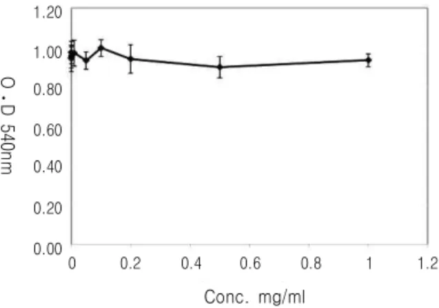
Fig. 1.Cytotoxicity of Polygonati rhizoma (Pr) extract on 3T3-L1 cells. Cells were exposed indicated concentration of Pr extract for 72 h. Data displayed reflect the mean SEM of three independent experiments
다시 1시간 동안 고정하였다. formalin 용액을 제거 하고 60% isopropanol로 세척한 후 실온에서 건조 시켰다. Oil Red O working solution을 가하여 10분 간 반응시킨 후 DW를 이용하여 5회 세척하였다. 염 색된 세포는 현미경 사진을 찍었고, 100% isopropanol 을 가하여 세포 내에 축적된 lipid를 용출하여 500 nm에서 흡광도를 측정하였다.
5) RNA extraction and Reverse-transcription and PCR
RNA는 TRIzol reagent (Invitrogen, Carlsbad, CA) 을 사용하여 분리하였고, 1 mg의 RNA를 MMLV reverse transcriptase와 random oligo (dT) primers (Invitrogen)를 이용하여 역전사 반응을 진행하여 cDNA를 준비하였다. PCR은 cDNA를 주형으로 하 여 다음의 primers를 이용하여 진행하였다. LPL, 5’-ATCCATGGATGGACGGTAACG-3’ and 5’-CT- GGATCCCAATACTTCGACCA-3’, aP2, 5’-AAA- GACAGCTCCTCCTCGAAGGTT-3’ and 5’-TGAC- CAAATCCCCATTTACGC-3’, adipsin, 5’-CCTGA- ACCCTACAAGCGATG-3’ and 5’-GGTTCCACTT- CTTTGTCCTCG-3’, PPARg, 5’-TTTTCAAGGGT- GCCAGTTTCAATCC-3’ and 5’-AATCCTTGGCC- CTCTGAGAT-3’, GAPDH, 5’-CTGCACCACCA- ACTGCTTAGC-3’ and 5’-GGGCCATCCACAGT- CTTCTGG-3’. 증폭된 PCR product는 1.5% agarose
gel 전기영동을 진행하였고, chemiluminescent detection system (ChemiDoc XRS system, Bio-Rad Laboratories, USA)을 이용하여 사진을 얻고 분석하였다.
6) 통계 처리
실험 결과의 통계 처리는 각 실험 결과의 평균값 과 평균 오차를 이용하여 표시하였고, student t-test 를 통하여 유의성을 검정하였다.
성 적 1. 黃精추출물의 세포독성 측정
黃精 추출물이 3T3-L1 세포에 독성을 나타내는 농도를 확인하고자 0.1, 0.2, 0.5, 1, 10, 50, 100, 200, 500, 1000 ㎍/ml 농도에서 MTT assay를 이용 하여 측정하였다. 黃精추출물은 1000 ㎍/ml 농도에 서도 세포에 독성을 나타내지 않았다.(Fig. 1)
2. 3T3-L1 세포 분화에서 triglyceride (TG)의 축적 양 측정
3T3-L1 세포를 분화 유도한 후 축적된 TG의 양 을 측정하였다. 黃精추출물을 500 ㎍/ml농도로 처리 한 실험에서는 축적되는 TG의 양이 24.57% 감소되 었다. Kaempferol을 50 μM 처리했을 경우 축적되는 TG의 양은 76.14%로 黃精추출물에 비해 더 감소되
1.0
0.8
0.6
0.4
0.2
0.0
1.0
0.8
0.6
0.4
0.2
0.0
Lipid accumulation(O
⋅D
⋅⋅
⋅500nm)
DMSO Pol-ext
24.57%
DMSO Kaempferol
76.14%
P<0.001 P<0.05
Lipid accumulation(O
⋅D
⋅⋅
⋅500nm)
A B
Fig. 2.Polygonati rhizoma (Pr) extract and its component kaempferol reduces triglyceride accumulation in 3T3-L1 differentiation. 3T3-L1 cells were differentiated and treated with DMSO, Pr extract (Pol-ext, 0.5 mg/ml) and Kaempferol (Kae, 40 μM). 10 days following differentiation, lipid droplets in cells were stained with Oil Red O, eluted and measured. Data displayed reflect the mean SEM of three independent experiments
DMSOKaempferol
×40 ×200
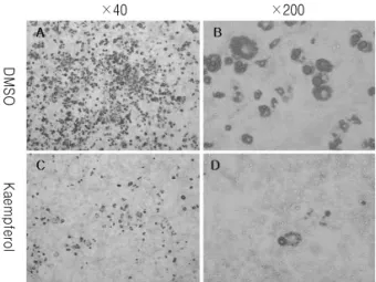
Fig. 3.Kaempferol reduces lipid accumulation in differentiated 3T3-L1 adipocytes. 3T3-L1 cells were differentiated and treated with DMSO, Kaempferol (40 μM). 10 days following differentiation, cells were stained with Oil Red O.
었다.(Fig. 2) 현미경을 이용하여 관찰한 결과 DMSO 대조군에 비해 kaempferol을 처리한 경우 분화가 일 어난 3T3-L1 세포의 수가 감소되어 있음을 확인할 수 있었다.(Fig. 3)
3. 지방세포 분화에 관여된 유전자의 mRNA 발현 측정
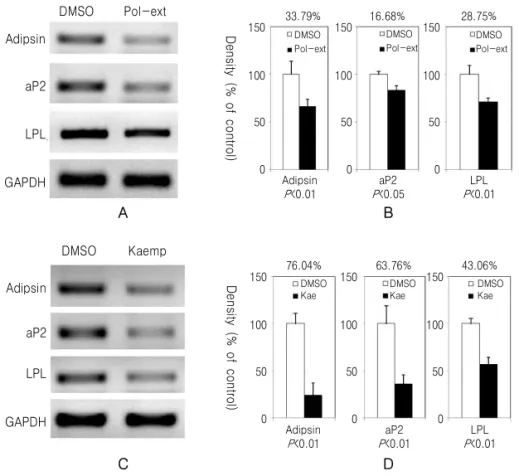
3T3-L1의 분화 유도 후 성숙된 지방세포에서 발 견되는 유전자의 발현을 확인하였다. Adipsin, adipocyte protein 2 (aP2), lipoprotein lipase (LPL) 유전자들
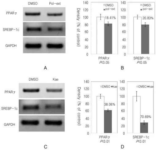
의 발현은 黃精추출물과 kaempferol을 처리한 실험 모두에서 감소하였으며 黃精추출물보다 kaempferol 을 처리한 실험에서 더 감소되었다(Fig. 4). PPARγ 와 SREBP-1c의 mRNA 발현도 DMSO를 처리한 대 조군에 비하여 감소되었다(Fig. 5).
고 찰
黃精은 性이 平하며 脾, 肺, 腎經에 歸經하여 補 中益氣, 安五臟 益脾胃 潤心肺, 塡精髓 助筋骨 등의
Adipsin
aP2
LPL
GAPDH
DMSO Pol-ext
33.79%
150 DMSO
Pol-ext
Density (% of control)
100
50
0
16.68% 28.75%
LPL P<0.01 aP2
P<0.05 Adipsin
P<0.01
DMSO Pol-ext DMSO
Pol-ext 150
100
50
0
150
100
50
0
A B
Adipsin aP2 LPL
GAPDH
DMSO Kaemp
76.04%
150 DMSO
Kae
Density (% of control)
100
50
0
63.76% 43.06%
LPL P<0.01 aP2
P<0.01 Adipsin
P<0.01
DMSO Kae DMSO
Kae 150
100
50
0
150
100
50
0
C D
Fig. 4.Polygonati rhizoma (Pr) extract and kaempferol reduce the expression of genes associated with 3T3-L1 cell differentiation. 3T3-L1 cells were differentiated and treated with DMSO, Pr extract (Pol-ext, 0.5 mg/ml) (A) and (B) and Kaempferol (Kae, 40 μM) (C) and (D). 10 days following differentiation of 3T3-L1, the mRNA expression of Adipsin, aP2, and LPL were monitored by RT-PCR (A) and (C). The expression of mRNA level was quantified and normalized with GAPDH by chemiluminescent detection system (B) and (D).
효능을 나타낸다. 임상에서 脾胃虛弱으로 小食하거 나 肺虛乾咳 病後 虛羸 體倦乏力 消渴 등의 치료에 활용되고 있다10). 실험 연구에 의하면 黃精추출물을 당뇨병에 유발된 쥐에게 장기 복용시켰을 때 혈당 및 체중이 감소되었고, 고지방 식이를 섭취시켜 고 지혈증이 유도된 쥐에게 장기 복용시켰을 때 체중 및 간 중량 감소 현상이 나타났고 혈중 콜레스테롤, 중성지방, 지방산의 수치가 감소되어 고지혈증이 완 화되었다11,13,14,18,19)
.
Flavonoid는 야채, 과일, 곡물, 차, 포도주 등 주로 식물이나 식물을 이용한 음식에서 발견되는 폴리페 놀 화합물로서 항산화, 항암, 항염증 작용에 관여한
다16,17). Kaempferol은 자연에서 발견되는 flavonoid
중 하나로서 항산화, 항염증, 항균 작용에 관여하고, 암세포의 apoptosis를 유도하여 항암작용에 관여한
다18-24). 또한 혈당의 감소, 동맥경화 수준의 감소,
허혈성심질환과 같은 관상동맥 질환에 대한 치료 효 과가 있다는 동물 모델 수준에서의 실험 결과들이 보고되어 있다1,25-27).
이러한 효능을 보이는 kaempferol은 우리 주변에 서 쉽게 볼 수 있는 호박, 당근, 무, 가지와 같은 채 소에 많이 함유되어 있고, 녹차 (Green tea) 및 홍차 (Black tea)에도 함유되어 있다. 또한 은행잎 (白果 葉, Ginkgo biloba), 솔잎 (松葉, Pinus densiflora Sieb.
et Zucc), 바디나물 (前胡, Angelicae decursivae radix), 둥글레 (黃精, Polygonati Rhizoma) 등의 한
DMSO Pol-ext
PPARγ
SREBP-1c
GAPDH
Density (% of control)
140 120 100 80 60 40 20 0
140 120 100 80 60 40 20 0
18.41% 20.83%
PPARγ P<0.05
SREBP-1c P<0.05 DMSO
pol-ext
DMSO pol-ext
A B
DMSO Kae
PPARγ
SREBP-1c
GAPDH
Density (% of control)
140 120 100 80 60 40 20 0
140 120 100 80 60 40 20 0 38.06%
70.69%
PPARγ P<0.01
SREBP-1c P<0.01
DMSO kae DMSO kae
C D
Fig. 5.Polygonati rhizoma (Pr) extract and kaempferol reduce expression of PPARγ and SREBP-1c in 3T3-L1 cell differentiation. 3T3-L1 cells were differentiated and treated with DMSO, Pr extract (Pol-ext, 0.5 mg/ml) (A) and (B) and kaempferol (Kae, 40 μM) (C) and (D). 10 days following differentiation, the mRNA expression of PPARγ and SREBP-1c were monitored by RT-PCR (A) and (C). The expression of mRNA level was quantified and normalized with GAPDH by chemiluminescent detection system (B) and (D).
약재에도 함유되어 있다28-30).
본 실험 결과 黃精의 이러한 효능은 실제 지방세포 분화에서 지방 축적을 24.57% 억제하는 것으로 나타 났다(Fig. 2A). 黃精의 활성물질 중 하나인 kaempferol 은 黃精추출물보다 더욱 지방 축적을 억제하여 76.14
%의 감소를 보였다(Fig. 2B).
지방전구세포에서 지방세포로 분화가 이루어지면 지방세포 특이적인 유전자의 발현이 나타난다. Adipsin, aP2, LPL 유전자는 성숙된 지방세포에서 발견되는 유전자로서 PPARγ와 SREBP-1c에 의해 조절되는 표적 유전자이다1,4,31). 이들은 지방세포 특이적인 지 방 축적 및 분비를 통한 항상성 유지, 당 대사 조절, 생체호르몬 분비를 통한 신호 전달 등의 기능을 유
지한다. 이와 같은 지방세포 특이적인 유전자는 분 화과정에서 작용하는 PPARγ, C/EBP family, SREBP family의 3가지 전사인자에 의해 조절된다. PPARγ 는 지방세포 분화에 필요한 유전자들의 발현을 조절 하는 핵심 전사인자로서 작용하며, 그 표적 유전자 로는 aP2와 adipsin 등이 있고 PPARγ에 의해 그 발 현이 증가된다1,32-34). 이 표적 유전자들은 黃精 추출 물과 kaempferol을 처리한 경우에 그 발현이 감소됨 을 확인하였다(Fig. 4).
PPARγ의 발현과 활성은 지방세포 분화에서 매우 중요한 부분으로 녹차, 포도 씨 사과, 딸기 등의 추 출물과 그 안에서 발견되는 flavonoids에 의해 발현 이 조절된다는 보고가 있으며 catechin, quercetin,
kaempferol 등에 의해 감소된다35-37). 黃精추출물 또 한 이러한 PPARγ의 발현을 감소시켰으며, kaempferol 에 의한 PPARγ의 발현 감소도 확인하였다(Fig. 5).
C/EBP family와 SREBP family는 PPARγ의 발현 과 활성을 조절하는 기능을 한다. SREBP family는 지질대사에서 중요한 기능을 하는 전사 인자로서, 지방산대사와 지질 생합성에 주로 관여하며 표적 유 전자로는 LPL, fatty acid synthase (FAS), stearoyl- CoA desaturase (SCD1) 등이 있고, 지질대사 과정 에서 나오는 산물에 의해 PPARγ의 활성을 증가시 킨다. C/EBP family는 분화 초기에 PPARγ의 발현 을 증가시키는 기능을 한다4,31,38,39)
.
3T3-L1 세포에 黃精추출물과 kaempferol을 처리 하여 분화 억제 효과가 나타났을 때 SREBP-1c의 발 현이 감소됨을 확인하였고, 그 표적 유전자인 LPL의 발현 억제도 확인하였다. 이와 같은 결과들은 黃精 이 지질대사에 관여하여 작용할 것이며, 약리 효능 이 PPARγ, SREBP family 등의 발현 조절 및 활성 조절에 의한 기전을 통하여 나타날 것임을 시사한 다. 또한 지방세포 분화 시 黃精에 의해 발현이 변 화되는 유전자들의 패턴 변화를 micro array 등의 방법을 이용하여 분석한다면 黃精의 작용 기전을 이 해하는데 도움이 될 것이다.
결 론
黃精과 그 성분인 kaempferol이 지질대사에 관여 하는 지와 그 분자적 기전을 규명하기 위하여 지방 세포의 분화에 억제 효과를 나타내는 지를 검토하고 분화에 관련된 유전자의 발현을 관찰하였다. 黃精과 kaempferol은 3T3-L1 지방전구세포의 분화 과정 중 에서 세포 내에 지방 축적을 억제하였고, 지방세포 분화를 알려주는 지표인 adipsin, LPL, aP2 유전자 의 발현을 억제하였다. 또한 黃精은 분화 과정 내에 서 핵심 전사인자인 SREBP-1c와 PPARγ의 발현을 감소시켰다. 이상의 결과로 黃精과 kaempferol은 PPARγ, SREBP family 등의 발현 및 활성 조절에 의한 기전을 통하여 지질대사에 관여할 것임을 시사
한다.
참고문헌
1. Tontonoz P, Hu E, Spiegelman BM. Regulation of adipocyte gene expression and differentiation by peroxisome proliferator activated receptor gamma. Curr Opin Genet Dev. 1995;5(5):571-6.
2. MacDougald OA, Lane MD. Transcriptional regulation of gene expression during adipocyte differentiation. Annu Rev Biochem 1995;64:345- 73.
3. Hamm JK, el Jack AK, Pilch PF, Farmer SR.
Role of PPAR gamma in regulating adipocyte differentiation and insulin-responsive glucose uptake. Ann N Y Acad Sci 1999;892:134-45.
4. Gregoire FM, Smas CM, Sul HS: Understanding adipocyte differentiation. Physiol Rev 1998;78 (3):783-809.
5. Park S, Hong SM, Ahn IS, Kim YJ, Lee JB.
Huang-Lian-Jie-Du-Tang Supplemented with Schisandra chinensis Baill. and Polygonatum odoratum Druce Improved Glucose Tolerance by Potentiating Insulinotropic Actions in Islets in 90% Pancreatectomized Diabetic Rats. Biosci Biotechnol Biochem. 2009. (in press)
6. 中醫硏究院 主編. 中醫症狀鑑別診斷學. 北京:人 民衛生出版社. 1987:43.
7. 張介賓. 景岳全書. 上海, 上海科學技術出版社.
1984:194.
8. 焦東海. 全國 第3届 肥胖病 學術交流會論文綜述.
中醫雜誌. 1992;3:47-8.
9. Heo SY, Kang HS. East-Wast Medical Discussion and Treatment of Obesity. J Oriental Rehabilitation Medicine. 1997;7(1):272-86.
10. Lee SI. Herbs. Seoul:Medical Herb Co. 1983:128 -9. (Korean)
11. Miura T, Kato A, Usami M, Kadowaki S, Seino Y. Effect of polygonati rhizoma on blood glucose and facilitative glucose transporter isoform 2
(GLUT2) mRNA expression in Wistar fatty rats.
Biol Pharm Bull. 1995;18(4):624-5.
12. Kato A, Miura T. Hypoglycemic activity of polygonati rhizoma in normal and diabetic mice.
Biol Pharm Bull. 1993;16(11):1118-20.
13. Roh SW, Kim JB. Effects of Polygonati Rhizoma on the Diet-induced Hyperlipidemia in Rats.
Korean J Oriental Physiology & Pathology.
2008;22(5):1147-51. (Korean)
14. Kato A, Miura T, Yano H, Masuda K, Ishida H, Seino Y. Suppressive effects of polygonati rhizoma on hepatic glucose output, GLUT2 mRNA expression and its protein content in rat liver.
Endocr J. 1994;41(2):139-44.
15. Miura T, Kato A. The difference in hypoglycemic action between polygonati rhizoma and polygonati officinalis rhizoma. Biol Pharm Bull. 1995;
18(11):1605-6.
16. Manach C, Scalbert A, Morand C, Remesy C, Jimenez L. Polyphenols. Food sources and bioavailability. Am J Clin Nutr. 2004;79(5):727- 47.
17. Spencer JP. Flavonoids: modulators of brain function? Br J Nutr. 2008;99E Suppl1:ES60-77.
18. Parveen Z, Deng Y, Saeed MK, Dai R, Ahamad W, Yu YH. Antiinflammatory and analgesic activities of Thesium chinense Turcz extracts and its major flavonoids, kaempferol and kaempferol- 3-O-glucoside. Yakugaku Zasshi 2007;127(8).
1275-9.
19. Singh R, Singh B, Singh S, Kumar N, Kumar S, Arora S. Anti-free radical activities of kaempferol isolated from Acacia nilotica (L.) Willd. Ex. Del.
Toxicol In Vitro. 2008;22(8):1965-70.
20. Gabrielska J, Soczynska-Kordala M, Przestalski S. Antioxidative effect of kaempferol and its equimolar mixture with phenyltin compounds on UV-irradiated liposome membranes. J Agric Food Chem. 2005;53(1):76-83.
21. Kang JW, Kim JH, Song K, Kim SH, Yoon JH,
Kim KS. Kaempferol and quercetin, components of Ginkgo biloba extract (EGb 761), induce caspase-3-dependent apoptosis in oral cavity cancer cells. Phytother Res. 2009. (in press) 22. Li W, Du B, Wang T, Wang S, Zhang J.
Kaempferol induces apoptosis in human HCT116 colon cancer cells via the Ataxia-Telangiectasia Mutated-p53 pathway with the involvement of p53 Upregulated Modulator of Apoptosis. Chem Biol Interact. 2009;177(2):121-7.
23. Kataoka M, Hirata K, Kunikata T, Ushio S, Iwaki K, Ohashi K, et al. Antibacterial action of tryptanthrin and kaempferol, isolated from the indigo plant (Polygonum tinctorium Lour.), against Helicobacter pylori-infected Mongolian gerbils. J Gastroenterol. 2001;36(1):5-9.
24. Park MJ, Lee EK, Heo HS, Kim MS, Sung B, Kim MK, et al. The anti-inflammatory effect of kaempferol in aged kidney tissues: the involvement of nuclear factor-kappaB via nuclear factor- inducing kinase/IkappaB kinase and mitogen- activated protein kinase pathways. J Med Food.
2009;12(2):351-8.
25. Lopez-Sanchez C, Martin-Romero FJ, Sun F, Luis L, Samhan-Arias AK, Garcia-Martinez V et al. Blood micromolar concentrations of kaempferol afford protection against ischemia/reperfusion- induced damage in rat brain. Brain Res. 2007;
1182:123-37.
26. Middleton E, Jr., Kandaswami C, Theoharides TC. The effects of plant flavonoids on mammalian cells: implications for inflammation, heart disease, and cancer. Pharmacol Rev. 2000;52(4):673-751.
27. Fang XK, Gao J, Zhu DN. Kaempferol and quercetin isolated from Euonymus alatus improve glucose uptake of 3T3-L1 cells without adipogenesis activity. Life Sci. 2008;82(11-12):615-22.
28. Lee CJ, Lee JH, Seok JH, Hur GM, Park Js J, Bae S, et al. Effects of betaine, coumarin and flavonoids on mucin release from cultured
hamster tracheal surface epithelial cells. Phytother Res. 2004;18(4):301-5.
29. Miean KH, Mohamed S. Flavonoid (myricetin, quercetin, kaempferol, luteolin, and apigenin) content of edible tropical plants. J Agric Food Chem. 2001;49(6):3106-12.
30. Park JS, Rho HS, Kim DH, Chang IS. Enzymatic preparation of kaempferol from green tea seed and its antioxidant activity. J Agric Food Chem.
2006;54(8):2951-6.
31. Fajas L, Schoonjans K, Gelman L, Kim JB, Najib J, Martin G, et al. Regulation of peroxisome proliferator-activated receptor gamma expression by adipocyte differentiation and determination factor 1/sterol regulatory element binding protein 1: implications for adipocyte differentiation and metabolism. Mol Cell Biol. 1999;19(8):5495-503.
32. Sears IB, MacGinnitie MA, Kovacs LG, Graves RA. Differentiation-dependent expression of the brown adipocyte uncoupling protein gene:
regulation by peroxisome proliferator-activated receptor gamma. Mol Cell Biol. 1996;16(7):
3410-9.
33. Spiegelman BM, Choy L, Hotamisligil GS, Graves RA, Tontonoz P. Regulation of adipocyte gene expression in differentiation and syndromes of obesity/diabetes. J Biol Chem. 1993;268(10):
6823-6.
34. Gerhold DL, Liu F, Jiang G, Li Z, Xu J, Lu M, et al. Gene expression profile of adipocyte differentiation and its regulation by peroxisome proliferator-activated receptor-gamma agonists.
Endocrinology. 2002;143(6):2106-18.
35. Lin J, Della-Fera MA, Baile CA. Green tea polyphenol epigallocatechin gallate inhibits adipogenesis and induces apoptosis in 3T3-L1 adipocytes. Obes Res. 2005;13(6):982-90.
36. Pinent M, Blade MC, Salvado MJ, Arola L, Hackl H, Quackenbush J, et al. Grape-seed derived procyanidins interfere with adipogenesis of 3T3-L1 cells at the onset of differentiation.
Int J Obes (Lond). 2005;29(8):934-41.
37. Hassan M, El Yazidi C, Landrier JF, Lairon D, Margotat A, Amiot MJ. Phloretin enhances adipocyte differentiation and adiponectin expression in 3T3-L1 cells. Biochem Biophys Res Commun.
2007;361(1):208-13.
38. Kim JB, Spiegelman BM. ADD1/SREBP1 promotes adipocyte differentiation and gene expression linked to fatty acid metabolism. Genes Dev.
1996;10(9):1096-107.
39. Brun RP, Kim JB, Hu E, Altiok S, Spiegelman BM. Adipocyte differentiation: a transcriptional regulatory cascade. Curr Opin Cell Biol. 1996;8 (6):826-32.