분화된 3T3-L1 세포에서 수세미오이 메탄올 추출물의 지방분해 효과
차승윤1․장자영1․이유현2․이규옥5․이호준3․황권택4․김용재5․전우진5․이정민6†
1경희대학교 의학영양학과, 2수원대학교 식품영양학과, 3한국식품연구원
4남부대학교 식품영양학과, 5전남대학교 식품영양학과, 6경희대학교 임상영양연구소
Lipolytic Effect of Methanol Extracts from Luffa cylindrica in Mature 3T3-L1 Adipocytes
Seung-Youn Cha1, Ja Young Jang1, Yoo-Hyun Lee2, Gyuok Lee5, Ho Joon Lee3, Kwon-Tack Hwang4, Yongjae Kim5, Woojin Jun5, and Jeongmin Lee6†
1Dept. of Medical Nutrition, Kyung Hee University, Gyeonggi 446-701, Korea
2Dept. of Food & Nutrition, University of Suwon, Gyeonggi 445-743, Korea
3Korea Food Research Institute, Gyeonggi 463-746, Korea
4Dept. of Food & Nutrition, Nambu University, Gwangju 506-706, Korea
5Dept. of Food & Nutrition, Chonnam National University, Gwangju 500-757, Korea
6Research Institute of Medical Nutrition, Kyung Hee University, Gyeonggi 446-701, Korea
Abstract
The effect of methanol extract from Luffa cylindrica (LCM) on lipid metabolism and underlying mechanisms of lipolysis in mature 3T3-L1 adipocytes were investigated. The intracellular lipid droplets were stained with Oil Red O dye and quantified. Compared to the control, lipid accumulation was significantly decreased by 19.4%
with the treatment of LCM at the concentration of 1000 μg/mL. Intracellular triglyceride (TG) level was also reduced by 21% at the concentration of 1000 μg/mL. To determine the mechanism for the reduction in TG content, levels of glucose uptake and glycerol release were measured. Incubation of the 3T3-L1 adipocytes with LCM did not affect the cellular uptake of glucose. However, the level of free glycerol released into the cultured medium drastically increased by 24.3% with the treatment of LCM. In subsequent measurements using quantitative real-time PCR, mRNA levels of hormone-sensitive lipase (HSL) and adipose triglyceride lipase (ATGL) except lipoprotein lipase (LPL) were significantly elevated at higher concentration. These results suggest that LCM partially stimulates the lipolysis through the induction of HSL and/or ATGL gene expression, resulting in the reduced lipid accumulation and increased glycerol release.
Key words: Luffa cylindrica, adipocytes, lipid accumulation, HSL, ATGL
†Corresponding author. E-mail: [email protected]
†Phone: 82-31-201-3838, Fax: 82-31-204-8119
서 론
비만은 지방의 축적과 에너지 저장을 위한 조절과정에서 주로 발생하게 된다(1). 지방세포 내에 과도한 지방의 축적 은 당뇨병, 고혈압, 심혈관 질환을 포함한 만성질병을 유발 하게 되는 원인으로 알려져 있다(2,3). 따라서 세포 내 지방 의 축적을 예방하거나 축적된 지방을 분해하도록 자극하는 방안에 대한 연구가 최근 많은 관심을 끌고 있다. 지방세포 내의 지방구(lipid droplet)는 지질의 대사 및 조절에 중요한 역할을 하는 것으로 알려져 있으며 지방구 내에 주로 존재하 고 있는 triglyceride의 분해와 이로 인한 glycerol의 유출은 세포 내 지방의 축적을 조절하는 중요한 기전으로 여겨져 왔다(4). 특히 lipase, TNF-α, perilipin A 등은 triglyceride
분해 과정의 조절에 중요한 역할을 담당하고 있다(5-7).
Hormone-sensitive lipase(HSL)는 지방분해 과정에서 매 우 중요한 lipase로 인식되어져 왔으며 insulin과 epinephrine 등의 호르몬 조절을 받는다. 분자생물학적 기전보고에 따르 면, triglyceride의 분해는 protein kinase A(PKA) 또는 G protein-coupled receptors와 cyclic AMP-activated extra- cellular signal-regulated kinase(ERK)에 의한 HSL의 인산 화 과정에 의한 것으로 알려져 있다(5). TNF-α는 주로 염증 관련 기전에 관여하는 것으로 알려져 있으며 NF-kB의 활성 화를 통해 유전자의 발현을 조절하는 것으로 알려져 있다 (6). NF-kB의 활성화는 비만의 유도와도 관련이 있을 것으 로 추론되기도 하지만 일부 사람과 동물을 대상으로 하는 실험에서 지방의 분해를 증가시킨다고 확인된바 있다(8). 최
근에는 adipose triglyceride lipase(ATGL)이 triglyceride에 특이적으로 작용하는 지방분해효소로 밝혀졌는데 지방분해 첫 번째 단계인 디글리세라이드의 생성에 작용하는 효소로 여겨지고 있다(9). 지방구의 막부위에 존재하고 있는 perilipin A 단백질은 지방분해를 억제하는 것으로 생각되고 있는데 그 기전으로는 HSL과 ATGL 등의 지방분해효소의 지방구 에 대한 접근을 방해함으로써 효소와 기질의 작용을 억제하 는 것으로 인식되고 있다(7).
수세미오이(Luffa cylindrica)는 박과식물로서 덩굴성 1년 생 초본이며 줄기는 오각형으로서 고온다습하고 보수력이 좋은 곳에서 자란다. 산업적으로는 세척용 수세미의 원료로 서 여름철 줄기를 채취하여 건조시켜 사용하고 있으며 감 기, 천식, 비염 등에 효능이 있는 것으로 알려져 있다(10).
주요 유효성분으로는 saponin, coumaric acid, α-spinasterol, stigmasterol 등을 포함하고 있으며 현재까지 항균성, 면역 조절 능력 등에 대한 연구가 진행된 바 있다. 본 연구에서는 아직까지 수행된 바가 없는 수세미오이의 항비만 효과를 평 가하기 위하여 인체적용시험에 앞서 in vitro 3T3-L1 지방 세포에서의 작용기전과 효능을 검증하고자 하였다.
재료 및 방법
재료 및 시약
수세미오이 동결건조 분말은 전라남도 장성군에서 수집 하여 물로 깨끗이 세척하고 얇게 썰어 freezer에서 -70oC까 지 급속 냉동시킨 후 동결건조기(freeze-dryer, Bondiro DC1316 Ilshin Lab Co., LTD., Yangju, Korea)에서 72시간 동안 건조하여 100 mesh 체로 입자를 고르게 하여 수세미오 이 분말을 제조하였다. 제조된 수세미오이 분말은 para film 으로 밀봉하여 -20oC에 냉동 보관하면서 사용하였다. Oil- Red O 염색시약, 글리세롤 측정 키트, MTT 시약은 Sigma Co.(St. Louis, MO, USA)에서 구입하였고, 세포 내 triglyc- eride 함량측정을 위한 AdipoRed Assay kit는 Cambrex Bioscience(Walkersville, MA, USA)에서, RNA 분리를 위 한 RNeasy mini kit, Taqman 중합효소는 Qiagen(Valencia, CA, USA)에서 구입하여 사용하였다. 3T3-L1 미분화된 지 방세포는 American Type Culture Collection(ATCC CL- 173, Manassas, VA, USA)로부터 구입하였고 지방세포의 분 화에 사용된 시약인 insulin, dexamethasone, 1-methyl 3- isobutylxanthin은 Sigma Co.로부터 구입하여 실험하였다.
수세미오이 메탄올 추출물의 제조
수세미오이 동결건조 분말 100 g을 메탄올 2 L와 섞어 교반기를 통해 상온에서 16시간 동안 침출하여 3,000 rpm에 서 20분간 원심분리한 후 Watman filter paper(No. 2)로 추 출액을 여과하였다. 이렇게 하여 얻어진 추출물을 회전 감압 증발기(Eyela, Tokyo Rikakikai Co., Tokyo, Japan)로 메탄
올을 증발시켜 감압농축한 후 다시 동결건조 하여 실험 때까 지 -20oC에 보관하였다. 메탄올 추출을 통해 얻어진 수세미 오이의 수득율은 약 9%로 나타났다.
3T3-L1 세포배양과 분화
3T3-L1 지방전세포의 배양과 유지는 10% bovine calf serum(FCS)을 넣은 Dulbecco's modified Eagle's medium (DMEM) 배지로 5% CO2, 37oC에서 60% 세포가 자랐을 때 계대배양을 하도록 하였다. 세포의 분화는 6-well plate에 well 당 1×10⁶ 세포를 분주하여 세포가 100% 밀집되게 배양 하였다(11). 2일 후 10% fetal bovine serum(FBS)와 MDI solution(0.5 mM IBMX, 0.5 μM dexamethasone, 10 μg/mL insulin)을 포함한 DMEM 배지를 3일 동안 처리하였고 다시 10% FBS와 10 μg/mL insulin을 포함한 DMEM을 3일 동안 처리하였고 그 이후는 10% FBS를 포함한 DMEM으로 배양 하며 세포 내 지방구의 형성을 근거하여 지방세포로 분화시 켰다. 모두 12일 동안의 분화를 통해 80% 이상의 세포가 분화되었음을 확인하였다.
MTT assay
배양 및 분화과정에서 생존하고 있는 세포의 수는 미토콘 드리아의 MTT(3-[4,5-2-yl]-2,5-diphenyl tetrazolium bro- mide)를 formazan dye로 전환하는 능력을 간접적으로 이용 하여 확인하였다(12). 우선 3T3-L1 세포를 12-well plate에 서 분화시킨 후 추가로 48시간 동안 배양하여 Hank's bal- anced salt solution에 용해시킨 50 mg/mL MTT 용액을 첨 가하여 37oC에서 4시간 더 배양하고 상층액을 2번 PBS로 세척한 후 1 mL DMSO를 첨가하여 염색을 유출시킨 다음 570 nm(reference wavelength; 665 nm)에서 흡광도를 측정 하였다.
Oil-Red O 염색과 triglyceride 함량의 측정
60-mm culture dish에서 분화된 3T3-L1 세포를 0, 100, 200, 400, 600, 1000 μg/mL 농도로 처리하여 48시간 동안 배양한 후 PBS로 2회 세척하고 1%(v/v) calcium chloride를 첨가시킨 4% formaldehyde로 세포를 30분간 상온에서 고정 시켰다. 고정된 세포를 건조시킨 후 absolute alcohol에 포화 시켜 0.4 μm filter로 여과한 Oil-Red O 염색시약을 세포에 40분간 처리한 후 PBS로 Oil-Red O 염색시약을 3번에 걸쳐 충분히 세척하였고 DMSO로 염색된 지방구를 유출시킨 후 560 nm에서 흡광도를 측정하였다.
세포 내 triglyceride 함량은 AdipoRed assay kit에 지시되 어 있는 정량법에 따라 수행하였는데 이 방법은 Nile Red 염색법으로서 triglyceride에 특이성이 높은 것으로 알려져 있다(12). 우선 분화된 3T3-L1 세포를 48시간 동안 수세미 오이 메탄올 추출물을 농도별로 처리한 후 PBS로 2회 세척 하고 20 μL의 AdipoRed 시약을 첨가하여 20분간 추가로 상 온에서 배양하였고 excitation 485 nm와 emission 572 nm에 서 형광신호를 측정하여 결과를 얻었다.
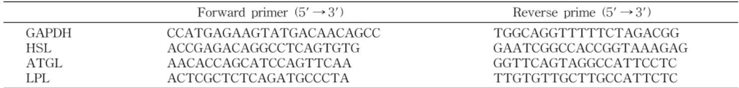
Table 1. Sequences for primer set for lipase amplification by RT-PCR
Forward primer (5' → 3') Reverse prime (5' → 3')
GAPDH HSL ATGL LPL
CCATGAGAAGTATGACAACAGCC ACCGAGACAGGCCTCAGTGTG AACACCAGCATCCAGTTCAA ACTCGCTCTCAGATGCCCTA
TGGCAGGTTTTTCTAGACGG GAATCGGCCACCGGTAAAGAG GGTTCAGTAGGCCATTCCTC TTGTGTTGCTTGCCATTCTC
Free glycerol의 측정
분화된 3T3-L1 세포에 시료를 처리한 후 48시간 배양한 배지를 eppendorf tube에 넣고 70oC에서 10분간 가열하여 세포로부터 유리된 효소들을 불활성화 시켰다. 50 μL의 배 지를 glycerol reagent에 첨가하여 1분간 반응시킨 후 1-mL cuvette에 옮겨 넣고 흡광도를 540 nm에서 측정하였다. 이 때 단백질량을 Bradford 방법으로 정량하여 세포수를 보정 하였다.
Reverse-transcriptase polymerase chain reaction
세포 내 총 RNA를 추출하고 정량하기 위하여 RNeasy Mini kit를 이용하였다. 1 μg의 총 RNA를 oligo(dT) primer 와 superscript II 역전사효소와 함께 반응시켜 1차 cDNA를 제작하였다. PCR 반응은 1 μg의 cDNA를 Gold Taq poly- merase를 첨가하여 32 cycles로 PCR 반응시킨 다음 1.5%
agarose gel에 전기영동한 후 EtBr로 염색하여 UV에서 DNA를 확인하였고 DNA의 정량적 분석은 Applied Bio- system Inc.(Foster city, CA, USA)에서 제공된 7500 Systems SD software version 3.1.을 이용하여 측정하였다.
PCR에 사용된 primer set은 Table 1에 나타내었다.
통계처리
실험결과는 평균(mean)±표준편차(SD)로 나타내었고 통 계적 유의성은 Duncan's multiple range test와 Student's t-test를 이용하여 그룹간의 차이를 통계처리한 후 p<0.05 수준에서 유의성을 검정하였다.
Fig. 1. Cell viability of mature 3T3-L1 adipocytes, deter- mined by MTT reagent, with treatment of Luffa cylindrica methanol extract at various concentrations. Significant differ- ences were determined by Duncan's multiple range test at p<0.05.
결과 및 고찰
세포독성시험
수세미오이 메탄올 추출물의 세포독성 범위를 분화된 3T3-L1 지방세포를 이용하여 MTT 시험법에 의해 측정하 였다(Fig. 1). 0 μg/mL에서 1200 μg/mL 농도범위 내에서 측정한 결과 1200 μg/mL의 농도까지 세포의 생장에 영향을 미치지 못하였다. 따라서 이후의 모든 실험에서는 보다 안전 한 농도 범위인 1000 μg/mL까지 사용하였다. 하지만 수세미 오이 메탄올 추출물의 경우 다른 천연물 또는 식품의 메탄올 추출물을 연구한 보고와 비교해볼 때(13-15) 유효범위가 대 체로 넓게 나타나고 있어 비교적 세포독성이 낮은 것으로 생각된다.
Oil-Red O 염색에 의한 세포 내 지방구와 triglyceride 함량 측정
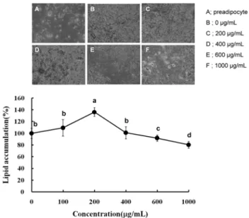
분화된 3T3-L1 지방세포에 수세미오이 메탄올 추출물을 0 μg/mL에서 1000 μg/mL까지 처리한 후 생성되었던 지방 구에 미치는 영향을 확인하기 위해 Oil-Red O 시약으로 염 색한 결과 600 μg/mL과 1000 μg/mL에서 각각 9.7%와 19.4%
의 감소를 나타내었다(Fig. 2). 특이한 점은 200 μg/mL에서 유의적인 상승이 나타났으나 600 μg/mL과 1000 μg/mL에서 다시 유의적인 감소가 나타났다. 200 μg/mL에서 일시적으 로 유의적인 증가를 보인 것이 농도에 따른 두 가지 효능이 나타나는 것으로 생각되지는 않는데 그 이유는 Fig. 3의 triglyceride의 함량이 농도에 따라 변화가 나타나지 않았기 때문이다. 따라서 저농도에서 나타난 일시적인 증가는 실험 적인 오차에 의한 것으로 판단된다. 이와는 달리 600 μg/mL 이상의 고농도에서 나타난 세포 내 지방축적의 억제는 Oil-Red O 염색에 대한 현미경적 관찰에서도 동일한 결과로 확인되었다(Fig. 2). 대부분의 지방구는 triglyceride와 per- ilipin A와 같은 단백질로 구성되어 있기 때문에 지방구의 감소를 보다 세밀하게 확인하기 위해 triglyceride의 함량을 형광염색법에 의해 측정하였다(Fig. 3)(16). Oil-Red O 염색 결과와 마찬가지로 600 μg/mL와 1000 μg/mL의 농도에서 지방구 내의 triglyceride의 함량이 각각 13.5%와 21% 감소 하는 것으로 나타났다. 지방구에 존재하는 triglyceride는 3T3-L1 지방전세포로부터 지방세포로 분화하는 단계에서 3가지 과정에 의해 생성된다. 첫째로 세포 내로 유입되는 포 도당의 대사과정, 둘째로 lipoprotein lipase에 의해 분해되어 유입된 모노글리세롤과 지방산, 그리고 세포 내 de novo 지
Fig. 2. Inhibitory effect of Luffa cylindrica methanol extract on the lipid accumulation in 3T3-L1 adipocytes. The intra- cellular lipid accumulation was quantified by Oil Red O staining and also optically observed by an inverted microscope (×100; A:
undifferentiated control, B: differentiated control or 0 μg/mL, C:
200 μg/mL, D: 400 μg/mL, E: 600 μg/mL, and F: 1000 μg/mL).
Values are expressed as the mean±SD of at least three in- dependent experiments, each performed in triplicate (n=3). Sig- nificant differences were determined by Duncan's multiple range test at p<0.05.
Fig. 3. Inhibitory effect of Luffa cylindrica methanol extract on intracellular triglyceride (TG) content in 3T3-L1 adi- pocytes. The intracellular TG content was fluorescently quanti- fied by AipoRed assay kit. Values are expressed as the mean±SD of at least three independent experiments, each performed in trip- licate (n=3). *Significant difference compared to control group at p<0.05.
방산 합성과정 등으로 인해 종합적으로 세포 내에 trigly- ceride가 생성되어 분화 마지막 단계까지 계속적으로 지방 구는 형성되게 되는데 시간이 지날수록 지방구끼리 결합하 여 그 크기가 점점 증가하게 된다. 본 연구에서는 수세미오 이 메탄올 추출물의 지방구 감소효과를 검증하기 위하여 형 성된 지방구를 분해 또는 감소시킬 수 있는 지 여부를 측정
한 것으로 다소 고농도인 600 μg/mL와 1000 μg/mL에서 그 효과가 입증되었다. 따라서 지방구의 감소에 대한 작용기작 을 두 가지 측면에서 고려하였는데 첫째, 지방구의 분해로 인한 감소와 둘째, glucose 유입 억제로 인한 지방구 합성의 감소 측면에서 검토하였다.
Glycerol release 측정
지방세포 내 축적된 지방구에 존재하는 triglyceride가 분 해되면 glycerol과 지방산으로 나누어지는데 인체의 경우 glycerol은 세포 외 혈액으로 유리되어 간으로 이송되게 된 다(17). 마찬가지로 3T3-L1 지방세포계에서는 유리된 gly- cerol의 함량이 지방구 내 triglyceride의 분해 정도를 간접적 으로 나타내는 측도가 된다. 본 연구에서 수세미오이 메탄올 추출물 0 μg/mL에서 1000 μg/mL에서 처리한 결과 1000 μg/
mL에서 유리된 glycerol의 정도가 24.3% 증가한 것을 확인 하였는데 이는 triglyceride의 분해로 인한 것으로 생각된다 (Fig. 4). 지방세포에서 triglyceride의 분해는 insulin res- ponsive lipase인 HSL에 의해 주로 매개되어지는 것으로 알 려져 있는데 세포 내 지방의 함량을 조절하는 중요한 단계로 여겨지고 있다. 본 연구에서는 유리된 glycerol이 HSL 등의 지방분해효소에 의한 것인지 판단하기 위해 mRNA의 발현 과 지방세포로 유입된 glucose의 양을 측정하였다.
Glucose uptake의 측정
지방세포에서 glucose uptake는 인슐린의 신호전달체계 에 의해 세포질에서 세포막으로 이동하는 glucose trans- porter-4(Glut-4)에 의해 이루어진다(18,19). 지방세포와 근 육조직에서 Glut isoform인 Glut-1과 Glut-4가 주로 작용하 여 glucose uptake에 관여한다. Glut-1은 지방세포, 근육세 포, 그리고 많은 다른 세포에도 존재하는데 인슐린의 존재에
Fig. 4. Stimulatory effect of Luffa cylindrica methanol ex- tract on glycerol release in 3T3-L1 adipocytes. The values were calculated as a percentage of free glycerol content of the differentiated control cells not treated with Luffa cylindrica meth- anol extract. Values are expressed as the mean±SD of at least three independent experiments, each performed in triplicate (n=3).
*Significant difference compared to control group at p<0.05.
Fig. 5. Inhibitory effect of Luffa cylindrica methanol extract on glucose uptake in 3T3-L1 adipocytes. The glucose uptake was fluorescently quantified by using 2-NBDG in the media con- taining no phenol red and glucose. Values are expressed as the mean±SD of at least three independent experiments, each per- formed in triplicate (n=3).
관계없이 세포막에 존재한다. 이와 달리 Glut-4는 세포질에 분포되어 있다가 인슐린의 존재 시 세포막으로 이동하여 glucose uptake를 용이하게 하는 것으로 알려져 있다(20-24).
지방세포로 유입된 포도당은 해당과정을 통해 triglyceride 의 전구물질인 glyceraldehyde-3-phosphate를 생성하게 되 고 다시 dihydroxyacetone-phosphate를 거쳐 glycerol로 전 환하게 되며 이것은 지방산과 결합하여 triglyceride로 합성 되어 지방구를 형성하게 된다. 따라서 지방세포 내로 포도당 의 유입은 지방세포의 분화와 세포내 지방의 축적에 중요한 역할을 한다고 하겠다. 본 연구에서 수세미오이 메탄올 추출 물의 지방대사에 미치는 영향을 확인하기 위해 실험한 결과 분화된 3T3-L1 지방세포에서 세포 내 포도당의 유입은 농 도에 따라 전혀 변화가 나타나지 않았다(Fig. 5). 특히 1000 μg/mL에서 감소하는 경향을 나타내었지만 통계적 유의성 은 나타나지 않았다. 이러한 결과는 수세미오이 메탄올 추출 물이 포도당의 대사와는 상관없는 다른 과정을 통해 세포 내 지방의 축적을 억제한 것으로 판단된다. 특히 포도당이 정상적으로 세포 내로 유입되는 과정을 통해 지방이 합성됨 에도 불구하고 지방의 축적이 감소하였다는 것은 세포 내 지방의 분해속도가 합성속도보다 높은 경우에 해당된다고 하겠다. 따라서 세포 내 지방분해에 관여하는 효소의 발현을 확인하고자 하였다.
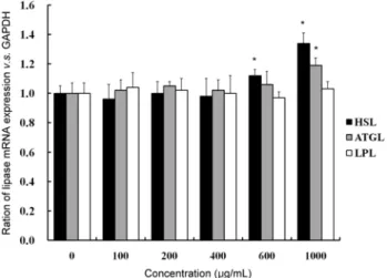
지방분해 효소의 mRNA 발현
지방대사에 관여하는 지방분해 효소는 LPL, HSL, 그리고 ATGL의 3가지 정도로 알려져 있다. LPL은 주로 지방조직, 심장조직, 그리고 골격근 주위의 모세혈관에 주로 분포하고 있는 세포 외 효소로서 chylomicron에 있는 triglyceride를 분해하여 monoglyceride와 두 사슬의 지방산으로 분리된 후 세포 내로 유입이 용이하게 함으로서 지방세포의 trigly- ceride 재생산에 도움을 주는 효소이다(25). 이와 반면 HSL
Fig. 6. Effect of Luffa cylindrica methanol extract on mRNA expressions of lipases in 3T3-L1 adipocytes. The values were calculated as a percentage of lipase mRNA expression versus GAPDH as an internal control. Values are expressed as the mean
±SD of at least three independent experiments, each performed in triplicate (n=3). *Significant difference compared to control group at p<0.05.
은 아드레날린계 호르몬의 작용에 의해 활성화된 protein kinase A(PKA)에 의해 인산화 됨으로써 활성화되어 세포 내에 존재하는 triglyceride를 분해하고 지방산과 글리세롤 을 생성하는 효소로 알려져 있다(5). 이때 생성된 glycerol은 세포 밖으로 유리되어진다. 최근 들어 ATGL 효소가 발견되 어 triglyceride의 분해 첫 단계인 diglyceride의 생성에 관여 하는 것으로 알려져 있다(26). 따라서 LPL, HSL 그리고 ATGL 효소간의 작용에 따라 세포 내 지방 축적과 분해가 일정 수준 조절을 받게 된다고 하겠다. 본 연구에서는 3T3-L1 지방세포로부터 LPL mRNA의 발현을 확인한 결과 수세미오이 메탄올 추출물의 처리에 따른 효과는 나타나지 않았다(Fig. 6). 이에 반해 HSL은 600 μg/mL과 1000 μg/
mL 농도에서, 그리고 ATGL은 1000 μg/mL의 농도에서 mRNA의 발현이 각각 13%, 37%와 24%로 유의적인 증가를 나타내었다. 이러한 결과는 동일 농도인 1000 μg/mL에서 HSL의 발현이 ATGL보다 더 많은 경향을 보여 영향성이 높은 것으로 생각되며 수세미오이 메탄올 추출물이 trigly- ceride에서 diglyceride로 분해되는 첫 단계에서 효과적으로 작용하는 것을 의미한다. 결론적으로 분화된 3T3-L1 지방 세포에서 수세미오이 메탄올 추출물의 지방축적 억제효과 는 HSL과 ATGL 지방분해 효소의 발현 증가에 의한 것으로 판단된다.
요 약
분화된 3T3-L1 지방세포에서 수세미오이의 메탄올 추출 물이 지방의 축적 및 분해에 미치는 영향을 확인하였다. 수 세미오이의 메탄올 추출물 안전범위(0~1000 μg/mL)에서 지방대사 관련한 여러 과정을 확인한 결과 600 μg/mL 이상 의 농도에서 세포 내 지방축적이 억제되었고 특히 지방구
내의 triglyceride 함량이 억제되는 것으로 나타났다. 이러한 결과에 대한 기전을 위해 포도당의 유입 억제로 인한 세포 내 triglyceride의 합성감소와 지방분해효소의 활성화로 인 한 세포 내 triglyceride의 분해 증가의 두 가지로 확인하였 다. 우선적으로 수세미오이의 메탄올 추출물의 포도당 유입 억제 효과를 실험하였지만 아무런 영향이 나타나지 않았다.
하지만 지방분해효소 중 LPL을 제외한 HSL과 ATGL의 유 전자 발현이 증가됨에 따라 세포 내 triglyceride이 지방산과 glycerol로 분해되었을 것으로 생각되며 이러한 결과는 세포 외로 유리된 glycerol의 함량이 수세미오이의 메탄올 추출물 1000 μg/mL의 농도에서 증가한 것을 통해 재확인할 수 있었 다. 하지만 600 μg/mL의 농도에서 triglyceride 분해는 촉진 되었으나 glycerol의 유리가 유의적으로 나타나지 않은 것은 세포 내에서 분해된 glycerol의 일부가 세포 내 생합성에 재 사용되었을 것으로 생각되며 실제 세포 외로 유리된 양을 확인하기 위해서는 보다 높은 농도가 필요한 것으로 여겨진 다. 전반적으로 본 실험에서는 수세미오이의 메탄올 추출물 이 분화된 3T3-L1 지방세포에서 지방구의 생성을 억제하였 으나 효과 농도가 다른 많은 천연물에 비해 다소 높게 나타 남으로써 제약으로서의 생산으로는 제한점이 있을 것으로 생각되는 반면 식품으로서 꾸준히 사용될 경우 항비만 소재 로서의 가능성이 있을 것으로 생각된다.
감사의 글
본 연구는 2008년도 농림기술관리센터(ARPC)연구비 지 원에 의해 수행된 결과의 일부이며, 이에 감사를 드립니다 (과제번호:108067-3)
문 헌
1. Spiegelman BM, Flier S. 1996. Adipogenesis and obesity;
rounding out the big picture. Cell 87: 377-389.
2. Kopelman PG. 2000. Obesity as a medical problem. Nature 404: 635-643.
3. Visscher TL. 2001. The public health impact of obesity.
Annu Rev Publ Health 22: 355-375.
4. Frayn KN, Karpe F, Fielding BA, Macdonald IA, Coppack SW. 2003. Integrative physiology of human adipose tissue.
Int J Obes Relat Metab Disord 27: 875-888.
5. Holm C. 2003. Molecular mechanisms regulating hormone- sensitive lipase and lipolysis. Biochem Soc Trans 31: 1120- 1124.
6. Halle M, Berg A, Northoff H, Keul J. 1998. Importance of TNF-alpha and leptin in obesity and insulin resistance: a hypothesis on the impact of physical exercise. Exerc Im- munol Rev 4: 77-94.
7. Akter MH, Yamaguchi T, Hirose F, Osumi T. 2008. Perilipin, a critical regulator of fat storage and breakdown, is a target gene of estrogen receptor-related receptor alpha. Biochem Biophys Res Commun 368: 563-568.
8. Baillie GS. 2009. Compartmentalized signaling: spatial reg- ulation of cAMP by the action of compartmentalized phos- phodiesterases. FEBS J 276: 1790-1799.
9. Fushiki H, Hayakawa Y, Gomori A, Seo T, Tewari S, Ozaki S, Yoshimoto R. 2010. In vivo imaging of obesity-induced inflammation in adipose tissue. Biochem Biophys Res Com- mun 391: 674-678.
10. Haemmerle G, Lass A, Zimmermann R, Gorkiewicz G, Meyer C, Rozman J, Heldmaier G, Maier R, Theussel C, Eder S, Kratky D, Wagner EF, Klingenspor M, Hoefler G, Zechner R. 2006. Defective lipolysis and altered energy me- tabolism in mice lacking adipose triglyceride lipase. Science 312: 734-737.
11. Tabata M, Tanaka S, Cho HJ, Uno C, Shimakura J, Ito M, Kamisako W, Honda C. 1993. Production of an anti-allergic triterpene bryonolic acid, by plant cell cultures. J Nat Prod 56: 165-174.
12. Gao CL, Zhu JG, Zhao YP, Chen XH, Ji CB, Zhang CM, Zhu C, Xia ZK, Peng YZ, Guo XR. 2010. Mitochondrial dys- function is induced by the overexpression of UCP4 in 3T3-L1 adipocytes. Int J Mol Med 25: 71-80.
13. Kasugai S, Hasegawa N, Ogura H. 1990. A simple in vito cytotoxicity test using the MTT (3-(4,5)-dimethylthiazol- 2-yl)-2,5-diphenyl tetrazolium bromide) colorimetric as- say: analysis of eugenol toxicity on dental pulp cells (RPC-C2A). Jpn J Pharmacol 52: 95-100.
14. Spiekermann P, Rehm BH, Kalscheuer R, Baumeister D, Steinbüchel A. 1999. A sensitive, viable-colony staining method using Nile red for direct screening of bacteria that accumulate polyhydroxyalkanoic acids and other lipid stor- age compounds. Arch Microbiol 171: 73-80.
15. Pavithra PS, Sreevidya N, Verma RS. 2009. Antibacterial and antioxidant activity of methanol extract of Evolvulus nummularius. Indian J Pharmacol 41: 233-236.
16. Chon JW, Sung JH, Hwang EJ, Park YK. 2009. Chlorella methanol extract reduces lipid accumulation in and in- creases the number of apoptotic 3T3-L1 cells. Ann N Y Acad Sci 1171: 183-189.
17. Liu F, Kim J, Li Y, Liu X, Li J, Chen X. 2001. An extract of Lagerstroemia speciosa L. has insulin-like glucose up- take-stimulatory and adipocyte differentiation-inhibitory activities in 3T3-L1 cells. J Nutr 131: 2242-2247.
18. Ray H, Pinteur C, Frering V, Beylot M, Large V. 2009.
Depot-specific differences in perilipin and hormone-sensi- tive lipase expression in lean and obese. Lipids Health Dis 8: 58.
19. Knutson VP. 2009. The release of lipoprotein lipase from 3T3-L1 adipocytes is regulated by microvessel endothelial cells in an insulin-dependent manner. Endocrinology 141:
693-701.
20. Ranganathan G, Unal R, Pokrovskaya I, Yao-Borengasser A, Phanavanh B, Lecka-Czernik B, Rasouli N, Kern PA.
2006. The lipogenic enzymes DGAT1, FAS, and LPL in adi- pose tissue: effects of obesity, insulin resistance, and TZD treatment. J Lipid Res 47: 2444-2449.
21. Bogan JS, Mckee AE, Lodish HF. 2001. Insulin-responsive compartments containing GLUT4 in 3T3-L1 and CHO cells:
regulation by amino acid concentrations. Mol Cell Biol 21:
4785-4806.
22. Czech MP, Corvera. 1999. Signaling mechanisms that regu- late glucose transport. J Biol Chem 274: 1865-1868.
23. Holman GD, Cushman SW. 1996. Subcellular trafficking of GLUT4 in insulin target cells. Semin Cell Dev Biol 7: 259- 268.
24. Pessin JE, Thurmond DC, Elmendorf JS, Coker KJ, Okada S. 1999. Molecular basis of insulin-stimulated GLUT4 vesi- cle trafficking: location! location! location! J Biol Chem 274:
2593-2596.
25. Rea S, James DE. 1997. Moving GLUT4: the biogenesis and trafficking of GLUT4 storage vesicles. Diabetes 46: 1667- 1677.
26. Jiang B, Yang Y, Jin H, Shang W, Zhou L, Qian L, Chen M. 2008. Astragaloside IV attenuate lipolysis and improves insulin resistance induced by TNF α in 3T3-L1 adipocytes.
Phytother Res 22: 1434-1439.
27. Moreno-Aliaga MJ, Matsumura F. 1999. Endrin inhibits
adipocyte differentiation by selectively altering expression pattern of CCAAT/enhancer binding protein-α in 3T3-L1 cells. Mol Pharmacol 56: 91-101.
28. Zimmermann R, Strauss JG, Haemmerle G, Schoiswohl G, Birner-Gruenberger R, Riederer M, Lass A, Neuberger G, Eisenhaber F, Hermetter A, Zechner R. 2004. Fat mobi- lization in adipose tissue is promoted by adipose trigly- ceride lipase. Science 306: 1383-1386.
(2010년 4월 15일 접수; 2010년 5월 12일 채택)