Biomedical Science Letters 2014, 20(2): 62~69 eISSN : 2288-7415
Anti-Adipogenic Activity of Ailanthoidol on 3T3-L1 Adipocytes
Ju-Hyung Park
1, Jong-Gab Jun
2and Jin-Kyung Kim
1,†1
Department of Biomedical Science, Catholic University of Daegu, Gyeongsan-Si 712-702, Korea,
2
Department of Chemistry and Institute of Natural Medicine, Hallym University, Chuncheon 200-702, Korea
Previous our study demonstrated that ailanthoidol (3-deformylated 2-arylbenzo[b]furan), a neolignan from Zanthoxylum ailanthoides or Salvia miltiorrhiza Bunge, is a novel anti-inflammatory agent. In this investigation, we examined the anti-adipogenic effect of ailanthoidol. Our data showed that ailanthoidol suppressed lipid droplet formation and adipocyte differentiation in 3T3-L1 cells. Treatment of the 3T3-L1 adipocytes with ailanthoidol resulted in an attenuation of the releases of leptin and interleukin-6. The expression of peroxisome proliferator-activated receptor (PPAR)γ and CCAAT/
enhancer-binding protein (C/EBP)α, the central transcriptional regulators of adipogenesis, was decreased by treatment with ailanthoidol. Additionally, ailanthoidol treatment increased the phosphorylation levels of 5' adenosine monophosphate- activated protein kinase. These results suggest that ailanthoidol effectively suppresses adipogenesis and that it exerts its role mainly through the significant down-regulation of PPARγ and C/EBPα expression. Our findings provide important insights into the mechanisms underlying the anti-adipogenic activity of ailanthoidol.
Key Words: Adipogenesis, Ailanthoidol, C/EBPalpha, PPARgamma
INTRODUCTION
Adipogenesis is the process by which an undifferentiated preadipocyte is converted to a fully differentiated adipocyte (Otto and Lane, 2005) and is closely related to the etiology of obesity and obesity-related metabolic disorders (Spiegelman et al., 1993). Since adipocyte differentiation plays a key role in fat mass growth, regulation of adipogenesis is a potential strategy for obesity prevention (Lee et al., 2012).
Obesity is a growing epidemic worldwide and significantly increases the risk of a number of chronic diseases such as insulin resistance, diabetes mellitus, coronary heart disease, hypertension (World Health Organisation, 2011). It is associated with an imbalance between energy intake and
expenditure and excess accumulation of adipose tissue. The increase in adipose tissue mass is caused by enlargement of adipocytes induced by lipid accumulation and an increase in the total number of adipocytes due to adipogenesis (Poulos et al., 2010).
The differentiation of preadipocytes into adipocytes involves the stimulation of a cascade of transcriptional events that includes expression of CCAAT/enhancer-binding protein (C/EBP)β and C/EBPδ, which together induce expression of peroxisome proliferator-activated receptor (PPAR)γ and C/EBPα (White and Stephens, 2010). The expression of both C/EBPα and PPARγ is increasing from undetectable levels in preadipocytes to detectable levels 2 days after differentiation induction and to full expression about 5 days after initiation of the differentiation program (White and Stephens, 2010). The activation of C/EBPα and PPARγ leads to terminal differentiation through trans- activation of adipocyte-specific genes such as fatty acid binding proteins aP2, and fatty acid synthase (FAS) (Farmer, 2006). Furthermore, 5' adenosine monophosphate-activated protein kinase (AMPK) is a key factor that controls cellular
Original Article
*Received: March 25, 2014 / Revised: May 14, 2014 Accepted: May 24, 2014
†Corresponding author: Jin-Kyung Kim. Department of Biomedical Science, College of Natural Science, Catholic University of Daegu, 13-13 Hayang-Ro, Gyeongsan-Si, Gyeongsangbuk-Do 712-702, Korea.
Tel: +82-53-850-3774, Fax: +82-53-850-3774 e-mail: [email protected]
○CThe Korean Society for Biomedical Laboratory Sciences. All rights reserved.
energy homeostasis and metabolism. AMPK also attenuates PPARγ and C/EBPα expression to inhibit fat accumulation during adipogenesis (Rossmeisl et al., 2004), (Hwang et al., 2009).
There is strong interest in developing new anti-adipogenic agents from plants used in traditional medicine. Ailanthoidol (3-deformylated 2-arylbenzo[b]furan, Fig. 1A), a neolignan from Zanthoxylum ailanthoides or Salvia miltiorrhiza Bunge, is used in Chinese traditional herbal medicine. Our previous study demonstrated that ailanthoidol has anti-inflammatory activity in vitro and in vivo (Kim and Jun, 2011). In addition to anti-inflammatory effect, Lee et al. reported ailanthoidol exhibited a radical quenching property by a 1,1-diphenyl- 2-picryrel-hydrazyl radical scavenging assay as well as anti-tumor activity using a 12-0-tetradecanoylphorbol-13- acetate-induced skin cancer model (Lee et al., 2006). Since the effects of ailanthoidol on adipogenesis are unknown, the present study was designed to evaluate the anti-adipogenic action of ailanthoidol in vitro system.
MATERIALS AND METHODS Chemicals and reagents
The Dulbecco's modified Eagle's medium (DMEM), fetal bovine serum (FBS), penicillin, and streptomycin used in this study were obtained from Hyclone (Logan, UT, USA).
Bovine Serum (BCS) was obtained from GIBCO (Grand Island, NY, USA). Free glycerol reagent, 3-isobutyl-1- methylxanthine (IBMX), dexamethasone and insulin were obtained from Sigma (St. Luis, MO, USA). Tumor necrosis factor (TNF)-α, interleukin (IL)-6 enzyme-linked immuno- sorbent assay (ELISA) kit obtained from eBioscience (San Diego, CA, USA). Adiponectin and leptin ELISA kit obtained from Biosensis (Thebarton, Australia) and KOMABIOTECH (Seoul, Korea), respectively.
Cell culture and differentiation
Mouse 3T3-L1 fibroblast cells were obtained from the Korean Cell Bank (Seoul, Korea) and cultured in DMEM containing 10% BCS, 100 U/mL penicillin and 100 μg/mL streptomycin at 37℃ in 5% CO
2.
3T3-L1 differentiation has been achieved as previously
described (Chavey et al., 2003; Rhyu et al., 2014). Briefly, to induce differentiation, 2-day postconfluent 3T3-L1 pre- adipocytes (designated 'day 0') were stimulated with differentiation medium (DM) containing 10% FBS, 10 μg/
ml insulin, 0.5 mM IBMX and dexamethasone for 2 days (day 2). Cells were then maintained in a 10% FBS/DMEM medium with 5 μg/mL insulin for another 2 days (day 4) and then cultured in 10% FBS/DMEM medium for an additional 4 days (day 8), at which time more than 90% of cells became mature adipocytes with lipid-filled droplets.
Cell viability
The effects of ailanthoidol on the viability of 3T3-L1 were tested using the CellTiter 96
®AQ
ueousOne Solution Assay of cell proliferation (Promega, Madison, WI), which
Fig. 1. Effects of ailanthoidol on 3T3-L1 cells. (A) Chemical structure of ailanthoidol. (B) 3T3-L1 cells were treated with the indicated concentrations of ailanthoidol for 24 h, and proliferation was determined as described in Materials and Methods. The results are reported as mean ± SEM of three independent experiments in triplicate.A
B
uses colorimetry to count the number of viable cells. 3T3- L1 cells were plated at a density of 1 × 10
4cells in 96- well flat-bottom plate, and ailanthoidol were added to each plat at indicated concentrations. After a 24 h incubation period, the number of viable cells was counted according to the manufacturer's instructions.
Oil Red-O staining
Oil Red-O staining was performed on day 8. 3T3-L1 adipocyte cells were washed with phosphate buffered saline (PBS) and fixed with 10% formalin. After Oil Red-O stain, cells were photographed using a phase-contrast microscope (Leica DMI 4000B, GmbHWetzlar, Germany) in com- bination with a digital camera at 200 × magnification. The lipid droplets were dissolved in isopropanol and measured at 490 nm.
Lipolysis
Glycerol release into the culture medium was used to assess changes in lipolysis levels (Green et al., 2004).
Mature 3T3-L1 adipocytes (day 8) were treated with the indicated dose of ailanthoidol for 48 h. The medium was collected, centrifuged and the supernatant was used to measure glycerol release using free glycerol reagent (Sigma) in a spectrophotometer with the wavelength set at 540 nm.
Adipokine and cytokine measurements
The amount of TNF-α, IL-6, adiponectin and leptin in the cell culture supernatant was measured using by ELISA assay. After 8 days of adipogenic differentiation with the presence or absence of ailanthoidol, the culture supernatant was collected and assayed according to the manufacturer's instructions.
Western blotting analysis
Differentiated 3T3-L1 cells with or without various concentration of ailanthoidol washed twice with ice-cold phosphate-buffered saline (PBS) and then lysed in ice-cold PRO-PREP
TMProtein Extraction Solution (iNtRON Bio- technology, Seongnam-Si, Korea). Proteins were separated by 10% sodium dodecyl sulfate-polyacylamide gel electro- phoresis (SDS-PAGE) and the separated proteins were
electrophoretically transferred onto nitrocellulose membranes.
The membrane was blocked with 3% skim milk in Tris- buffered saline/Tween 20 solution. The blots were incubated with the PPARγ α (Santa Cruz Biotechnology, Santa Cruz, CA, USA), C/EBPα (Santa Cruz Biotechnology), phospho- AMPKα Rabbit mAb (Cell Signaling Technology, Danvers, MA, USA) and β-actin (Sigma-Aldrich). Immunoreactive bands were detected by incubating the samples with horseradish peroxidase-conjugated secondary antibodies and visualized using a WEST-ZOL plus Western Blot Detection System (iNtRON Biotechnology).
Statistical analysis
The data are depicted as the means ± SEM. The values were evaluated by one-way analysis of variance (ANOVA) with Bonferroni multiple comparison post tests using the GraphPad Prism 4.0 software (GraphPad software Inc., San Diego, CA). Null hypotheses of no difference were rejected if P-values were less than 0.05.
RESULTS Effects of ailanthoidol on cell viability
The 5-(3-carboxymethoxyphenyl)-2-(4,5-dimethylthiazoly) -3-(4-sulfophenyl)tetrazolium, inner salt (MTS) assay was performed to assess the effect of the ailanthoidol on 3T3-L1 cell viability. As shown in Fig. 1B, ailanthoidol up to 20 μM showed no significant effect on viability after 24 h treatment. Since ailanthoidol showed no cytotoxicity with concentrations up to 20 μM in 3T3-L1 cells, we used within 20 μM ailanthoidol for rest of the experiments.
Effects of ailanthoidol on fat accumulation
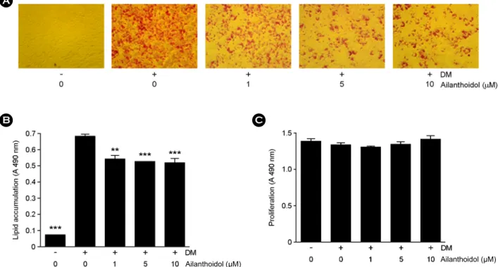
The effect of ailanthoidol on preventing lipid accumulation was examined by Oil Red-O staining of 3T3-L1 adipocytes.
The results represent lipid droplet accumulation, because triglycerides in adipocytes stain with Oil-Red-O staining solution apart from free fatty acids and phospholipids. To differentiate from 3T3-L1 to maturated adipocytes, we used insulin, dexamethasone, and IBMX. As shown in Fig.
2A, ailanthoidol reduced lipid accumulation, indicated by
decreased Oil Red-O staining. Adipocytes treated with the
10 μM ailanthoidol showed reduced lipid droplet size compared with that in DM cells. The OD values of Oil red O decreased from 78.0%, to 75.4%, and 74.2% in the 1 μM, 5 μM, 10 μM of ailanthoidol, respectively (Fig. 2B). In order to test the possibility that ailanthoidol treatment affect the viability of differentiated 3T3-L1 cells, we measured the proliferation at day 8. As shown in Fig. 2C, ailanthoidol did not affect the viability of differentiated 3T3-L1 cells suggesting that ailanthoidol inhibited the differentiation of preadipocytes into adipocytes without cytotoxicity.
Effects of ailanthoidol on lipolysis
The lipolysis of fully differentiated adipocytes was examined to investigate whether ailanthoidol reduce lipid content by increasing lipolysis. At a concentration of 5 and 10 μM, ailanthoidol increased lipolysis to 22 and 25%, respectively (Fig. 3).
Fig. 2. Effects of ailanthoidol on the accumulation of lipid drop in 3T3-L1 adipocyte. (A) Confluent 3T3-L1 preadipocytes differentiated into adipocytes in medium containing different concentrations of ailanthoidol for 8 days and morphological changes were photographed based on staining lipid accumulation by Oil-Red-O on differentiation day 8. (B) The quantity of extracted intracellular Oil-Red-O, without or with various concentrations of ailanthoidol. The results are reported as mean ± SEM of four independent experiments in triplicate. (C) Confluent 3T3-L1 preadipocytes were incubated with ailanthoidol (0~10 μM) for 8 days. Cell viability after treatment with ailanthoidol was determined by the MTS assay. Statistical significance is based on the difference when compared with differentiated 3T3-L1 cells (**P
< 0.01, ***P < 0.001).
Fig. 3. Effects of ailanthoidol on 3T3-L1 adipocyte lipolysis.
Fully differentiated adipocytes (on day 8) were cultured with in- dicated concentration of ailanthoidol for 48 h. The conditioned medium was removed from each well and assayed for glycerol content. The results are reported as mean ± SEM of four indepen- dent experiments in triplicate. Statistical significance is based on the difference when compared with differentiated 3T3-L1 cells (*P < 0.05).
A
Ailanthoidol (μM)
B C
Ailanthoidol (μM)
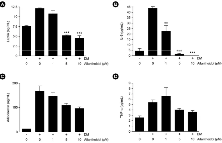
Effects of ailanthoidol on adipokine release
To determine whether the release of adipokines is modulated by ailanthoidol, we examined the levels of leptin, IL-6, adiponectin and TNF-α in ailanthoidol-treated 3T3-L1 adipocytes. Differentiated 3T3-L1 cells exposed to ailanthoidol at concentration of 1, 5 and 10 μM displayed a dose-dependent inhibited production of leptin (12%, 57%
and 62%, respectively) and IL-6 production (47%, 93%
and 100%, respectively) (Fig. 4A and 4B). In the presence of up to 10 μM of ailanthoidol, the adiponectin and TNF-α of differentiated 3T3-L1 cells were not significantly lower than non-treated cells (Fig. 4C and 4D).
Effects of ailanthoidol on the expression of adipocyte- specific transcription factors
To identify a possible mechanism by which adipogenesis
is reduced, we performed an experiment to determine the effects of ailanthoidol on the expression of PPARγ and C/EBPα, major transcription factors regulating adipogenesis.
As shown in Fig. 5, ailanthoidol treatment resulted in a dose-dependent suppression of PPARγ and C/EBPα at the protein levels. At a concentration of 10 μM, ailanthoidol decreased the expression of PPARγ and C/EBPα by 19%
and 63%, respectively, in 3T3-L1 cells.
We also determined whether ailanthoidol affected the activation. AMPK activity was measured by the amount of phosphorylation at AMPK threonine 172 (pAMPK). Treat- ment with ailanthoidol reduced pAMPK protein expression.
The data above suggest the involvement of the AMPK pathway in ailanthoidol-induced anti-adipogenic activity in 3T3-L1 adipocytes.
Fig. 4. Effects of ailanthoidol on adipokine release in 3T3-L1 adipocytes. 3T3-L1 adipocytes were treated with indicated concentration of ailanthoidol in differentiation medium from days 0 to 8 of adipogenesis. The amount of leptin (A), IL-6 (B), adiponetin (C) and (D) TNF-α were determined in cell culture medium. The results are reported as mean ± SEM of four independent experiments in triplicate.
Statistical significance is based on the difference when compared with differentiated 3T3-L1 adipocytes (**P < 0.01, ***P < 0.001).
A B
D C
DISCUSSION
Obesity has dramatically increased in most developing nations and is a prevalent condition related to metabolic disorders worldwide. Because the currently available anti- obesity drugs are plagued by numerous adverse effects (Li and Cheung, 2011), there is renewing interest in natural products as therapeutics since they are considered safer than synthetic compounds (Vermaak et al., 2011). The ability to inhibit adipogenesis is used to evaluate phytochemicals for anti-obesity potential (Hsu and Yen, 2008). In this study, we evaluated the effects of ailanthoidol on adipocyte
differentiation as well as its inhibitory mechanisms on adipogenesis in mouse 3T3-L1 cells.
Ailanthoidol, at most of the concentrations tested, decreased lipid accumulation. At the molecular level, expression of PPARγ and C/EBPα decreased after treatment.
PPARγ, a transcription factor of the nuclear receptor super-
family, is the master regulator of adipogenesis since it is
both necessary and sufficient for adipogenesis (Rosen and
Spiegelman, 2000). The expression of PPARγ alone induces
adipogenesis in fibroblasts (Tontonoz et al., 1994). Most
regulators of adipogenesis seem to function by activating
or inhibiting PPARγ expression (Rosen and MacDougald,
2006). PPARγ induces the expression of C/EBPα binding
Fig. 5. Effects of ailanthoidol on the expression of adipocyte-specific transcription factors of adipogenic differentiation. 3T3-L1 cells were treated with indicated concentration of ailanthoidol in differentiation medium from days 0 to 8 of adipogenesis. Total protein was isolated from 3T3-L1 cells at day 8 and immunoblotted for pAMPK, PPARγ, C/EBPα, and β-actin as designated. The bands were quantified using NIH image analysis software and their relative intensity was expressed as fold against the image of the untreated cells. Quantification of protein levels expressed as mean ± SEM of three independent experiments in each column. Statistical significance is based on the difference when compared with differentiated 3T3-L1 adipocytes (*P < 0.05, **P < 0.01, ***P < 0.001).to its promoter region (Rosen et al., 2002), and the ailanthoidol induced reduction of C/EBPα expression may be the result of a decrease in PPARγ expression. C/EBPα is a member of the C/EBP family basic-leucine zipper class of transcription factors, and forms a positive feedback loop with PPARγ to reinforce the expression of adipocyte- specific genes (Rosen et al., 2002).
Our results also demonstrated the level of phosphorylated AMPK was induced by ailanthoidol treatment. AMPK is a metabolic regulator that acts as a cellular fuel gauge in eukaryotes and a well-characterized target if anti-obesity and anti-diabetic treatment (Hardie, 2008). AMPK activation requires phosphorylation at threonine 172 (Carling, 2004) and its substrates include a number of biosynthetic enzymes such as acetyl-CoA carboxylase, FAS, glycerol-3-phosphate acyltransferase, and 3-hydroxy-3-methylglutaryl-CoA re- ductase. Phosphorylation of these proteins reduces their activity and leads to decreased energy consumption in their respective biosynthetic pathways as well as increased fatty acid oxidation to increase energy production (Hardie, 2008), (Kahn et al., 2005). That is, activated AMPK decreases lipogenesis, increases fatty acid oxidation, and increases lipolysis in adipocytes. Although the mechanism by which ailanthoidol inhibits the phosphorylation of AMPK is beyond the scope of the present research, ailanthoidol may exert its anti-adipogenic effect via the inhibition of AMPK activation.
Our data showed that the releases of adipokines, leptin and IL-6, were decreased in ailanthoidol-treated 3T3-L1 cells compared with untreated control cells. Adipose tissue synthesizes and secretes a number of factors, such as leptin, IL-6, resistin and adiponectin (Tilg and Moschen, 2006).
Among these factors, leptin is important because of its key roles in energy balance and leptin expression is a late indicator of adipocyte maturation and a key mediator of adipose tissue endocrine function. In addition, IL-6 is a major circulating cytokine and released from both macro- phages and adipocytes (Purohit et al., 1995). IL-6 is higher in obese patients and in patients with diabetes (Pradhan et al., 2001). In the obese state, however, it is plausible that IL-6 released from an expanded adipose tissue mass could contribute to certain aspects of the associated patho-
physiology, including a proinflammatory state predisposing to atherosclerosis. Thus reducing secretion of IL-6 in adipo- cytes is important to control obesity. In sum, ailanthoidol is a possible agent to regulate obesity since it blocked IL-6 and leptin secretion.
In conclusion, this study demonstrated a potent anti- adipogenic effect of ailanthoidol on cellular and molecular levels, implicating a potential application of this cost- effective natural product in the prevention of obesity.
Acknowledgements
This work was supported by the National Research Foundation of Korea (NRF) grant funded by the Korea government (MEST) (No. 2011-0028637).
REFERENCES
Carling D. The AMP-activated protein kinase cascade-a unifying system for energy control. Trends Biochem Sci. 2004. 29: 18 -24.
Chavey C, Mari B, Monthouel MN, Bonnafous S, Anglard P, Van Obberghen E, Tartare-Deckert S. Matrix metalloproteinases are differentially expressed in adipose tissue during obesity and modulate adipocyte differentiation. J Biol Chem. 2003.
278: 11888-11896.
Farmer SR. Transcriptional control of adipocyte formation. Cell Metab. 2006. 4: 263-273.
Green A, Rumberger JM, Stuart CA, Ruhoff MS. Stimulation of lipolysis by tumor necrosis factor-alpha in 3T3-L1 adipocytes is glucose dependent: implications for long-term regulation of lipolysis. Diabetes. 2004. 53: 74-81.
Hardie DG. AMPK: a key regulator of energy balance in the single cell and the whole organism. Int J Obes. 2008. 32: S7-12.
Hsu CL, Yen GC. Phenolic compounds: evidence for inhibitory effects against obesity and their underlying molecular signaling mechanisms. Mol Nutr Food Res. 2008. 52: 53-61.
Hwang JT, Kwon DY, Yoon SH. AMP-activated protein kinase: a potential target for the diseases prevention by natural occurring polyphenols. N Biotechnol. 2009. 26: 17-22.
Kahn BB, Alquier T, Carling D, Hardie DG. AMP-activated protein kinase: ancient energy gauge provides clues to modern under- standing of metabolism. Cell Metab. 2005. 1: 15-25.
Kim JK, Jun JG. Ailanthoidol suppresses lipopolysaccharide-
stimulated inflammatory reactions in RAW264.7 cells and endotoxin shock in mice. J Cell Biochem. 2011. 112: 3816- 3823.
Lee EJ, Kang M, Kim YS. Platycodin D inhibits lipogenesis through AMPKα-PPARγ2 in 3T3-L1 cells and modulates fat accumulation in obese mice. Planta Med. 2012. 78: 1536 -1542.
Lee YJ, Kao ES, Chu CY, Lin WL, Chiou YH, Tseng TH.
Inhibitory effect of ailanthoidol on 12-O-tetradecanoyl- phorbol-13-acetate-induced tumor promotion in mouse skin.
Oncol Rep. 2006. 16: 921-927.
Li MF, Cheung BM. Rise and fall of anti-obesity drugs. World J Diabetes. 2011. 2: 19-23.
Otto TC, Lane MD. Adipose development: from stem cell to adipocyte. Crit Rev Biochem Mol Biol. 2005. 40: 229-242.
Poulos SP, Dodson MV, Hausman GJ. Cell line models for differentiation: preadipocytes and adipocytes. Exp Biol Med.
2010. 235: 1185-1193.
Pradhan AD, Manson JE, Rifai N, Buring JE, Ridker PM.
C-reactive protein, interleukin 6, and risk of developing type 2 diabetes mellitus. JAMA. 2001. 286: 327-334.
Purohit A, Ghilchik MW, Duncan L, Wang DY, Singh A, Walker MM, Reed MJ. Aromatase activity and interleukin-6 pro- duction by normal and malignant breast tissues. J Clin Endocrinol Metab. 1995. 80: 3052-3058.
Rhyu J, Kim MS, You MK, Bang MA, Kim HA. Pear pomace water extract inhibits adipogenesis and induces apoptosis in 3T3-L1 adipocytes. Nutr Res Pract. 2014. 8: 33-39
Rosen ED Spiegelman BM. Molecular regulation of adipogenesis.
Annu Rev Cell Dev Biol. 2000. 16: 145-171.
Rosen ED, Hsu CH, Wang X, Sakai S, Freeman MW, Gonzalez FJ, Spiegelman BM. C/EBPalpha induces adipogenesis through PPARgamma: a unified pathway. Genes Dev. 2002. 16: 22 -26.
Rosen ED, MacDougald OA. Adipocyte differentiation from the inside out. Nat Rev Mol Cell Biol. 2006. 7: 885-896.
Rossmeisl M, Flachs P, Brauner P, Sponarova J, Matejkova O, Prazak T, Ruzickova J, Bardova K, Kuda O, Kopecky, J.
Role of energy charge and AMP-activated protein kinase in adipocytes in the control of body fat stores. Int J Obes Relat Metab Disord. 2004. 28: S38-44.
Spiegelman BM, Choy L, Hotamisligil GS, Graves RA, Tontonoz P. Regulation of adipocyte gene expression in differentiation and syndroms of obesity/diabetes. J Biol Chem. 1993. 268:
6823-2866.
Tilg H, Moschen AR. Adipocytokines: mediators linking adipose tissue, inflammation and immunity. Nat Rev Immunol. 2006.
6: 772-783.
Tontonoz P, Hu E, Graves RA, Budavari AI, Spiegelman BM.
mPPARgamma2: tissue-specific regulator of an adipocyte enhancer. Genes Dev. 1994. 8: 1224-1234.
Vermaak I, Viljoen AM, Hamman JH. Natural products in anti- obesity therapy. Nat Prod Rep. 2011. 28: 1493-1533.
White UA, Stephens JM. Neuropoietin activates STAT3 indepen- dent of LIFR activation in adipocytes. Biochem Biophys Res Commun. 2010. 395: 48-50.
World Health Organisation. Global status report on noncommuni- cable diseases 2010. Chapter 1. Burden: mortality, morbidity and risk factors. World Health Organization. 2011. 9-31.