This research was carried out with support of“Cooperative Research program for Agriculture Science & Technology Development (Project NO.PJ0113052016)” Rural Development Administration.
접수일 : 2019년 6월 26일, 수정일 : 2019년 7월 15일, 채택일 : 2019년 7월 18일
†
Corresponding author : Wookyoung Kim, Department of Food Science and Nutrition, Dankook University, 119 Dandae-ro, Dongnam-gu, Cheonan 31116, Korea
Tel : 82-41-550-3471, Fax : 82-41-559-7955, E-mail : [email protected], ORCID : https://orcid.org/0000-0002-8652-5339
옥수수수염 추출물이 SW480 Colon Cancer Cell에서 NF-κB와 염증성 사이토카인 발현에 미치는 영향
최현지·김선림1·강현중2·김명환3·김우경†
단국대학교 식품영양학과·
1농촌진흥청 국립식량과학원 중부작물부 중부작물과·
2
농촌진흥청 국립농업과학원 농업생명자원부 생물안정과·
3단국대학교 식품공학과
The Effect of Saccharin on the Gene Expression of NF-κB and Inflammatory Cytokines in LPS-Stimulated SW480 Colon Cancer Cells
Hyunji Choi·Sunlim Kim1·Hyeonjung Kang2·Myunghwan Kim3·Wookyoung Kim†
Dept. of Food Science and Nutrition, Dankook University, Cheonan 31116, Korea
1
Central Area Crop Breeding Division, Department of Central Area Crop Science, National Institute of Crop Science, Rural Development Administration, Suwon 16429, Korea·
2
Biosafety Division, National Institute of Agriculture Science, Rural Development Administration, Jeonju 54874, Korea
3
Dept. of Food Engineering, Dankook University, Cheonan 31116, Korea
ABSTRACT
There have been no published studies concerning the anti-inflammatory effects of corn silk on colon cancer cells. Thus, this study was conducted to investigate the effect of corn silk extract containing high levels of maysin on in- flammation and its mechanism of action in colon cancer cells. SW 480 human colon cancer cells were treated with 1 μg/mL of lipopolysaccharide (LPS) to induce inflammation, and next they were treated with different concentrations of corn silk ex- tract (0, 5, 10 and 15 μg/mL). The concentrations of nitric oxide (NO) were determined. The mRNA expressions of inducible nitric oxide synthase (iNOS), cyclooxygenase-2 (COX-2), tumor necrosis factor α (TNF-α), inter- leukin-1beta (IL-1β) and interleukin-6 (IL-6), were determined. Western blot analysis was performed to determine the protein expressions of nuclear factor-kappa B (NF-κB) and mitogen-activated protein kinases, and the latter consists of extracellular signal-related kinase (ERK), c-jun NH2-terminal kinase (JNK) and p38 MAP kinase (p38).
The concentration of NO and the mRNA expression of iNOS were significantly and dose-dependently decreased in the corn silk-treated groups (P<0.05). The mRNA expression of TNF-α, IL-1β and IL-6 were significantly in- creased in the LPS-treated group (P<0.05), but these expressions were significantly and dose-dependently de- creased in the corn silk treated groups (P<0.05). The protein expressions of NF-κB (in a dose-dependent fash- ion), ERK (at 10 and 15 μg/mL), JNK (at 15 μg/mL) and p38 (at 10 and 15 μg/mL) were significantly de- creased with corn silk treatments (P<0.05). In conclusion, corn silk extract containing high levels of maysin seems to inhibit the LPS-induced inflammatory responses in SW480 colon cancer cells via the NF-κB pathway.
Key words : corn silk, inflammation, maysin, colon cancer cell, NF-κB
서 론
2016년 국가암등록통계에 따르면 남녀 전체에서 가장 많이 발생한 암은 위암이었고, 그 다음으로는 대장암, 갑상선암, 폐암, 유방암, 간암 순으로 많이 나타났다. 남자는 위암, 폐암, 대장암 순이었고, 여 자는 갑상선암, 유방암, 대장암 순으로 높았다 (National Cancer Center 2016). 우리나라에서 2000년 이후로 폐암, 간암 및 위암의 사망률은 지속적으로 감소하는 것과 다르게 대장암의 연령 표준화 사망 률은 점점 증가하고 있다(Jung 등 2017). 대장암의 발병요인으로는 유전적 요인보다는 환경적 요인이 크게 작용하며, 적은 신체 활동, 붉은 고기와 가공 육의 섭취, 음주와 비만 등이 발병 위험을 높이는 것으로 보고되고 있다(Tan & Chen 2016). 부적절한 식생활과 비만은 체내 염증을 유발하고, 체내 염증 증가는 대장암 발병과 진행에 중요한 역할을 하는 것으로 보고되었다(Cho 등 2016).
염증이란 세균 감염, 조직의 손상에 대한 인체의 국소적인 반응으로 발적, 종창, 발열 및 통증 등의 증상이 나타나게 된다(Coussens & Werb 2002). 급성 적인 염증에서는 면역세포들이 다양한 방법으로 세 균을 제거하고 체내의 환경을 정상상태로 돌리는 과정이 일어난다. 그러나 이 과정에서 대식세포의 활성화가 과도하게 일어나면 염증매개물질 분비를 통해 과도한 염증반응을 유도하게 된다. 이러한 염 증반응이 지속되거나 반복되면 다양한 사이토카인 (cytokine)들이 과다 분비되어 대식세포의 기능이 손 상되고, 혈관 내피 기능 이상, 산화 스트레스 및 만 성 염증을 유발하고, 일부는 암으로도 진행된다고 보고되어 있다(Schäffler 등 2006; Lin & Karin 2007;
Park & Spurlock 2012). 대표적인 염증성 사이토카인 으로는 TNF-α, IL-1, IL-6 등이 있으며, IL-1은 대부 분의 세포에 존재하며 T세포와 B세포의 자극제 역 할을 하는 선천성 면역반응의 중요한 조절인자이다 (Dinarello 2013). IL-6은 염증반응의 초기에 관여하는 데, 염증 초기에 필요한 단백질 합성을 유도하고 B세포
가 항체 생성 세포로 분화하도록 한다(Lin 등 2000).
TNF-α의 분비 증가는 여러 염증 관련 질환을 유발 한다고 알려져 있다(Ruan 등 2002; Andrade-Oliveira 등 2015; Castoldi 등 2016). 염증반응이 없는 암 환 자들보다 전신적인 염증반응을 보이는 암 환자의 경우 암을 치료하기 위한 수술이나 약물을 복용한 후에 암이 재발하거나 부작용이 생기는 경우가 더 많다고 보고되고 있다(Yoon 2012). 따라서 염증을 억제하는 것은 암의 발병뿐만 아니라 암의 치료에 도 도움을 줄 수 있을 것으로 알려져 있다.
옥수수는 벼과에 속하는 1년 초본으로 남미가 원 산지인 세계 식용작물 중 하나이다. 과실은 식용이 나 동물사료로 사용되어 왔지만 부산물인 옥수수수 염(Zeamays L.)은 최근까지도 사용목적이 없어 폐기 되어 왔다(Hasanudin 등 2012). 예로부터 옥수수수염 은 옥발, 옥미발, 옥촉서예라고도 불리웠고 황색-담 갈색을 띄며 비뇨계 감염과 질병 치료에 사용되어 베트남, 중국, 남미 등지에서는 요도결석, 신장염, 당뇨병 치료 및 이뇨제로 사용되어 왔다(Grases 등 1993; Min 등 2010). 옥수수수염에는 메이신(maysin) 을 비롯한 apimaysin, methoxyamaysin 등의 물질들이 특이적으로 존재한다. 특히, 메이신은 옥수수수염에 함유된 대표적인 생리활성물질로 옥수수의 해충인 회색담배나방 유충(corn earworm)의 생육을 억제하 고, 종양 세포주에 대한 세포독성 효과 및 라디칼 소거활성 등이 보고되었다(Byrne 등 1996). 농촌진흥 청은 옥수수수염에서 메이신 등 생리활성물질을 분 리하는 기술 개발에 성공하였고, 메이신의 생리활성 스크리닝 및 효능 검증에 대한 연구들을 보고하였 다(Kim 등 2014).
옥수수수염 추출물 또는 메이신의 처리가 대식세 포에서 유도성 산화질소 생성효소(inducible nitric oxide synthase, iNOS)와 산화질소(nitric oxide, NO)의 생성을 억제한다고 보고되었다(Kim 등 2004). 또한 옥수수수염 추출물이 in vitro, in vivo 실험에서 신경 세포의 독성 및 염증을 감소시켜 뇌신경을 보호하 고 사이토카인 생성을 억제하였다고 보고하였다
(Kim 등 2004; Kim 등 2005). 그러나 옥수수수염 추 출물이 염증에 미치는 영향은 매우 제한적으로 보 고되고 있고, 암세포에서의 항염증 효과에 대한 연 구는 아직 없는 실정이다. 따라서 본 연구는 옥수수 수염 추출물이 대장암세포인 SW480 세포에서의 항 염증 효과와 그 기전을 관찰하는 것을 목적으로 실 행되었다.
연구방법
1. 실험재료세포배양에 필요한 Dulbecco’s Modified Eagle’s Medium/Nutrient Mixture F-12(DMEM/F-12), fetal bo- vine serum(FBS), penicillium, streptomycin, phophatate buffered saline(PBS), trypsin-EDTA는 Welgene(Daegu, Korea)에서, isopropanol은 Samchun(Pyeongtaek, Korea)에 서, transferrin은 Gibco BRL(NY, USA)에서, bovine se- rum albumin(BSA), selenium, Griess reagent, Tri re- agent와 chloroform은 Sigma(St. Louis, USA)에서, anti- body(β-actin, NF-κB, ERK, JNK, p38)는 Cell Signaling(Danvers, USA)에서 구입하였다. 옥수수수염 추출물(corn silk extract, CSE)은 농촌진흥청에서 제 공받았다. 추출물 제조를 위해 사용된 옥수수수염에 는 메이신 함량이 2,783 mg/100 g이었으며, 1 kg의 옥수수수염에서 3.5 g의 추출물을 얻었으며, 옥수수 수염 추출물의 메이신 함량은 400 mg/1,000 mg 옥 수수수염 추출물이다.
2. 세포배양
인체 대장암 세포주인 SW480은 한국 세포주 은 행(Korea Cell Line Bank, Seoul, Korea)에서 구입하여 실험하였다. SW480은 10% FBS, 100 units/mL penicillium, 100 μg/mL streptomycin을 첨가한 DMEM/F-12를 배양액으 로 습윤한 5% CO2, 37oC incubactor에서 배양하고 세
포의 밀도가 70∼80%가 되면 trypsin-EDTA를 처리하 여 계대배양하였다. 모든 실험은 FBS가 함유되지 않은 serum free medium(SFM, 0.01% BSA, 5 μg/mL transferrin, 5 ng/mL selenium, 100 units/mL pen- icillium-100 μg/mL streptomycin)으로 진행하였다.
3. 세포증식능 실험
옥수수수염 추출물이 대장암세포 SW480의 증식 에 미치는 영향을 알아보기 위해 tetrazolium-based colorimetric assay(MTT assay)를 실시하였다. 24 well plate에 세포를 1.5×10⁴ cells/mL로 분주하고 48시간 배양 후 SFM으로 교환한 다음 24시간 배양하고 옥 수수수염 추출물을 농도별(0, 5, 10, 15 μg/mL)로 SFM에 희석하여 처리하였다. 옥수수수염 추출물 을 농도별로 분주한 plate를 습윤한 5% CO2, 37oC incubactor에서 시간별(0, 24, 48, 96시간)로 배양하였 다. 각 배양시간이 끝나면 MTT assay로 세포증식을 측정하였다.
옥수수수염 추출물이 암세포에서 염증에 미치는 영향을 알아보기 위해 LPS를 처리하여 염증을 유발 하였다. 세포증식 실험과 같은 방법으로 24 well plate에 세포를 1.5×10⁴ cells/mL로 분주하고 48시간 배양 후 SFM으로 교환한 다음 24시간 배양하고 LPS를 1 μg/mL로 처리하고 옥수수수염 추출물은 0, 5, 10, 15 μg/mL로 처리하여 24시간 습윤한 5%
CO2, 37oC incubactor에서 배양하고 MTT assay를 하 였다.
4. 염증 관련 실험
1) NO 생성량 측정
NO의 농도는 배양액의 nitrite 농도를 Griess re- agent를 사용하여 측정하였다. 대장암세포를 100 mm dish에 1.5×10⁵ cells/mL 농도로 10 mL 분주하고, 48시간 배양 후 SFM으로 교환하였다. 24시간 배양 후 LPS 는 1 μg/mL, 옥수수수염 추출물은 0, 5, 10, 15 μg/mL
Table 1. Primer sequences used for real time PCR.
Gene
1)Primer Sequence
2)COX-2 Forward 5’-TCCTTGCTGTTCCCACCCATG-3’
Reverse 5’-CATCATCAGACCAGGCACCAG-3’
iNOS Forward 5’-CGGTGCTGTATTTCCTTACGAGGCGAA-3’
Reverse 5’-GGTGCTGTCTGTTAGGAGGTCAAGTAA-3’
TNF-α Forward 5’-CAGAGGGAAGAGTTCCCCAG-3’
Reverse 5’-CCTTGGTCTGGTAGGAGACG-3’
IL-1β Forward 5’-GTAGCCCACGTCGTAGCAAA-3’
Reverse 5’-CCCTTCTCCAGCTGGGAGAC-3’
IL-6 Forward 5’-CCAGTACCCCCAGGAGAAGA-3’
Reverse 5’-CAGCTCTGGCTTGTTCCTCA-3’
β-actin Forward 5’-AGGTCATCACTATCGGCAAT-3’
Reverse 5’-ACTCATCGTACTCCTGCTTG-3’
1)
COX-2: cyclooxygenase-2, iNOS: inducible nitric oxide synthase, IL-1β: interleukin 1 beta, IL-6: interleukin 6, TNF-α: tumor necrosis factor α, β-actin: beta-actin
2)
T: thymine, A: adenine, C: cytosine, G: guanine
로 처리하여 24시간 습윤한 5% CO2, 37oC incubactor 에서 배양하였다. 배양이 끝나면 상층액 100 μL를 96 well plate에 분주하고 Griess reagent를 첨가하여 상온에서 10분 동안 반응시킨 후, ELISA microplate reader(infinite 200, Tecan, Groedig, Austria)를 이용하 여 540 nm에서 흡광도를 측정하였다.
2) 사이토카인(TNF-α, IL-1β, IL-6), iNOS 및 COX-2 mRNA 발현
대장암세포를 100 mm dish에 1.5×10⁵ cells/mL 농 도로 10 mL 분주하고, 48시간 배양 후 SFM으로 교 환하였다. 24시간 배양 후 LPS는 1 μg/mL, 옥수수 수염 추출물은 0, 5, 10, 15 μg/mL로 처리하여 24시 간 습윤한 5% CO2, 37oC incubator에서 배양하였다.
배양이 끝나면 세포를 수집하여 PBS로 2회 세척 후 Tri reagent를 이용하여 RNA를 추출하였다. 추출한 RNA sample은 ELISA microplate reader(infinite 200, Tecan, Groedig, Austria)로 260 nm, 280 nm에서 흡광 도를 측정하여 농도를 계산하였다. cDNA로 역전사 시키기 위하여 SuperScript kit(invitrogen, Carisbad, USA)를 사용하였다. 5 μg으로 정량한 total RNA를 tube에 준비하고 Oligo DT 1 μL를 포함한 총량이 12 μL가 되도록 DEPC water를 첨가하여 70oC에서 10분 배양하고 reaction mixture(5X first standard buffer 4 μL, 0.1M DTT 2 μL, 10 mM NTP 1 μL) 7 μL 를 첨가해 72oC에서 5분 배양한 후, superscript Ⅱ reverse transcriptase 0.5 μL씩 첨가해 42oC에서 1시 간 45분, 70oC에서 15분 배양하였다. 여기에 RNase H 0.5 μL를 넣어 37oC에서 1시간 배양하고 DEPC water 80 μL를 가하여 실험 시까지 −20oC에 보관 하였다.
Real time PCR을 실행하기 위해 cDNA sample 2 μL에 SYBP Green Master Mix(Apllied biosystems, CA, USA) 10 μL, forward/reverse primer(Table 1)를 각각 1 μL와 Nuclease free water 6 μL를 넣고 Applied Biosystems StepOne(Apllied biosystems, CA, USA) software v2.1을 사용하여 95oC에서 10분 반응
시킨 후 95oC에서 15분, 60oC에서 1분 반응시키고, 95oC에서 15분, 60oC에서 1분, 95oC에서 15분으로 40 cycles을 반응시켰다. 실험결과는 Applied Biosystems StepOne(Apllied biosystems, CA, USA) software v2.1에 내장되어 있는 프로그램을 이용하여 △△CT방법으 로 계산하였고 3번 반복 실험을 진행하였다.
3) NF-κB와 MAPKs(ERK, JNK, p38) 단백질 발현 실험
세포는 total RNA를 추출하기 위한 실험과 같은 방법으로 배양하였다. LPS와 옥수수수염 추출물을 함께 처리하여 24시간 배양한 후 dish의 medium을 제거하고 cold PBS로 세척하고 1 mL의 PBS를 넣고 cell scraper로 세포를 수집하였다. 1X RIPA lysis buf- fer(Cell signaling, Danvers, USA) 400 μL를 첨가하 여 세포 균질액을 얻은 후 실험 시까지 −70oC에 보 관하였다. Bio-rad protein assay kit(Bio Rad, Hercules, USA)를 이용하여 단백질을 정량하여 50 μg의 단백 질을 SDS-PAGE(sodium dodecyl sulfate-polyacrylamide gel electrophoresis)로 분리하고 membrane에 옮겼다.
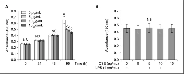
Figure 1. Effects of corn silk extract (CSE) on cell proliferation in SW480 cells. The effect of corn silk extract (CSE) on viable cell numbers were estimated by the MTT assay (A). The effect of Effects of corn silk extract (CSE) on viable cell numbers were estimated by the MTT assay. SW480 cells were plated in 24 well plates at a density of 1.5×10
4cells/mL. After 48 h, the medium was replaced with serum-free media (SFM) to induce serum starvation for the next 24 h. After an overnight in- cubation, the cells were treated with various concentrations of corn silk extract (0, 5, 10, 15 μg/mL). An MTT assay was performed at every 0, 24, 48, and 96 h during experimental treatments. The effect of corn silk extract (CSE) and LPS on viable cell numbers after 24 h incubation was measured by the MTT assay (B). The cells were treated with various con- centrations of corn silk extract (0, 5, 10, 15 μg/mL) in the presence or absence of LPS to induce inflammation (1 μg/mL) for 24 h and then MTT assay was performed. Each graph represents the mean±SE of three independent experiments. a>b:
The different letters indicate significant differences among group at α=0.05 as determined by Duncan’s multiple range test.
LPS, lipopolysaccharide.
그 후 5%의 nonfat dry milk나 BSA에 일차 antibody (β-actin, ERK, JNK, p38, NF-kB)를 1:1,000으로 희 석하여 4oC에서 overnight하였다. 다음날 TBST(Tris buffer saline-Tween; 200 mM Tris, 1.37M Nacl, pH 7.6, 0.1% Tween 20)로 3번 세척한 후 anti-mouse나 rabbit IgG로 3시간 실온에서 배양 후 다시 TBST로 3번 세척하였다. 발색시약을 이용하여 20초∼1분간 발 색시킨 후 X-Omat Film(Kodak)으로 현상하여 imag- ing program인 Image J launcher(provided by NCBI)를 사용하여 밴드의 밀도를 측정하였다.
5. 통계처리
본 연구에서 얻어진 모든 자료는 SPSS statistics (ver.23, SPSS Inc., Chicago, IL, USA)을 사용하여 평 균과 표준오차를 계산하였고, ANOVA 분석 후 Duncan’s multiple range test를 이용하여 각 군 간의 유의성을 P<0.05 수준에서 검증하였다. 모든 실험
은 독립적으로 3회 이상 실시하였다.
결 과
1. 암세포 증식능력 측정옥수수수염 추출물의 대장암세포 증식 억제 효과 를 알아보기 위한 MTT assay 결과는 Fig. 1과 같다.
옥수수수염 추출물을 농도별(0, 5, 10, 15 μg/mL)로 첨가하여 0, 24, 48, 96시간 배양한 결과, 배양시간 48시간까지는 증식 억제 효과가 없었으나, 96시간 배양하면 농도 의존적으로 세포증식이 억제되었다 (Fig. 1A)(P<0.05). 염증을 유발한 후 옥수수수염 추 출물이 암세포에서 염증과 관련한 인자들에 미치는 영향을 관찰하기 위해 세포에 LPS를 처리하였다. 염 증을 유발하기 위한 LPS 처리가 세포증식에 영향이 없는 조건에서 실험을 진행하기 위해 여러 조건에
Figure 2. Effects of corn silk extract (CSE) on NO concentration (A) and the mRNA expression of iNOS (B) in LPS-stimulated SW480 cells. After SW480 cell was divided by 1.5×10
5cells/mL in a 100 mm dish and incubated for 48 h, the medium was re- placed with SFM. SFM was treated with 1 μg/mL of LPS and corn silk extract concentration of 0, 5, 10, 15 μg/mL and in- cubated another 24 h in a 37
oC 5% CO
2incubator. After incubation, 100 μL of the supernatant was dispensed into a 96-well plate. Griess reagent was added to determine NO concentration (A). At the end of incubation, the cells were col- lected, washed twice with PBS, and extracted with Tri reagent to obtain total RNA from sample. Total RNA was isolated and real-time PCR was performed for iNOS (B). Each graph represents the mean±SE of three independent experiments.
a>b: The different letters indicate significant differences among group at α=0.05 as determined by Duncan’s multiple range test. NO, nitric oxide; iNOS, inducible nitric oxide.
서 예비실험을 한 결과 LPS와 옥수수수염 추출물을 함께 처리한 후 24시간 배양 시에 세포증식에 영향 이 없었으므로 다른 실험에서의 배양조건을 이와 같이 하였다(Fig. 1B).
2. NO 생성과 iNOS mRNA 발현
옥수수수염 추출물이 염증상태를 억제하는지를 알아보기 위해 NO의 생성과 NO를 생성하는 유도효 소인 iNOS의 mRNA 발현을 측정한 결과, LPS만을 처리한 군에서 NO의 생성과 iNOS mRNA 발현이 유의적으로 증가하였고, 옥수수수염 추출물을 처리 하였을 경우 농도 의존적인 유의적인 감소를 보였 다(P<0.05)(Fig. 2).
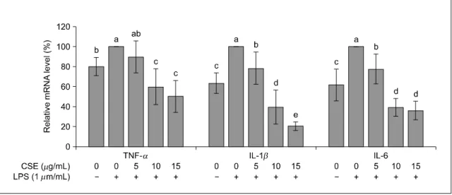
3. 사이토카인(TNF-α, IL-1β, IL-6)과 COX-2 mRNA 발현
옥수수수염 추출물이 사이토카인 TNF-α, IL-1β,
IL-6과 COX-2의 mRNA 발현에 미치는 영향을 알아 보기 위해 real-time PCR을 시행한 결과는 Fig. 3, 4 와 같다. TNF-α, IL-1β, IL-6의 mRNA 발현은 LPS 만을 처리한 군이 유의적으로 증가하였고 옥수수수 염 추출물을 첨가하면 TNF-α와 IL-1β는 농도 의 존적으로 발현이 감소하였고, IL-6은 농도가 증가하 면 발현이 유의적으로 감소하였으나(P<0.05) 처리 농도 10과 15 μg/mL 에서는 차이가 없었다(Fig. 3).
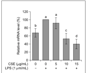
COX-2의 mRNA의 발현도 LPS만을 처리한 군에서 유의적으로 증가하였고, 옥수수수염 추출물을 처리 한 군에서는 10과 15 μg/mL에서 유의적으로 감소 하였다(P<0.05)(Fig. 4).
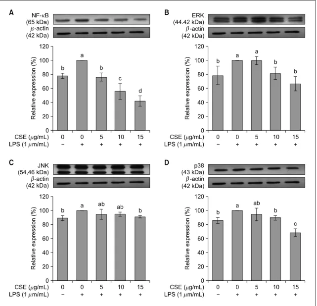
4. NF-κB와 MAPKs(ERK, JNK, p38) 단백질 발현
SW480 대장암세포에서 염증과 관련된 단백질인 NF-κB 및 MAPKs(ERK, JNK, p38)의 단백질 발현 을 실험한 결과는 Fig. 5와 같다. NF-κB의 발현은 LPS만 처리 시 증가하였다가 옥수수수염 추출물을
Figure 3. Effects of corn silk extract (CSE) on the mRNA expression of TNF-α, IL-1β and IL-6 in LPS-stimulated SW480 cells. After SW480 cell was divided by 1.5×10
5cells/mL in a 100 mm dish and incubated for 48 h, the medium was replaced with SFM. SFM was treated with 1 μg/mL of LPS and corn silk extract concentration of 0, 5, 10, 15 μg/mL and incubated an- other 24 h in a 37
oC, 5% CO
2incubator. At the end of incubation, the cells were collected, washed twice with PBS, and extracted with Tri reagent to obtain total RNA from sample. Total RNA was isolated and real-time PCR was performed.
Each graph represents the mean±SE of three independent experiments. a>b: The different letters indicate significant dif- ferences among group at α=0.05 as determined by Duncan’s multiple range test. TNF-α, tumor necrosis factor α; IL-1β, interleukin 1 beta; IL-6, interleukin 6.
처리하면 농도에 따라 농도 의존적으로 감소하였다 (P<0.05). ERK, JNK, p38도 LPS만 처리 시 발현이 유의적으로 증가하였으나, ERK는 옥수수수염 추출물 10과 15 μg/mL에서 유의적인 감소가 있었고(P<0.05), JNK는 옥수수수염 추출물 15 μg/mL에서만 유의적 인 감소가 있었으며(P<0.05), p38은 10과 15 μg/mL 에서 유의적으로 감소하였다(P<0.05).
고 찰
옥수수수염의 주요 생리활성물질인 메이신은 플 라보노이드의 일종으로 산화에 안정적이고 항비만 효과와 항산화 효과가 있다고 보고되었다(Grases 등 1993; Byrne 등 1996; Min 등 2010). 그러나 암세포 에서 옥수수수염 추출물의 항염증 효과 및 기전에 관한 연구가 매우 제한적이다. 따라서 본 연구에서 는 대장암세포인 SW480 세포에서 옥수수수염 추출 물의 항염증 효과를 조사하고 그 기전을 알아보는
것을 목적으로 하였다. 본 연구에서 사용한 SW480 대장암세포는 대장암의 구조적 및 생리학적 특성 연구에 가장 널리 사용되는 세포이다(Cha 등 2017).
염증 유발을 위해 사용한 LPS는 그람음성의 세포벽 에 있는 내독소로써 다양한 염증전(proinflammatory) 생성을 특징으로 한다(Triantafilou & Triantafilou 2005).
SW480 대장암세포에 LPS를 처리하면 NADPH 산화 효소(NADPH oxidase)에 의하여 대장암세포 분열능 력이 감소하므로(Cha 등 2017), SW480 대장암세포 에 LPS 첨가로 인한 세포 성장에 대한 독성 정도를 확인하기 위하여 MTT assay를 하였다. 그 결과 LPS 처리 후 24시간 배양이 세포증식에 유의적 차이가 없는 것으로 나타나 이후 모든 실험은 LPS와 옥수 수수염 추출물의 처리시간을 24시간으로 시행하였 다.
본 연구에서 옥수수수염 추출물 처리군에서 농도 의존적으로 NO 생산과 iNOS의 mRNA 발현이 유의 적으로 감소하였다. NO는 활성산소의 일종으로 NO 합성효소(NO synthase, NOS)에 의하여 L-아르지닌
Figure 4. Effects of corn silk extract (CSE) on the mRNA ex- pression of COX-2 in LPS-stimulated SW480 cells.
After SW480 cell was divided by 1.5×10
5cells/mL in a 100 mm dish and incubated for 48 h, the medium was replaced with SFM. SFM was treated with 1 μg/mL of LPS and corn silk extract concentration of 0, 5, 10, 15 μg/mL and incubated another 24 h in a 37
oC, 5% CO
2incubator. At the end of incubation, the cells were collected, washed twice with PBS, and extracted with Tri reagent to obtain total RNA from sample. Total RNA was isolated and real-time PCR was performed. a>b: Each graph represents the mean±SE of three independent experiments. a>b:
The different letters indicate significant differences among group at α=0.05 as determined by Duncan’s multiple range test. COX-2, cyclooxygenase-2.
(L-arginine)으로부터 생성된다(Moncada 1991). NOS의 일종인 iNOS는 자극을 받으면 NO를 과도하게 생성 시켜 염증반응과 염증매개체의 증가로 염증이 심화 되어 조직의 손상, 유전자 변이 및 신경 손상을 발 생시킨다(Won 등 2006). NO 과다 생성은 대식세포 에서 사이토카인 생성을 증가시키고, COX-2 발현을 증가시켜 염증을 일으킨다고 보고되었다(Habib 등 1997; Tsujii 등 1997). 그러므로 본 실험결과 옥수수 수염 추출물은 iNOS 발현을 감소시켜 NO의 생성을 억제하여 염증상태를 완화하는 것을 확인할 수 있 었다.
암세포에서 발현되는 주요 사이토카인에는 TNF-α, IL-1β, IL-6 등이 있으며, 암세포의 생성, 발달, 전 이, 신생혈관 형성 등과 같이 암의 발달단계마다 중 요한 역할을 한다(Lin & Karin 2007). TNF-α는 염 증을 유발하는 종양괴사인자로 과다 생성되면 염증 뿐 아니라 조직 파괴, 장기 손상 등의 병적인 반응 을 악화시킨다. IL-1β는 IL-1의 한 종류로 다양한 암에서 생성되어 암세포의 전이와 혈관생성유전자, 성장인자의 발현을 유도하여 암의 과정을 조절하는 중요한 인자로 알려져 있다. IL-6는 자가분비인자로 써 다양한 암에서 세포사멸을 억제하고 암세포의 성장을 촉진시키고 암세포의 침윤과 전이에 중요한 역할을 한다(Lin & Karin 2007). 본 연구에서 SW480 대장암세포에 LPS만을 첨가하였을 때 TNF-α, IL-1 β와 IL-6 mRNA 발현은 유의하게 증가하였으나, 옥 수수수염 추출물을 처리하면 모든 사이토카인의 mRNA 발현은 옥수수수염 추출물 처리 농도 10 μg/mL 이상에서 유의적으로 감소하였다. 단핵구 세포에 서 9∼250 μg/mL 농도의 옥수수수염 추출물 첨가 가 TNF-α 생성을 농도 의존적으로 억제하였고 (Habtemariam 1998), 카르기닌으로 유도된 쥐 흉막염 (pleurisy) 모델에서 옥수수수염 추출물의 첨가가 TNF-α, IL-1β, VEGF-α 및 IL-17 농도를 유의적으 로 감소시켰고 이 결과는 옥수수수염 추출물의 항 염증 효과가 염증성 사이토카인의 생산을 억제시켰 기 때문이라고 보고하였다(Wang 등 2012). 본 연구
에서도 대장암세포에서 옥수수수염 추출물은 염증 성 사이토카인의 분비를 억제하므로 염증상태를 완 화할 수 있는 것으로 사료된다.
COX-2는 프로스타글란딘 E2(prostaglandin E2, PGE2)와 같은 아이코사노이드(eicosanoid)의 생성을 유도하는 효소로(Yan 등 2003; Lee 등 2004; Khan 등 2014) COX-2에 의해 생성되는 PGE2는 모세혈관 투과성 및 혈류를 증가시켜 발적, 부종 등을 유발하 고, 중추신경계에 작용하여 통증을 유발하는 등 염 증 과정에 전반적으로 관여한다고 알려져 있다 (Legler 등 2010; Reader 등 2011). 본 연구에서 옥수 수수염 추출물은 10 μg/mL 이상의 농도에서 COX-2 의 mRNA 발현을 감소시켜 염증반응을 억제시킨다 는 것을 보여주고 있다.
염증이 유발되면 전사인자인 NF-κB나 MAPKs가
Figure 5. Effects of corn silk extract (CSE) on protein expression of NF-κB (A), ERK (B), JNK (C), p38 (D) in LPS-stimulated SW480 cells. After SW480 cell was divided by 1.5×10
5cells/mL in a 100 mm dish and incubated for 48 h, the medium was re- placed with SFM. SFM was treated with 1 μg/mL of LPS and corn silk extract concentration of 0, 5, 10, 15 μg/mL and in- cubated another 24 h in a 37
oC, 5% CO
2incubator. At the end of incubation, the cells were collected, washed twice with PBS. Protein expression was measured by western blot assay. β-actin was used as an internal control in a densitometric analysis. Each graph represents the mean±SE of three independent experiments. a>b: The different letters indicate sig- nificant differences among group at α=0.05 as determined by Duncan’s multiple range test. Nuclear factor-kappa B, NF-κB; extracellular signal-related kinase, ERK; c-jun NH2-terminal kinase, JNK; p38 MAP kinase, p38.
활성화되어 사이토카인 생성을 증가시킨다고 보고 되고 있다(Baeuerle & Henkel 1994; Hardwick 등 2001; Bonizzi & Karin 2004; Lea 등 2014). 염증이 유발되면 NF-κB는 핵막을 통해 핵 안으로 들어가
핵 안에서 여러 사이토카인과 염증 유도효소의 발 현을 증가시키는 역할을 한다(Baeuerle & Henkel 1994; Bonizzi & Karin 2004). 염증반응의 또 다른 경 로로 MAPKs가 활성화되면 세포의 성장, 분열, 스트
레스나 사이토카인에 의한 세포반응을 조절한다고 보고되어 있으며, ERK, JNK 및 p38 등 최소한 3가지의 신호경로가 있다고 알려져 있다(Hardwick 등 2001;
Lea 등 2014). Quassin, picrasmin 등을 포함한 소태나 무의 추출물이나(Ryu 등 2011) 라이코펜(Cha 등 2017), 당근 추출물 등(Lee 등 2013) 다양한 천연 화 합물이 NF-κB 활성 억제 및 MAPKs의 인산화를 유도하여 사이토카인의 생성을 억제하는 것이 보고 되었다. 옥수수수염 추출물에 대한 연구에서도 옥수 수수염 물추출물을 처리하면 HepG2/NF-κB 세포와 마우스에서 LPS 유도에 의한 NF-κB의 발현이 유 의적으로 감소하여 사이토카인의 분비를 억제하였 다고 보고하였다(Ho 등 2017). 본 연구에서도 옥수 수수염 추출물은 대장암세포에서 NF-κB와 MAPKs 단백질 발현 모두 처리 농도 15 μg/mL에서 유의적 인 억제가 있었다. 이는 대장암세포에서 NF-κB와 MAPKs 경로를 통한 사이토카인 생성 억제가 염증 억제 기전으로 관여하는 것으로 생각된다.
결론적으로 본 실험에서 옥수수수염 추출물은 SW480 대장암세포에서 iNOS mRNA 발현 억제에 의한 NO 생성 감소, TNF-α, IL-1β와 IL-6과 같은 사이토카인들의 mRNA 발현을 감소시켰을 뿐 아니 라 COX-2의 mRNA 발현도 억제시키는 항염증 효과 가 있었으며, 이는 NF-κB와 MAPK 발현을 억제하 는 기전에 의한 것임을 확인할 수 있었다.
요약 및 결론
옥수수수염에는 메이신을 비롯한 apimaysin, me- thoxmaysin 등의 생리활성 물질들이 함유되어 있다.
본 연구는 SW480 대장암세포에서 옥수수수염 추출 물의 항염증 효과와 그 기전을 확인하는 것을 목적 으로 실행되었다. 연구결과는 다음과 같다.
1. 옥수수수염 추출물을 SW480 대장암세포에 농도 별(0, 5, 10, 15 μg/mL)로 처리하여 배양하면 배 양시간 48시간까지는 옥수수수염 추출물은 세포
증식에 영향이 없었으나, 96시간 배양하면 농도 의존적으로 세포증식을 억제시켰다(P<0.05). 염증 유 발이 세포증식에 영향을 주지 않는 조건을 설정 하기 위한 실험에서 염증을 유발하기 위한 LPS 처리 후 24시간 배양하였을 때 세포증식에 영향 이 없었으므로 이후 모든 실험을 이와 같은 배양 조건으로 시행하였다.
2. 염증 유발을 위해 LPS를 처리한 군에서 NO 생성 과 iNOS mRNA 발현이 유의적으로 증가하였고, 옥수수수염 추출물을 함께 처리하면 NO 생성과 iNOS mRNA 발현이 농도 의존적으로 감소하였 다.
3. TNF-α, IL-1β와 IL-6의 mRNA 발현은 LPS만 처 리한 군에서 유의적으로 증가하였고, 옥수수수염 추출물을 처리하면 TNF-α와 IL-1β는 농도 의존 적으로 발현이 감소하였고, IL-6은 농도가 증가하 면 발현이 유의적으로 감소하였으나 10과 15 μg/mL 에서는 차이가 없었다.
4. COX-2의 mRNA 발현은 LPS만을 처리한 군에서 유의적으로 증가하였고, 옥수수수염 추출물 10과 15 μg/mL에서 유의적으로 감소하였다.
5. NF-κB mRNA 발현은 LPS만 처리 시 증가하였 다가 옥수수수염 추출물을 처리하면 농도에 따라 농도 의존적으로 유의적으로 감소하였다. ERK, JNK와 p38 mRNA 발현도 LPS만 처리 시 유의적 으로 증가하였으나, ERK는 옥수수수염 추출물 10과 15 μg/mL에서 유의적인 감소가 있었고(P<0.05), JNK는 옥수수수염 추출물 15 μg/mL에서만 유의 적인 감소가 있었으며(P<0.05), p38은 10과 15 μg/mL에서 유의적으로 감소하였다(P<0.05).
이상의 결과를 종합해보면 대장암세포에서 옥수수 수염 추출물은 iNOS 발현 억제에 의한 NO 생성을 감소 시켰고, COX-2 발현을 억제하였으며, TNF-α, IL-1β와 IL-6와 같은 사이토카인의 생성을 감소시키는 항염증 효과가 있었다. 또한 이러한 항염증효과는 NF-κB와 MAPK 발현 억제에 의한 것임을 확인할 수 있었다.
ORCID
최현지: https://orcid.org/0000-0003-4758-0527 김선림: https://orcid.org/0000-0003-2800-4487 강현중: https://orcid.org/0000-0002-0595-3025 김명환: https://orcid.org/0000-0001-8128-1534 김우경: https://orcid.org/0000-0002-8652-5339
REFERENCES
Andrade-Oliveira V, Câmara NO, Moraes-Vieira PM (2015):
Adipokines as drug targets in diabetes and underlying disturbances. J Diabetes Res 2015:681612
Baeuerle PA, Henkel T (1994): Function and activation of NF-kappa B in the immune system. Annu Rev Immunol 12(1):141-179
Bonizzi G, Karin M (2004): The two NF-kappaB activation pathways and their role in innate and adaptive immunity.
Trends Immunol 25(6):280-288
Byrne PF, McMullen MD, Snook ME, Musket TA, Theuri JM, Widstrom NW, Wiseman BR, Coe EH (1996): Quantitative trait loci and metabolic pathways: genetic control of the concentration of maysin, a corn earworm resistance factor, in maize silks. Proc Natl Acad Sci USA 93(17):8820-8825 Castoldi A, Naffah de Souza C, Câmara NO, Moraes-Vieira
PM (2016): The macrophage switch in obesity development.
Front Immunol 6:637
Cha JH, Kim WK, Ha AW, Kim MH, Chang MJ (2017):
Anti-inflammatory effect of lycopene in SW480 human colorectal cancer cells. Nutr Res Pract 11(2):90-96 Cho YA, Lee J, Oh JH, Shin A, Kim J (2016): Dietary
inflammatory index and risk of colorectal cancer: a case-control study in Korea. Nutrients 8(8):E469
Coussens LM, Werb Z (2002): Inflammation and cancer.
Nature 420(6917):860-867
Dinarello CA (2013): Overview of the interleukin-1 family of ligands and receptors. Semin Immunol 25(6):389-393 Grases F, March JG, Ramis M, Costa‐Bauzá A (1993): The
influence of Zea mays on urinary risk factors for kidney stones in rats. Phytother Res 7(2):146-149
Habib A, Bernard C, Lebret M, Creminon C, Esposito B, Tedgui A, Maclouf J (1997): Regulation of the expression of cyclooxygenase-2 by nitric oxide in rat peritoneal macrophages. J Immunol 158(8):3845-3851
Habtemariam S (1998): Extract of corn silk (stigma of Zea mays) inhibits the tumour necrosis factor-alpha and bacterial lipopolysaccharide-induced cell adhesion and ICAM-1 expression. Planta Med 64(4):314-318
Hardwick JC, van den Brink GR, Offerhaus GJ, van Deventer SJ, Peppelenbosch MP (2001): NF-kappaB, p38 MAPK and JNK are highly expressed and active in the stroma of human colonic adenomatous polyps. Oncogene 20(7):819-827 Hasanudin K, Hashim P, Mustafa S (2012): Corn silk (Stigma
maydis) in healthcare: a phytochemical and pharmacological review. Molecules 17(8):9697-9715
Ho TY, Li CC, Lo HY, Chen FY, Hsiang CY (2017):
Corn silk extract and its bioactive peptide ameliorated lipopolysaccharide-induced inflammation in mice via the nuclear factor-B signaling pathway. J Agric Food Chem 65(4):759-768
Jung KW, Won YJ, Oh CM, Kong HJ, Lee DH, Lee KH (2017): Cancer statistics in Korea: incidence, mortality, survival, and prevalence in 2014. Cancer Res Treat 49(2):292-305
Khan SA, Ali A, Khan SA, Zahran SA, Damanhouri G, Azhar E, Qadri I (2014): Unraveling the complex relationship triad between lipids, obesity, and inflammation. Mediators Inflamm 2014:502749
Kim KA, Choi SK, Choi HS (2004): Corn silk induces nitric oxide synthase in murine macrophages. Exp Mol Med 36(6):545-550
Kim KA, Shin HH, Choi SK, Choi HS (2005): Corn silk induced cyclooxygenase-2 in murine macrophages.
Biosci Biotechnol Biochem 69(10):1848-1853
Kim SL, Kim MJ, Lee YY, Jung GH, Son BY, Lee JS, Kwon YU, Park YI (2014): Isolation and identification of flavonoids from corn silk. Korean J Crop Sci 59(4):435-444 Lea MA, Pourat J, Patel R, desBordes C (2014): Growth
inhibition of colon cancer cells by compounds affecting AMPK activity. World J Gastrointest Oncol 6(7):244-252 Lee DJ, Park SM, Hwangbo M, Jung TY, Kim SC, Jee SY
(2013): Roots of Daucus carota sativa abrogates acute phase of inflammation by the inhibition of NO and pro-
inflammatory cytokine production. J Korean Orient Ophthalmol Otorhinolaryngol Dermatol 26(2):45-57
Lee KM, Kang BS, Lee HL, Son SJ, Hwang SH, Kim DS, Park JS, Cho HJ (2004): Spinal NF-kB activation induces COX-2 upregulation and contributes to inflammatory pain hypersensitivity. Eur J Neurosci 19(12):3375-3381
Legler DF, Bruckner M, Uetz-von Allmen E, Krause P (2010):
Prostaglandin E2 at new glance: novel insights in functional diversity offer therapeutic chances. Int J Biochem Cell Biol 42(2):198-201
Lin WW, Karin M (2007): A cytokine-mediated link between innate immunity, inflammation, and cancer. J Clin Invest 117(5):1175-1183
Lin Y, Lee H, Berg AH, Lisanti MP, Shapiro L, Scherer PE (2000): The lipopolysaccharide-activated toll-like receptor (TLR)-4 induces synthesis of the closely related receptor TLR-2 in adipocytes. J Biol Chem 275(32):24255-24263 Min OJ, Sharma BR, Park CM, Rhyu DY (2010): Effect of
myadis stigma water extract on adipogenesis and blood glu- cose in 3T3-L1 adipocytes and db/db mice. Korean J Pharmacogn 42(2):201-208
Moncada S, Palmer RM, Higgs EA (1991): Nitric oxide:
physiology, pathophysiology, and pharmacology. Pharmacol Rev 43(2):109-142
National Cancer Center (2016). Cancer registration statistics.
Available from: http://www.ncc.re.kr/cancerStatsList.ncc.
Accessed June 10, 2019
Park EJ, Spurlock M (2012): Effect of triacsin C on LPS -induced inflammation in 3T3-L1 adipocytes. J Korean Soc Food Sci Nutr 41(2):283-288
Reader J, Holt D, Fulton A (2011): Prostaglandin E2 EP receptors as therapeutic targets in breast cancer. Cancer Metastasis Rev 30(3-4):449-463
Ruan H, Hacohen N, Golub TR, Van Parijs L, Lodish HF
(2002): Tumor necrosis factor-alpha suppresses adipocyte- specific genes and activates expression of preadipocyte genes in 3T3-L1 adipocytes: nuclear factor-kappaB activa- tion by TNF-alpha is obligatory. Diabetes 51(5):1319-1336 Ryu IH, Cho HB, Kim SB, Seo YJ, Choi CM (2011): The in-
hibitory effect of Picrasmae Lignum on inflammatory responses. J Korean Oriental Obstet Gynecol 24(1):1-14 Schäffler A, Müller-Ladner U, Schölmerich J, Büchler C
(2006): Role of adipose tissue as an inflammatory organ in human diseases. Endocr Rev 27(5):449-467
Tan J, Chen YX (2016): Dietary and lifestyle factors associated with colorectal cancer risk and interactions with microbiota:
fiber, red or processed meat and alcoholic drinks. Gastrointest Tumors 3(1):17-24
Triantafilou M, Triantafilou K (2005): The dynamics of LPS recognition: complex orchestration of multiple receptors. J Endotoxin Res 11(1):5-11
Tsujii M, Kawano S, DuBois RN (1997): Cyclooxygenase-2 ex- pression in human colon cancer cells increases metastatic potential. Proc Natl Acad Sci USA 94(7):3336-3340 Wang GQ, Xu T, Bu XM, Liu BY (2012): Anti-inflammation
effects of corn silk in a rat model of carrageenin-induced pleurisy. Inflammation 35(3):822-827
Won JH, Im HT, Kim YH, Yun KJ, Park HJ, Choi JW, Lee KT (2006): Anti-inflammatory effect of buddlejasaponin IV through the inhibition of iNOS and COX-2 expression in RAW 264.7 macrophages via the NF-kappaB inactivation.
Br J Pharmacol 148(2):216-225
Yan H, Kermouni A, Abdel-Hafez M, Lau DC (2003): Role of cyclooxygenases COX-1 and COX-2 in modulating adipo- genesis in 3T3-L1 cells. J Lipid Res 44(2):424-429 Yoon SW (2012): Systemic inflammatory response as a prog-
nostic factor in patients with cancer. J Korean Orient Oncol 17(1):1-7