Journal of Bacteriology and Virology 2014. Vol. 44, No. 1 p.95 – 101 http://dx.doi.org/10.4167/jbv.2014.44.1.95
Anti-inflammatory Effect of Plocamium telfairiae Extract in
LPS-stimulated Bone Marrow-derived Macrophages and Dendritic Cells
Eileen Shrestha1,2, Sohyun Kim1,2, Doobyeong Chae1,2, Zahid Manzoor1,2, Eun-Sook Yoo1,2, Hee-Kyoung Kang1,2, Jin-Won Hyun1,2, Mi-Hee Ko3 and Young-Sang Koh1,2*
1Jeju National University School of Medicine, Jeju; 2Institute of Medical Science, Jeju National University, Jeju; 3Jeju Biodiversity Research Institute, Jeju Technopark, Jeju, Korea
Marine algae are rich sources of various biologically active compounds with potential pharmaceutical properties. In the present study, we investigated the inhibitory effects of Plocamium telfairiae extract (PTE) on proinflammatory cytokine production in bone marrow-derived macrophage (BMDMs) and dendritic cells (BMDCs). PTE pre-treatment in LPS-stimulated BMDMs and BMDCs showed a strong inhibition on interleukin (IL)-12 p40, IL-6, and tumor necrosis factor (TNF)-α production as compared to non-treated controls. PTE pre-treatment showed significant inhibition on phosphorylation of mitogen-activated protein kinases and degradation of inhibitor of kappa B (IκBα). Taken together, these results suggest that PTE may have potential anti-inflammatory property and hence, warrant further studies concerning the potentials of PTE for medicinal purpose.
Key Words: Plocamium telfairiae, Interleukin, Proinflammatory cytokine, Inflammation
INTRODUCTION
Inflammation is the protective response of a host towards microbial infection or tissue damage (1, 2). In acute inflammation, innate immune system plays an important role mediated by phagocytes together with dendritic cells and macrophages (3). Pathogen associated molecular patterns (PAMPs), conserved in microorganisms, are recognized by pathogen recognition receptors (PRRs) such as Toll-like receptors (TLRs) (4, 5).
Lipopolysaccharide (LPS), a bacterial endotoxin, is the potent microbial initiator of inflammation (6, 7). Recognition
of LPS by TLR4 activates mitogen-activated protein kinases (MAPKs) and nuclear factor kappa (NF-κB)-light-chain -enhancer of activated B cells resulting in the expression of proinflammatory cytokine genes (4).
MAPKs belong to a large family of serine/threonine protein kinases which play a key role in cell survival/
apoptosis, proliferation, differentiation and inflammatory responses (8, 9). Extracellular signal-regulated kinases (ERKs), c-jun N-terminal kinase (JNK) and p38 are the main classes of MAPKs. Inhibition of MAPKs such as ERK and JNK has been reported to prevent the production of proinflammatory cytokines (10). LPS plays a major role in pathogenesis of sepsis (endotoxic shock) by releasing
95
Received: January 20, 2014/ Revised: February 4, 2014/ Accepted: February 7, 2014
*Corresponding author: Young-Sang Koh. Department of Microbiology and Immunology, Jeju National University School of Medicine, 102 Jejudaehakno, Jeju 690-756, Korea.
Phone: +82-64-754-3851, Fax: +82-64-702-2687, e-mail: [email protected]
**This work was supported by the National Research Foundation of Korea Grant funded by the Korean Government (MEST) (NRF-C1ABA001- 2011-0021037).
○CCThis is an Open Access article distributed under the terms of the Creative Commons Attribution Non-Commercial License (http://creativecommons.org/license/by-nc/3.0/).
Communication
diverse range of proinflammatory mediators (11). Sup- pression of proinflammatory cytokine overproduction may improve the survival in endotoxic shock (12). Hence, therapies targeting inhibition of MAPKs and NF-κB might be effective in the treatment of inflammatory diseases such as sepsis.
Marine algae have been recognized as an important source of different minerals and bioactive compounds with potential pharmaceutical properties (13). Plocamium telfairiae is a red alga which is distributed along the coastline of Korea. Methanolic extracts from P. telfairiae have been previously reported as chemopreventive or chemotherapeutic agent in colon carcinoma cells (14). Moreover, P. telfairiae possess both lipophilic content and antioxidant capacity (15). To the best of our knowledge, the present study is the first report on anti-inflammatory effect of Plocamium telfairiae extract (PTE) on primary murine BMDMs and BMDCs.
MATERIALS AND METHODS Preparation of Plocamium telfairiae extract (PTE) Plocamium telfairiae were collected on Jeju Island, Korea. The collected specimen has been deposited at the herbarium of Jeju Biodiversity Research Institute (JBRI).
The materials for extraction were cleaned, dried at room temperature for one week and ground into a fine powder.
The dried alga was extracted with 80% ethanol (EtOH; 2 ℓ) at room temperature for 24 h and then evaporated under a vacuum. P. telfairiae ethanol extract (PTE) was suspended in water (4 ℓ).
Mice
Six-week-old female C57BL/6 mice were purchased from Orient Bio Inc. (Seongnam, Korea) and maintained under specific pathogen-free conditions. All mice were maintained and used in accordance with institutional and National Institutes of Health guidelines. All animal pro- cedures were approved by and performed according to the guidelines of the Institutional Animal Care and Use Committee of Jeju National University (#2010-0028).
Cell cultures and measurement of cytokine production To grow bone marrow-derived macrophage (BMDMs) and dendritic cells (BMDCs), wild-type six-week-old female C57BL/6 mice were used as previously described (16).
Briefly, bone marrow cells were cultured in RPMI 1640 (BD, Grand Island, NY, USA) medium containing granulocyte- macrophage colony-stimulating factor for dendritic cells.
For macrophages, bone marrow cells were cultured in DMEM medium containing macrophage colony-stimulating factor. On day 6 of incubation, BMDMs and BMDCs were harvested and seeded in 48-well plates at a density of 1 × 10⁵ cells/0.5 ml, and then treated with the PTE for 1 h before stimulation with LPS (10 ng/ml). Supernatants were harvested after 18 h of stimulation. The concentrations of murine IL-12 p40, IL-6, and TNF-α in the culture super- natants were determined by ELISA (BD PharMingen, San Jose, CA, USA, R&D system, MN, USA).
Cell viability assay
The cell viability was evaluated by standard procedure of 3-(4, 5-dimethyl-2, 5 thiazolyl)-2, 5 diphenyl tetrazolium bromide (MTT) assay (17). Briefly, the cells were seeded at a density of 1 × 10⁵ cells/0.5 ml in 48 well culture plates and incubated at 37℃, 5% CO2 for 1 h. Then, cells were treated with different concentrations of extract for 1 h, followed by stimulation with LPS (10 ng/ml) for 18 h. Then, 0.2 mg MTT (Sigma, MO, USA) was added to each well and the plates were incubated for 4 h at 37℃. The plate was centrifuged and the supernatants were removed, and the formazen crystals were dissolved with 250 μl dimethyl sulphoxide (DMSO) (Amresco, Solon, OH, USA). A wave- length of 540 nm was used to measure the absorbance.
Western blot analysis
This was performed using standard techniques as pre- viously described (18). In brief, BMDMs were dispensed to 60-mm culture dishes (Nunc, Roskilde, Denmark) at 4
× 106 cells/dish and cultured for 24 h at 37℃. The cells were pre-treated with or without PTE (25 μg/ml) for 1 h before treatment with LPS (10 ng/ml) for the indicated
time points. The cells were collected and then lysed in lysis buffer (PRO-PREP lysis buffer; iNtRON Biotechnology, Seongnam, Korea). A protein sample (30 μg) was separated by electrophoresis in 10% SDS-polyacrylamide gels and transferred to a polyvinylidene fluoride membrane (Bio- Rad, Hercules, CA, USA). The membrane was incubated with corresponding primary antibodies of MAPK and IκBα (Cell Signaling Technology, Danvers, MA, USA) and immunoactive bands were detected as previously described (19).
Data analysis
All experiments were performed at least three times, and the data are presented as mean ± standard deviation (SD).
One-way analysis of variance (ANOVA) was used for
comparison between the treated and the control groups. p <
0.05 was considered statistically significant.
RESULTS AND DISCUSSION
Effects of PTE on cell viability in BMDMs and BMDCs
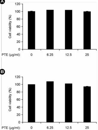
To confirm the anti-inflammatory effects of PTE, colorimetric MTT assay was used for cell viability. The results showed little or no effect on cell viability of BMDMs (Fig. 1A) and BMDCs (Fig. 1B) until 25 μg/ml. Hence, we used non-toxic concentrations of PTE for further study.
Effects of PTE on the production of IL-12 p40, IL-6 and TNF-α in LPS-stimulated BMDMs and BMDCs
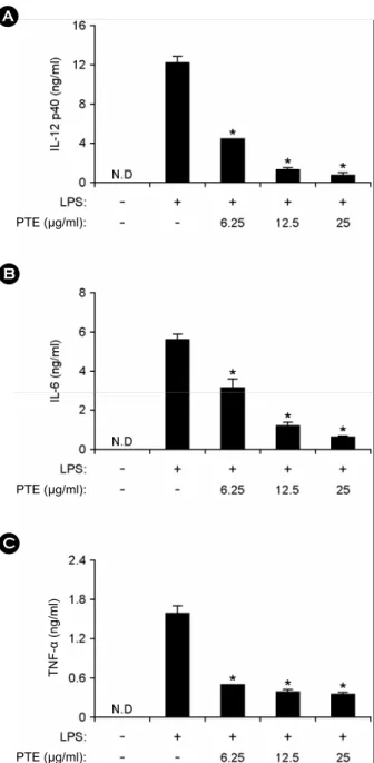
Dendritic cells and macrophages are the potent antigen presenting cells, involved in the production of pro- inflammatory cytokines such as IL-12 p40, IL-6 and TNF-α (20). To determine anti-inflammatory activity of extract, PTE was examined for the inhibitory effects on LPS- stimulated IL-12 p40, IL-6 and TNF-α production in BMDMs and BMDCs by using ELISA assay. Treatment of BMDMs and BMDCs with LPS alone resulted in significant production of IL-12 p40, IL-6 and TNF-α (Fig. 2 and Fig. 3).
PTE pre-treatment showed strong inhibition of IL-12 p40, IL-6 and TNF-α production in LPS-stimulated BMDMs (Fig. 2). Similarly, it strongly inhibited IL-12 p40, IL-6 and TNF-α production in LPS-stimulated BMDCs (Fig. 3).
Hence, these results show that PTE has an inhibitory effect on proinflammatory cytokine production in LPS-stimulated BMDMs and BMDCs.
IL-12 is a critical molecular component linking innate and adaptive immune responses (21). IL-12 plays a key role in type 1 T helper cell (Th1) mediated autoimmune diseases (22). Therefore, suppression of IL-12 production by PTE may help to prevent IL-12 associated autoimmune diseases. IL-6 plays an important role in different biological activities including hematopoiesis and inflammation (23).
However, overproduction of IL-6 is linked to inflammatory bowel disease (24). In the present study, pre-treatment of PTE showed a strong inhibition of IL-6 production in B
A
PTE (μg/ml):
PTE (μg/ml):
Figure 1. Effects of PTE on cell viability in BMDMs and BMDCs. BMDMs (A) and BMDCs (B) were treated with the indicated concentrations of PTE for 1 h before stimulation with LPS (10 ng/ml). The cell viability was measured using MTT assay.
Data are representative of three independent experiments. PTE, Plocamium telfairiae extract.
LPS-stimulated BMDMs and BMDCs. So, down regulation of IL-6 production by PTE may have potentials in anti- inflammatory diseases. TNF-α has a vital role in cell
differentiation, apoptosis and immunoregulatory activities (25). However, overproduction of TNF-α is associated with inflammatory disorders and sepsis (26, 27). Here, PTE pre- treatment showed significant inhibition of TNF-α production
PTE (μg/ml):
PTE (μg/ml):
PTE (μg/ml):
B
C A
TNF-α (ng/ml)
Figure 2. Effects of PTE on IL-12 p40, IL-6, and TNF-α production in LPS-stimulated BMDMs. BMDMs were treated with PTE at the indicated doses for 1 h before stimulation with LPS (10 ng/ml). The concentrations of murine IL-12 p40 (A), IL-6 (B) and TNF-α (C) released into the culture medium were determined by ELISA. Data are representative of three independent experi- ments. PTE, Plocamium telfairiae extract; N.D., not detectable. *p
< 0.05 versus PTE-untreated cells in the presence of LPS.
A
TNF-α (ng/ml)
PTE (μg/ml):
PTE (μg/ml):
PTE (μg/ml):
B
C
Figure 3. Effects of PTE on IL-12 p40, IL-6 and TNF-α production in LPS-stimulated BMDCs. BMDCs were treated with PTE and the concentrations of murine IL-12 p40 (A), IL-6 (B) and TNF-α (C) measured as described in Fig. 2. Data are representative of three independent experiments. PTE, Plocamium telfairiae extract; N.D., not detectable. *p < 0.05 versus PTE-untreated cells in the presence of LPS.
which indicates that it is likely to be a potential candidate
for treating TNF-α associated diseases. Effects of PTE on the phosphorylation of MAPK and degradation of IκBα by LPS-stimulated BMDMs
Activation of MAPK and NF-κB pathways by TLR4 leads to the production of pro-inflammatory cytokines (4). There-
IκBα
IκBα/p38
B A
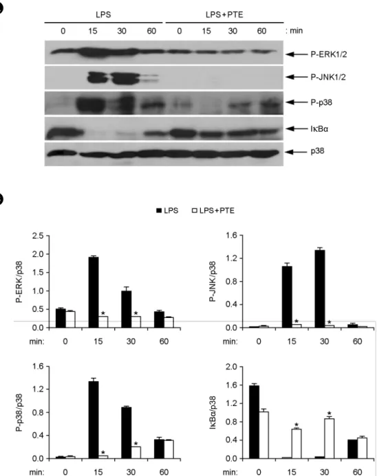
Figure 4. Effects of PTE on the phosphorylation of MAPKs and degradation of IκBα in LPS-stimulated BMDMs. (A) BMDMs were pre-treated with or without PTE (25 μg/ml) for 1 h and then stimulated with LPS (10 ng/ml) for the indicated time periods. Cell lysates were analyzed by western blot analysis. Total p38 MAPK was taken as the loading control. Data are representative of three independent experiments. (B) Phosphorylation of ERK, JNK and p38 and expression of IκBα protein was quantified using scanning densitometry, and the band intensities were normalized by that of total p38 protein. PTE, Plocamium telfairiae extract. *p < 0.05 versus PTE-untreated cells in the presence of LPS.
fore, we examined the effects of PTE on phosphorylation of MAPK and NF-κB activation in LPS-stimulated BMDMs by western blot analysis (Fig. 4). Here, LPS stimulation of BMDMs significantly increased the phosphorylation of all three MAPKs; ERK1/2, JNK1/2 and p38. Phosphorylation of ERK1/2, JNK1/2 and p38 was detected between 15 to 30 min of LPS stimulation (Fig. 4A and B). Phosphorylation of MAPK went back to baseline levels by 60 min of LPS stimulation. PTE pre-treatment showed inhibition of ERK1/
2, JNK1/2 and p38 phosphorylation (Fig. 4A and B).
NF-κB is an important transcription factor involved in the expression of various cytokines. Stimulation of cells by LPS leads to degradation of IκB and translocation of NF-κB into the nucleus (28, 29). We studied the effects of PTE on NF-κB activation indirectly by degradation of IκBα in LPS-stimulated BMDMs. LPS was able to induce degradation of IκBα within 15 min of stimulation (Fig. 4A and B). However, PTE pre-treatment blocked the IκBα degradation in LPS-stimulated BMDMs (Fig. 4A and B).
Hence, these data show that PTE pre-treatment inhibited LPS-stimulated MAPK phosphorylation and NF-κB acti- vation. Taken together, these results suggest that inhibition of pro-inflammatory cytokine production by PTE may be associated with blockage of MAPK and NF-κB activation.
In conclusion, this study indicates that PTE has an inhibitory effect on production of proinflammatory cytokines and TLR4-mediated MAPK and NF-κB activation. Further studies are warranted for detailed mode of actions of the pure biologically active compounds from PTE.
REFERENCES
1)Medzhitov R. Origin and physiological roles of in- flammation. Nature 2008;454:428-35.
2)Barton GM. A calculated response: control of in- flammation by the innate immune system. J Clin Invest 2008;118:413-20.
3) Akira S, Uematsu S, Takeuchi O. Pathogen recognition and innate immunity. Cell 2006;124:783-801.
4) Takeuchi O, Akira S. Pattern recognition receptors and inflammation. Cell 2010;140:805-20.
5) Mogensen TH. Pathogen recognition and inflammatory signalling in innate immune defenses. Clin Microbiol Rev 2009;22:240-73.
6)Cohen J. The immunopathogenesis of sepsis. Nature 2002;420:885-91.
7) Guha M, Mackman N. LPS induction of gene expression in human monocytes. Cell Signal 2001;13:85-94.
8) Thalhamer T, McGrath MA, Harnett MM. MAPKs and their relevance to arthritis and inflammation. Rheuma- tology 2008;47:409-14.
9) Manzoor Z, Koh YS. Mitogen-activated protein kinases in inflammation. J Bacteriol Virol 2012;42:189-95.
10) Weiss T, Shalit I, Blau H, Werber S, Halperin D, Levitov A, et al. Anti-inflammatory effects of moxifloxacin on activated human monocytic cells: inhibition of NF- kappaB and mitogen-activated protein kinase activation and of synthesis of proinflammatory cytokines.
Antimicrob Agents Chemother 2004;48:1974-82.
11)O'Sullivan AW, Wang JH, Redmond HP. NF-kappaB and p38 MAPK inhibition improve survival in endotoxin shock and in a cecal ligation and puncture model sepsis in combination with antibiotic therapy. J Surg Res 2009;152:46-53.
12)Shaked G, Czeiger D, Dukhno O, Levy I, Artru AA, Shapira Y, et al. Ketamine improves survival and suppresses IL-6 and TNF alpha production in a model of gram-negative bacterial sepsis in rats. Resuscitation 2004;62:237-42.
13) Blunt JW, Copp BR, Hu WP, Munro MH, Northcote PT, Prinsep MR. Marine natural products. Nat Prod Rep 2009;26:170-244.
14) Kim JY, Yoon MY, Cha MR, Hwang JH, Park E, Choi SU, et al. Methanolic extracts of Plocamium telfairiae induce cytotoxicity and caspase-dependent apoptosis in HT-29 human colon carcinoma cells. J Med Food 2007;
10:587-93.
15)Huang HL, Wang BG. Antioxidant capacity and lipophilic content of seaweeds collected from the Qingdao coastline. J Agric Food Chem 2004;52:4993-7.
16) Koo JE, Hong HJ, Dearth A, Kobayashi KS, Koh YS.
Intracellular invasion of Orientia tsutsugamushi activates inflammasome in ASC-dependent manner. PLoS One 2012;7:e39042.
17) Chae D, Manzoor Z, Kim SC, Kim S, Oh TH, Yoo ES, et al. Apo-9'-fucoxanthinone, isolated from Sargassum muticum, inhibits CpG-induced inflammatory response by attenuating the mitogen-activated protein kinase pathway. Mar Drugs 2013;11:3272-87.
18) Manzoor Z, Kim S, Chae D, Yoo ES, Kang HK, Hyun JW, et al. Sea lettuce (Ulva fasciata) extract has an inhibitory effect on pro-inflammatory cytokine pro- duction in CpG-stimulated bone marrow-derived macrophages and dendritic cells. Food Sci Biotechnol 2013;22:781-6.
19) Manzoor Z, Mathema VB, Chae D, Kang HK, Yoo ES, Jeon YJ, et al. Octaphlorethol A inhibits the CpG- induced inflammatory response by attenuating the mitogen-activated protein kinase and NF-κB pathways.
Biosci Biotechnol Biochem 2013;77:1970-2.
20)Kawai T, Akira S. The role of pattern-recognition receptors in innate immunity: update on Toll-like receptors. Nat Immunol 2010;11:373-84.
21) Trinchieri G. Interleukin-12 and the regulation of innate resistance and adaptive immunity. Nat Rev Immunol
2003;3:133-46.
22)Adorini L. Interleukin-12, a key cytokine in Th1- mediated autoimmune diseases. Cell Mol Life Sci 1999;
55:1610-25.
23)Kishimoto T. IL-6: from its discovery to clinical applications. Int Immunol 2010;22:347-52.
24) Mudter J, Neurath MF. IL-6 signaling in inflammatory bowel disease: pathophysiological role and clinical relevance. Inflamm Bowel Dis 2007;13:1016-23.
25) Baud V, Karin M. Signal transduction by tumor necrosis factor and its relatives. Trends Cell Biol 2001;11:372 -7.
26)Bradley JR. TNF-mediated inflammatory disease. J Pathol 2008;214:149-60.
27)Rigato O, Ujvari S, Castelo A, Salomão R. Tumor necrosis factor alpha (TNF-alpha) and sepsis: evidence for a role in host defense. Infection 1996;24:314-8.
28) Beinke S, Ley SC. Functions of NF-κB1 and NF-κB2 in immune cell biology. Biochem J 2004;382:393-409.
29) Baeuerle PA, Baltimore D. NF-κB: ten years after. Cell 1996;87:13-20.