Kor. J. Pharmacogn.
46(4) : 309∼ 314 (2015)
309
뽕잎 n-부탄올 추출물의 치주인대세포에서 LPS로 유도된 염증성 사이토카인의 억제와 골 형성 분화 조절
최정이1·김대근2·김은희3·이정근1*
1
아주대학교 의과대학,2
우석대학교 약학대학,3
강릉영동대학교n-Butanol Extract of Mulberry Leaves Suppresses LPS-induced Inflammatory Cytokines and Modulates Osteogenic Differentiation in
Periodontal Ligament Cells
Jeong Lee Choi1, Dae Keun Kim2, Eun Hee Kim3, and Jeong Keun Lee1*
1
Division of Oral and Maxillofacial Surgery, Department of Dentistry, Ajou University School of Medicine, Suwon 206, Korea2
College of Pharmacy, Woosuk University, Jeonju 565-701, Korea3
Department of dental Hygiene Gangneung Yeongdong University, 357 Gongiero Gangneung-si Kangwon-Do 210-792, KoreaAbstract − Periodontitis is a chronic inflammatory disease that is known to have the characteristics of destructed periodontal tissue. Anti-oxidant and anti-inflammatory effects of mulberry leaves in periodontal tissue is not well known until now. We investigated the effects of n-butanol extract of mulberry leaves on the lipopolysaccharide (LPS)-induced proinflammatory cyto- kines, such as IL-1β, IL-6, IL-8 and modulates osteogenic differentiation in periodontal ligament cells. The expression levels of Runx2, ALP and mRNA were increased by n-butanol extract of mulberry leaves at the concentration of 100 μg/ml in peri- odontal ligament cells. n-Butanol extract of mulberry leaves extract reduced the range of pathophysiological processes, such as inflammation and increase in the level of osteogenic-related genes. These findings suggest that n-butanol extract of mulberry leaves has therapeutic effects on periodontitis and periodontal tissue regeneartion.
Keywords − Melberry leaves, LPS, Inflammation, Periodontal ligament cells, Cell differentiation
치주질환은 치근면에 부착된 치주병인균의 독소에 의해 진행되어 치은에 국한되는 치은염(gingivitis)과, 치주인대, 치조골로 확산되어 치아와 치아 지지조직의 점진적 손상을 유발 하여 결국 치아를 상실하게 되는 치주염(periodontitis) 으로 구분된다. 이러한 치주염은 구강 내 대표적인 염증질 환으로, 치주질환의 원인균으로 알려진 그람음성세균의 세 포외막에 존재하는 내독소인 LPS가 숙주세포인 대식세포를 자극하여 interleukins, tumor necrosis factor-α(TNF-α), prostaglandins 와 같은 염증매개물질들을 분비 하여 치주조 직의 파괴를 일으키는 것으로 알려져 있다.
1-3)
이와 같이 만 성적으로 진행되는 치주염증 질환은 지속적인 염증 반응으 로 인하여 치주인대 및 치조골의 파괴로 연결되고 결국은 치아손실이 야기되므로 결합조직인 치주인대의 작용은 매 우 중요하다.4-6)
선행연구들에서 치은섬유모세포나 치주인대세포에 치주염 관련 세균 LPS를 처리한 경우 IL-1β, IL-6, IL-8의 발현이 증가됨을 보고하였고
7)
이에 따른 병인균의 억제와 염증매개물질의 성장억제를 위한 화학요법제8-11)
및 천연추출 항균제 및 항염증제에 대한 연구가 이루어져 왔 으나 내성균 발현, 과민반응, 위장장애, 치아 표면 착색, 박 리성 치은염 유발 등의 부작용으로12,13)
인하여 기존의 화학 제들과 같은 부작용이 없는 새로운 제제를 필요로 하고 있 다. 이와 관련한 천연물의 효과에 대하여 살펴보면 후박, 옥 수수, 은행엽추출물에서의 항균,14)
항염효과를, 홍화씨 추출 물이 치주인대세포와 조골유사세포의 골광물화 작용에 미 치는 효과15)
등 치주병인균에 대한 실험적 평가가 보고되었다.뽕잎은 뽕나무과 뽕나무속에 속하는 식물로서, 학명은 Morus alba L 로서 콩 다음으로 단백질 함량이 높을 뿐 아 니라 칼슘, 칼륨, 철 등외 50 여종의 무기성분이 함유되어 있어 미네랄과 섬유소의 함량이 현저히 높은 것으로 알려 져 있다. 뿐만 아니라 항산화,
16)
항염,17)
항당뇨, 고혈압억*교신저자(E-mail):[email protected] (Tel): +82-31-219-5333
제,
18)
등 뽕잎의 생리활성 효과에 대하여 많은 연구가 보고 되었으나 치주조직에서의 연구는 아직 보고 된 바가 없다.따라서 본 연구에서는 뽕잎의 치주인대세포에 대한 항산화, 항염증효과와 치주조직의 재생을 유도하는 골 대체물질로 서의 효과를 알아보고자 LPS 처리된 치주인대세포에서 뽕 잎을 처리하여 항산화효과와 염증성 사이토카인들의 발현 억제 등 골모세포로의 분화를 유도한 후 뽕잎에 의한 골모 세포 표지유전자 발현 효과를 알아보고자 하였다.
재료 및 방법
실험재료 − 본 실험에 사용된 뽕잎(Mulberry leaves)은 2012년 전북 완주군에서 채취하여 우석대학교 김대근 교수 가 확인하였고 표품은 우석대학교 약학대학 생약표본실에 보관하였다(WSU-12-15).
시약 및 기기 − Dulbecco's modified eagles medium (DMEM), fetal bovine serum(FBS), a-Minimum essential medium(a-MEM), 등은 Gibco laboratories제품을, 세포배양 용기는 Corning Inc.제품을 사용하였다. 추출 및 분획용 시 약은 1급 용매를 사용하였으며, column용 시약 등은 1급 용 매를 재증류하여 사용하거나, 특급시약을 사용하였다.
추출 및 분획 − 신선한 뽕잎을 음건 세절한 다음 건조하 여 얻은 시료 약 1.9 kg을 ethanol로 가끔 진탕하면서 5시간 씩 50
o
C에서 3회 온침 추출하였다. 그 추출액을 수욕상에서 감압농축하여 ethanol 엑스 약 290 g을 얻었으며, 이 ethanol 엑스를 증류수로 현탁시키고 상법에 따라 동량의 n-hexane (46 g), methylene chloride(3.8 g), ethyl acetate(2.4 g) 및 n- butanol(12.9 g)의 순으로 용매 분획하여 각각의 분획물을 얻 었다.세포 배양 − 치주인대세포 periodontal ligament cells(PDL) 은 서울대학교치과대학 치주과(Seoul,Korea)에서 공급받았 다. PDL cell은 1% antibiotics, 10% FBS가 함유된 DMEM 에서 37
o
C, 5% CO2
를 유지하면서 배양하였다. 골 세포분 화는 배양 시 10 mM-glycerophosphate와 50 μg/ml L- ascorbic acid를 첨가하였으며 2-3일 간격으로 배지를 교환 하였다. LPS에 의한 염증반응을 유도하기 위해 100 ng/ml E. coli LPS(Invivogen;San Diego, CA, USA)와 10 ng/ml CD14(R&D systems; Minneapolis, MN, USA)를 섞어서 처 리하였다. 뽕잎은 100 μg/ml의 농도로 사용하였다.ROS 생성률 측정 − 치주인대 세포를 96 well plate에 well당 5×10
4
cells/ml로 seeding하여 37o
C에서 하루 배양 후, serum을 제거한 배지로 6시간 배양 후 산화적 스트레스를 유발하기 위하여 100 ng/ml의 LPS를 주입하고 시간별로 배 양한 뒤 50 μM DCF-DA용액을 각 well에 주입하여 37o
C 에서 30분간 배양 후, FLUOstar OPTIMA(BMG lavtech, Ortenberg, Germany)로 excitation-480 nm, emission-535 nm에서 측정하였다.
Quantitative Real-Time PCR에 의한 염증성 Cytokine 과 골모세포 표지유전자 발현 의 측정 − 배양이 끝난 세포 로부터 easy- BLUE
TM
을 이용하여 total RNA를 분리하였 으며, 1 μg의 total RNA로부터 1U superscript reverse transcriptase를 이용하여 cDNA를 합성하고, 이 중 10%의 cDNA를 이용하여 2U의 Takara Taq, PCR buffer, 0.8 mM dNTP, 100 pM의 primer가 혼합된 용액 50 μl에서 PCR을 시행하였다. PCR은 95o
C, 30초 denaturation/ 45.5o
C, 30초 annealing/72o
C, 1분 extention으로 이루어진 과정을 32 cycle 을 시행하였다. PCR이 끝난 후 qRT-PCR은 바이오니아사 (Bionner, Korea)의 AccuPower®
GreenStarTM
qPCR PreMix 및 ExicyclerTM
96 Real-Time Quantitative Thermal Block 를 사용하여 실시하였다. qRT-PCR 측정은 ABI PRISM®
Biosystems 7300/7500Real-Time PCR system을 사용하여 melting curve 분석은 58~95o
C에서 시행하였다. 이때 사용 된 primer서열은 Table I과 같다.통계분석 − 실험 결과들은 평균±표준편차로 제시하였고, Student′s t-test로 분석하여 p값이 0.05보다 적은 경우 통계 적으로 유의한 차이가 있는 것으로 판단하였다.
결과 및 고찰
치주질환의 대표적인 내독소로 작용하는 LPS는 그람음성 세균의 세포외막에 존재하며 macrophage 또는 monocyte와 같은 면역세포와 여러 조직 세포에서 TNF-α, IL-6, IL-1β 와 같은 pro inflammatory cytokine을 증가시키는 것으로 알 려져 있다. Hausamn 등
19)
은 LPS가 치조골의 흡수를 유도 하며 실험동물에 투여할 경우 여러 가지 독성작용 이외에 다양한 약리학적, 병리학적 및 면역학적 작용을 한다고 보 Table I. Primer sequences and expected size of amplified productsIL-1β 58
o
Cforward: 5'-GATCTGCGTCTGCGACAAAC reverse: 5'-CGCAGTTCTTGGTCTCGTCA IL-6
58
o
Cforward: 5'-GCCAAATATGTGTCTGTGACTCA reverse: 5'-GGGCGAGTAGGAGCAGTTG IL-8
58
o
Cforward: 5'-ATGACTTCCAAGCTGGCCGTGGCT reverse: 5'-TCTCAGCCCTCTTCAAAAACTTCTC Runx2
60
o
Cforward: 5'-CCGCACGACAACCGCACCAT reverse: 5'-CGCTCCGGCCCACAAATCTC ALP
60
o
Cforward: 5'-CTGCCATCCTGATGGCAATG reverse: 5'-AGACTGCGCCTGGTAGTTGTTG RANKL
55
o
Cforward: 5'-GTTTCCCATAAAGTGAGTCTGT-3' reverse: 5'-TTAAAAGCCCCAAAGTATGTT-3' GAPDH
52
o
Cforward: 5'-CGGAGTCAACGGATTTGGTCGTAT-3' reverse: 5'-AGCCTTCTCCATGGTGGTGAAGAC-3'
고 하였다. 치주인대의 형태학적, 조직학적 변화는 치주질 환인 염증에 의해 유도되고,
20,21)
치주인대의 이러한 변화는 치근흡수와 치조골 흡수를 야기 한다.22)
이러한 치주질환의 치료에는 병인균의 억제와 염증매개물질의 성장억제를 위 한 화학요법제 및 천연추출 항균제 및 항염증제가 사용되 고 있다. 이때, 항염증제에 대한 연구는 치주질환의 진행 및 치주조직파괴와 관련한 cytokine, prostaglandin, collagenase 등에 관한 보고로서, 각종의 염증성 cytokine이 분비되고 다 시 염증성 cytokine에 의해서 prostaglandin이나 collagenase 등이 활성화되어 조직을 파괴하는 일련의 과정을 억제 내 지는 중지하고자 함이었을 뿐, 치주조직 중의 하나인 치조 골이 파괴되는 것을 정지하거나 예방적인 차원에서 유용한 방안을 살펴본 연구는 매우 드문 실정이다. 또한 이러한 억 제제의 투여는 사용상의 번거로움과 낮은 치료효과 및 고 혈압, 자궁내막암, 유방암등의 부작용 등 위험도가 높은 합 병증을 유발하는 것으로 보고되었다. 최근 이러한 부작용을 최소화하기 위한 식물 유래의 폴리화합물 중 하나인 quercetin 과 대두 및 콩류를 이용한 조골세포 활성 검색에 대한 연구 등이 보고되고 있다.뽕나무 과에 속하는 식물로서 열대지방에 걸쳐 130여종 의 품종이 세계적으로 존재하는 뽕잎은 항 고혈압, 항 당뇨, 콜레스테롤저하, 발암억제, 및 체지방 축적 억제 등 성인병 에 효능이 있다고 보고되고 있으며 뽕잎에는 다른 식물에 비해 함유된 각종 생리 활성 성분을 크게 휘발성 성분과 비 휘발성 성분으로 나눌 수 있는데 휘발성 성분으로는 guaiacol, eugenol, methyl salicylate, benzaldehyde 및 phenyl acetaldehyde 등이 보고되고 있으며 비 휘발성 성분은 플라 보노이드가 주류를 이루어 그 함량이 매우 높고 종류 또한 다양하다. 즉 rutin, quercetin, isoquerc, itrin, astragalin, quercetin 3,7-diglucoside 및 quercetin 3-triglucoside가 존재 한다. 이러한 뽕잎의 플라보노이드 화합물의 생리활성은 천
연에 존재하는 다른 물질에 비하여 작용이 매우 다양하여 이뇨작용, 항 모세혈관 투과작용, 항 알레르기작용, 항세균 작용등의 특성이 알려져 있으며 quercetin 3-0-(6''-0-acetyl)- β-D-glucoside와 kaempferol 3-0-(6''-0-acetyl)-β-D-glucoside 는 충치제거에 관여하는 것으로 알려져 있다. 또한 특이한 플라보노이드 골격인 kuwanon G는 혈압강하 작용이 매우 뛰어난 것으로 보고되었으며 뽕잎의 methanol 추출물은 항 산화 활성이 강한 것으로 보고되고 있다. 최근 꾸지뽕 잎 에 탄올 추출물이 연골세포의 손상과 뼈조직의 손상을 포함하 여 골관절염을 유발한 rat에서 연골조직의 형태를 유지하여 항관절염 효과가 있음을 실험적으로 검증하여 항관절염 효 능을 지니는 기능성소재로써 개발 가능성이 있음이 보고되 었다.
23)
본 연구에서는 다른 연구팀에서의 뽕잎을 각 용매별 추출 을 하여 에틸 아세테이트와 부탄올 추출물에서 항산화 물 질을 분리하여 구조식을 밝히고 전자공여능으로 항산화능 의 검증이 보고 되었던 뽕잎 n-butanol 추출물을 처리하여 항산화, 항염증효과와 조골세포 활성을 확인하였다.
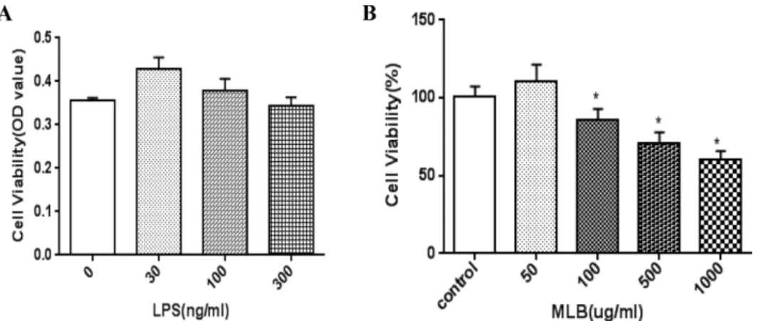
치주인대 세포(5×10
4
cells/ml)에 LPS(100 ng/ml)의 세포 독성(A)과 뽕잎 n-butanol 추출물(100 μg/ml)로 유발한 세포 독성(B)에 대한 결과를 살펴보았다. Fig. 1에서와 같이 치주 인대세포에서 LPS는 30 ng/ml에서 300 ng/ml까지 24시간 동안 독성이 보이지 않았으며, n-butanol 추출물은 100 μg/ml에서 1000 μg/ml까지 농도 의존적으로 독성이 나타나기 시작하였으나 100 μg/ml 단독으로 다시 확인하였을 때 독 성이 나타나지 않아 모든 실험을 진행하였다(Fig. 1). 치주 인대 내 ROS 농도의 측정은 DCF-DA의 산화에 의한 형광 감도의 변화로 측정하였다. LPS(100 ng/ml)를 처리하였을 때 발생한 ROS는 3시간 간격으로 측정 한 결과 시간이 지 남에 따라 모든 군에서 생성량이 증가하는 것을 확인하였 다. 이때 동일하게 LPS(100 ng/ml)와 함께 n-butanol 뽕잎추
Fig. 1. LPS induced cell toxicity (A) and effect of MLB (B) in PDL cells. PDL cells were seeded in triplicates in 96 well plate at 5×10
4
cells/well. Cells were either stimulated with various concentration of LPS (30~300 ng/ml) and MLB (50~1000 μg/ml). After 24 hours incubation at 37o
C, cell viability was determined by colorimetric assay using CCK-8.출물(100 μg/ml)을 함께 처리하여 항산화 효능이 나타나는 가를 확인하니 유의적으로 감소하는 것으로 산화적 손상에 의한 세포 보호 효과가 우수함을 확인할 수 있었다(Fig. 2).
LPS에 의해 유도된 IL-1β, IL-6, IL-8 발현에 n-butanol 뽕 잎추출물이 영향을 미칠 수 있는지 알아보기 위해, 치주인 대 세포에 LPS를 처리하고 24시간 후에 total RNA를 추출 하여 cDNA를 만들어 정량적 PCR을 시행해 각 사이토카인 의 발현 양을 확인하였다. IL-1β, IL-6, IL-8의 mRNA의 수 준을 관찰한 결과 대조군에 비해 LPS 처리 후 IL-1β, IL-6, IL-8의 발현이 모두 크게 증가하였다. n-butanol 뽕잎추출물 (100 μg/ml)을 24시간 전 처치하고 LPS와 함께 처리한 경 우에는 LPS에 의해 유도된 IL-1β, IL-6, IL-8 발현이 차단 되어 관찰한 시점에서 LPS 단독 처리군과 통계적으로 유의 한 차이가 있는 것으로 나타났다(Fig. 3). 치주인대 세포는
골모세포로 분화하여 치조골 형성에 관여 할 수 있을 뿐 아 니라, 세균에 노출 되었을 때 사이토카인들의 분비를 촉진 하여 치주조직의 염증반응에 관여하는 것으로 알려져 있다.
따라서 치주조직의 염증을 억제하기 위해서는 염증성 세포 들인 호중구, 대식세포, 비만세포, 등에서 생성하는 사이토 카인 뿐만 아니라 치주인대세포에서 분비하는 염증성 사이 토카인의 억제에 대한 중요성도 보고되었다.
24)
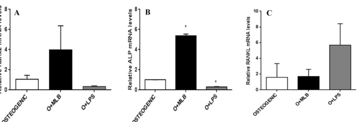
치주인대세 포를 골모세포로 분화 유도할 때 뽕잎의 n-butanol 추출물 을 처리하여 골형성 분화 표지자로 알려진 Runx2, ALP의 발현과 LPS가 파골세포 조절인자 RANKL의 발현을 조절 하는지 살펴보았다. 치주인대세포에서 뽕잎 n-Butanol 추출 물(100 μg/ml)을 처리하였을 때 골모세포 표지자인 Runx2, ALP 의 발현은 증가하였으나 LPS에 의해 각 발현이 감소 되었을 뿐 아니라 파골세포 조절인자인 RANKL의 발현이 크게 증가하였다(Fig. 4). LPS에 의한 RANKL의 발현증가 는 여러 연구들에서 LPS가 골모세포나 T 림프구에 작용하 여 RANKL의 생산을 촉진시키고 이에 따라 파골세포의 분 화를 촉진하거나,25-27)
파골세포로의 분화가 어느정도 진행 된 파골세포 전구세포에 직접 작용하여 분화를 촉진한다고하였다.
28,29)
단핵구 배양에 LPS를 처리하면 오히려 파골세포로의 분화를 억제하고 염증성 사이토카인의 생성과 염증 성 사이토카인인 IL-1β와 TNF-α는 RANKL에 독립적으로 파골세포 분화 및 활성을 촉진한다는 보고들이 있다
30)
. 이 것은 치주염 진행시 단핵구가 침윤된 초기에는 세균에 대 한 방어 작용이 우세하나 만성적으로 진행되면 활성화된 RANKL의 작용이 우세하여 단핵구가 파골세포로 분화하여 치조골의 흡수를 야기할 수 있음을 시사하는 바가 크다고 할 것이다. 치주염의 진행으로 RANKL이 다량 발현되어 파 골세포 조절인자들의 균형이 깨져 다른 염증성 사이토카인 의 작용과 더불어 치조골 흡수가 야기될 것으로 생각된다.Fig. 4에서와 같이 뽕잎 n-butanol 추출물(100 μg/ml)을 처리 Fig. 2. Effect of MLB on level of reactive oxygen species
in periodontal ligament cells treated with LPS. The data represent the mean±SD *P<0.05 significant difference compared with LPS control,
#
P<0.05 significant difference compared with LPS+MLB control.Fig. 3. MLB significantly supressed LPS-induced IL-1β, IL-6, and IL-8 expression. Periodontal ligament cells were incubated in the presence or absence of MLB (100 μg/ml), followed by treatment with LPS (100 ng/ml) for the indicated time for 24 h. Then, quan- titative RT-PCR was perfomed using total RNA. IL-1β, IL-6, and IL-8 mRNA levels were presented as the relative fold changes compared with that of control group (LPS(-)/MLB(-)). The data represent the mean±SD. *P<0.05 significant difference compared with control (LPS(-)/MLB(-)).
#
P<0.05 significant difference compared with LPS.함으로서 골모세포 표지자 Runx2, ALP 증가와 파골세포의 표지자 RANKL 이 억제 되면 각 조절인자들이 균형을 이 루어 치주조직의 파괴를 완화시키는 데 도움을 줄 수 있을 것으로 사료된다.
결 론
이상의 연구결과를 통하여 뽕잎이 사람 치주인대세포에 서 LPS에 유도된 ROS의 생성을 억제 하므로 항산화제로 서, 염증 사이토카인 발현을 억제하므로 염증 반응을 감소 시킬 수 있으며, 그 과정에 골모세포와 파골세포 표지자들 의 조절을 통하여 치조골 흡수를 감소시키는데 중요한 역 할을 할 수 있을 것임을 시사하였다. 이러한 연구결과는 치 주질환에 치료효과를 나타낼 가능성이 있음을 시사하였지 만 추후 치주염 동물 모델을 이용한 추가 연구가 진행되어 야 될 것으로 사료된다.
인용문헌
1. Jiang, Y., Mehta, C. K., Hsu, T. Y. and Alsulaimani, F. F.
(2002) Bacteria induce osteoclastogenesis via an osteoblast- independent pathway. Infect. Immun. 70: 3143-3148.
2. Nakashima, T., Kobayashi, Y., Yamasaki, S., Kawakami, A., Eguchi, K., Sasaki, H. and Sakai, H. (2000) Protein expres- sion and functional difference of membrane-bound and sol- uble receptor activator of NF-kappaB ligand: modulation of the expression by osteotropic factors and cytokines. Biochem.
Biophys. Res. Commun. 7: 768-775.
3. Graves, D. T., Oates, T. and Garlet, G. P. (2011) Review of osteoimmunology and the host response in endodontic and periodontal lesions. J. Oral Microbiol. 3: 1-15.
4. Snyderman, R. (1971) Periodontal disease: Amodel for the
study of inflammation. J. Infect. Dis. 123: 676-677.
5. Page, R. C. (1991) The role of inflammatory mediators in the pathogenesis of periodontal disease. J. Periodontal Res. 230- 242.
6. Wilson, M. (1995) Biological activities of lipopolysaccharide from oral bacteria and their relevance to the pathogenesis of chronic periodontitis. Sci. Pog. 78: 19-34.
7. Yamaji, Y., Kubota, T., Sasaguri, K., Sato, S., Suzuki, Y., Kumada, H. and Umemoto, T. (1995) Inflammatory cytokine gene expression in human periodonal ligament fibroblasts stimulated with bacterial lipopolysaccharides. Infect. Immun.
63: 3576-3581.
8. Emingil, G., Atilla, G., Sorsa, T., Luoto, H., Kirilmaz, L. and Baylas, H. (2004) The effect of adjunctive low-dose dox- ycycline therapy on clinical parameters and gingival crevic- ular fluid matrx metalloproteinase-8 levels in chronic periodontitis. J Periodontol. 75: 106-15.
9. Choi, D. H., Moon, I. S., Choi, B. K., Park, J. Won., Kim, Y.
S., Choi, S. H. and Kim, C. K. (2004) Effect of sub-anti- microbial dose doxycycline therapy on crevicular fluid MMP-8, and gingival tissue MMP-9, TIMP-1 and IL-6 levels in chronic periodontitis. J. Periodont Res. 39: 20-26.
10. Jang, H. S., Park, M. G., Kook, J. K., Kim, H. S. and Kim, B. O. (2005) Antimicrobial effect of cefixime on 6 species of periodotopathogens. J. Korean Acad. Periodontol. 35: 401- 411.
11. Park, S. P., Chung, H. J., Kim, Y. J. and Kim, O. S. (2004) Triclosan inhibition of prostaglandin E2 production in human gingival fibroblast. J Korean Acad. Periodontol. 34: 345-356.
12. Helgeland, K., Heyden, G. and Rolla, G. (1971) Effect of chlorhexidine on animal cells in vitro. Scan J. Dent. Res. 79:
209-215.
13. Pucher, J. J. and Daniel, J. C. (1993) The effects of chlor- hexidine digluconate on human 3ibroblasts in vitro. J. Peri- odont. 62: 526-532.
Fig. 4. n-Butanol mulberry leaves enhanced osteogenesis in PDL cells and suppressed expression of bone marker genes by LPS in osteogenic differentiation condition. Quantitative RT-PCR for mRNA levels on day 7 of induction PDL cells were cultured for 7 days in the presence of 1 μg/ml of LPS and 100 μg/ml of n-butanol mulberry leaves.
14. Kim, T. I., Choi, E. J., Chung, C. P., Han, S. B. and Ku, Y.
(2002) Antimicorbial effect of Zea mays L. and Magnoliae cortex extract mixtures on periodontal pathogen and effect on human gingival fibroblast cellular activity. J. Korean Acad.
Periodontol. 32: 249-255.
15. Kang, J. G., You, H. K. and Shin, H. S. (1998) Effect of extract of seeds of Carthamus tinctorius L. on mineralization in periodontal ligament cells and osteoblastic cells. J. Korean Acad. Periodontol. 28: 475-489.
16. Choi, S. R., You, D. H., Kim, J. Y., Park, C. B., Kim, D. H.
and Ryu, J. (2009) Antioxidant activity of methanol extracts from Curdrania tricuspidata Breau according to harvesting parts and time. Korea J. Medical Crop Sci. 17: 115-120.
17. Oh, P. S. and Lim, K. T. (2011) Anti-inflammatory effect of glycoprotein isolated from Curdrania tricuspidata Breau:
involvment of MAPK/NF-kB signaling. Immunol. Invest. 40:
76-91.
18. Kang, D. G., Hur, T. Y. and Lee, G. M. (2002) Effects of Curdrania tricuspidata water extract on blood pressure and renal functions in NO-dependent hypertention. Life Sci. 70:
2599-2609.
19. Hausmann, E. and Luderitz, O. (1975) Structural requirement for bone resorption by endotoxin and lipoteichoic acid. J.
Dent. Res. 54: 94-99.
20. Nishimura, K., Noguchi, Y., Shigeyama, Y., Naito, M., Fuka- zawa, E. and Yamaoka, A. (1991) An ultrastructural study comparing new gingival tissue attachment on chemically exposed fibrils and retained periodontal ligament. J. Osaka Dent. Univ. 25: 63-75.
21. Kai, K. (1989) SEM study of vascular architecture of peri- odontal ligament under chronic marginal periodontitis.
Kanagawa Shigaku 24: 273-1989.
22. Moskow, B. S. (1992) A histomorphologic study of the effects of periodontal inflammation on the maxillary sinus mucosa. J. Periodontol. 63: 674-681.
23. Nam, D. E., Kim, O. K. and Lee, J. (2013) Therapeutic effect
of Curdrania tricuspidata leaf extract on osteoarthritis. J.
Korean Soc. Food Nutr. 42: 697-704.
24. Jonsson, D., Nebel, D., Bratthall, G. and Nilsson, B. O. (2011) The human periodontal ligament cell: a fibroblast-like cell acting as an immune cell. J. Periodontal Res. 46: 153-157.
25. Teng, Y. T., Nguyen, H., Gao, X., Kong, Y. Y., Gorc zynski, R. M., Sinh, B., Ellen, R. P. and Penninger, J. M. (2000) Functional human T-cell immunity and osteoprotegerin ligand control alveolar bone destruction in periodontal infec- tion. J. Clin. Invest. 106: R59-R67.
26. Kikuchi, T., Matsuguchi, T., Tsuboi, N., Mitani, A., Tanaka, S., Matsuoka, M., Yamamoto, G., Hishikawa, T., Noguchi, T.
and Yoshikai, Y. (2001) Gene expression of osteoclast dif- ferentiation factor is induced by lipopolysaccharide in mouse osteoblasts via toll-like receptors. J. Immunol. 166: 3574- 3579.
27. Jiang, Y., Mehta, C, K., Hsu, T, Y. and Alsulaimani, F. H.
(2002) Bacteria induce osteoclastogenesis via an osteoblast- independent pathway. Infect. Immun. 70: 6 3143-3148.
28. Kanichiro, K., Naoyuki, T., Eijiro, J., Nobuyuki, U., Masa- michi, T., Shigeru, K., Nobuaki, N., Masahiko, K., Kyoji, Y., Nobuyuki, S., Hisataka, Y., Tomonori, M., Kanji, H., Mar- tind., T. J. and Tatsuo, S. (2000) Tumor necrosis factor α stimulates osteoclast differentiation by a mechanism inde- pendent of the Odf/Rankl-Rank interaction. J. Exp. Med. 191:
275-286.
29. Zou, W. and Bar-Shavit, Z, (2002) Dual modulation of osteo- clast differentistion by lipopolysaccharide. J. Bone Miner.
Res. 17: 1211-1218.
30. Kanami, I., Nobuyuki, U., Kanichiro, K., Koji, S., Xiaotong, L., Masamichi, T., Nobuo, O., Tatsuji, N. and Naoyuki, T.
(2003) Lipopolysaccharide promotes the survival of osteo- clasts via toll-like receptor 4, but cytokine production of osteoclasts in response to lipopolysaccharide is different from that of macrophages. J. Immunol. 170: 3688-3695.
(2015. 11. 14 접수; 2015. 11. 19 심사; 2015. 11. 24 게재확정)