Received August 27, 2018, Revised September 20, 2018, Accepted September 20, 2018 Corresponding author: Na Young Jo
Department of Acupuncture & Moxibustion Medicine, Je-Cheon Hospital of Traditional Korean Medicine, Semyung University, Semyung-ro 66, sinwoul-dong, Jecheon 27136, Korea
Tel: +82-43-649-1825, Fax: +82-43-645-1382, E-mail: [email protected] This paper was supported by the Semyung University Research Grant of 2017.
CCThis is an open access article distributed under the terms of the Creative Commons Attribution Non-Commercial License (http://creativecommons.org/licenses/ by-nc/4.0) which permits unrestricted non-commercial use, distribution, and reproduction in any medium, provided the original work is properly cited.
The Effect of Woogakseungmatang Extract on NO Production in LPS- Stimulated RAW 264.7 Cells
Na Young Jo
Department of Acupuncture & Moxibustion Medicine, Je-Cheon Hospital of Traditional Korean Medicine, Semyung University
우각승마탕이 LPS로 유도된
RAW 264.7 세포에서 NO 생산에 미치는 영향
조나영
세명대학교 제천한방병원 침구의학과
Objectives : Woogakseungmatang is a prescription medication mainly used to treat facial paralysis in Korean medicine. The purpose of this study is to investigate the effects of Woogakseungmatang on anti-inflammation and anti-oxidation. Methods : Woogakseungmatang was extracted using hot water. Cytotoxicity was assessed using the 3-(4,5-dimethylthiazol- 2-yl)-2,5-diphenyltetrazolium bromide(MTT) method; nitric oxide(NO) production and Prostaglandin E
2(PGE
2) production in RAW cells treated with Woogakseungmatang were investigated; and the cytokine changes associated with inflammation were examined. The antioxidant capacity of Woogakseungmatang was measured using the 1,1-diphenyl-2-picrylhydrazyl (DPPH) method. Results : RAW cells treated with Woogakseungmatang showed 90% cell viability at a 100-μg/ml concentration. NO production was decreased by 15% at a 100-μg/ml concentration. PGE
2production was decreased by 18% at a 100-μg/ml concentration. Interleukin 1β(IL-1β), interleukin 6(IL-6), and tumor necrosis factor-α(TNF-α) were significantly reduced at 100 μg/ml compared with those in the control group. The DPPH free radical scavenging capability was more than 50% at 100 μg/ml.
Conclusions : Woogakseungmatang showed only a slight anti - inflammatory effect at 100 μg/ml and it was difficult to confirm the concentration-dependent anti-inflammatory effect. Therefore, this study means to confirm the potential anti-inflammatory effects of Woogakseungmatang. Based on this research, more systematic and diverse studies should be conducted.
Key words : Woogakseungmatang, Seogakseungmatan g, Anti-inflammation, Antioxidant, Macrophage, Korean Medicine
Introduction
An inflammatory reaction is a defensive reaction against a harmful stimulus to the living body. It is a defense mechanism of a living body that localizes the influence of an external
stimulus to the cells and tissues of the living body to restore
and maintain a damaged region to normal
1). However, if a
defense mechanism is not performed smoothly in the human
body and the inflammation reaction continues, it is a main
factor in various inflammatory diseases, such as skin diseases,
Table 1. Compositions of Woogakseungmatang. Prescription was based on Donguibogam. 6 g of Rhinocerotis Cornu was replaced with 48 g of Buffle Cornu
Components herbs of Woogakseungmatang Content
Buffle Cornu 48 g
Cimicifugae Rhizoma 5 g
Sapshnikoviae Radix 4 g
Osterici Radix 4 g
Cnidii Rhizoma 3 g
Aconiti Koreani Tuber 3 g
Angelicae Dahuricae Radix 3 g
Scutellariae Radix 3 g
Glycyrrhizae Radix 2 g
Total 75 g
asthma, cancer, coronary artery disease, and rheumatoid arthritis
2). Therefore, the fast and effective defense against and elimination of inflammatory substances are essential for the prevention and treatment of various inflammatory diseases.
Woogakseungmatang consists of the substitution of Seogak ( Rhinocerotidae unicornis L. ) for Woogak ( Bubalus bubalis L. ) among the constituents of Seogakseungmatang. Seogak is the horn of a rhinoceros that has been cut and dried; the rhinoceros is an endangered species, and rhinos are protected by the Convention on International Trade in Wildlife Species, so these horns have been replaced with those of water buffalo.
Woogak is used to treat headaches, high fever, and skin diseases in Korean medicine; Woogak has similar efficacy to Seogak. In a recent clinical study, the use of Woogak rather than Seogak was effective for fever and pediatric fever;
however, when Woogak is used instead of Seogak, its capacity must be increased. Generally, 30∼120 g should be added to the daily intake capacity and boiled in water. In clinical practice, it is common to prescribe Woogak instead of Seogak
3).
In Donguibogam, Seogakseungmatang is used medicinally when the muscles are paralyzed and painful. It can be applied to the forehead, chin, and cheeks to treat facial pain and the paralysis of facial muscles. Therefore, Seogakseung- matang mainly uses facial pain for the same idiopathic paralysis
4).
Facial paralysis is the most common disease of the brain;
it is caused by paralysis of the muscles of the eyes and mouth, and the symptoms can make a patient appear crooked on one side. The causes of this disease include thoughtfulness, overwork, and cold exposure. Symptoms can include senile neuropathy, sensory depression, abnormal sensation in the trigeminal nerve, facial numbness, pain, auditory irritability, larynx, and tinnitus
5). In the case of facial paralysis with severe pain, anti-inflammatory drugs are prescribed to reduce the inflammatory response
6). In Korean medicine, Woogak- seungmatang is specifically prescribed for painful facial paralysis; therefore, it is necessary to study the anti- inflammatory effect of Woogakseungmatang.
Previous studies on Seogakseungmatang have been used in cases of trigeminal neuralgia
6)and antithrombotic studies
7). Studies on the anti-inflammatory effects of Woogakseungmatang are severely limited; we studied 3-(4,5-dimethylthiazol- 2-yl)-2,5-diphenyltetrazolium bromide (MTT), nitric oxide (NO), Prostaglandin E
2(PGE
2), and cytokine to determine the anti-inflammatory and 1,1-diphenyl-2-picrylhydrazyl (DPPH) effects of Woogakseungmatang.
Materials and Methods
1. Materials
The medicinal materials used in the experiments were purchased from OmniHub (Kyeongbuk, Korea). The prescription’s composition was in accordance with Donguibogam (Table 1).
Five batches of the prescription were placed in 3 L of water and subjected to hot water extraction for four hours. The filtrate was first filtered and then poured through filter paper.
The filtrate was concentrated under reduced pressure in a
rotary vacuum evaporator, and the concentrated solution was
lyophilized in a free dryer for seven days. After drying, 26.53
g of concentrated product was obtained (yield: 7.07%). The
obtained powder was stored at −80°C in a cryogenic freezer
and diluted to the required concentration according to the
experiment.
2. Cell culture
The RAW 264.7 cells used in the experiments were purchased from Korean Cell Line Bank. Separated cells were suspended in 20 ml of Dulbecco’s Modified Eagle’s Medium(DMEM) containing 10% fetal bovine serum (FBS) and 1% penicillin and cultured in a cell incubator (37°C, 5% CO
2).
There were 10 or fewer subcultures. C ells were subjected to adaptation times for 24 hours before the samples were treated.
3. Cytotoxicity
Cells were plated at 1×10
4cells/well in a 96-well plate and cultured for 24 hours. The extracts were treated at 10, 25, 50, 100, and 200 μg/ml for 24 h. After incubation, 20 μl of MTT(5 mg/ml) solution was added and incubated in a cell incubator(37°C, 5% CO
2) for 4 hours. The supernatant of the sample was removed, 100 μl of dimethyl sulfoxide (DMSO) was added, and it was shaken for 30 minutes. After the reaction, the absorbance at 570 nm was measured using an ELISA reader and the cell survival rate of the control group was expressed as a percentage.
4. NO production
Ninety six well plates at 1×10
4cells/well were cultured for 24 hours. After cultivation, the cells were replaced with fresh culture medium. The cells were treated with 1 μg/ml of LPS. The extracts were treated at 10, 25, 50 and 100 μg/
ml. And cultured for 24 hours in a cell culture incubator. The supernatant of the sample and the Griess reagent were mixed in the same ratio. The mixed sample was shaken for 5 minutes. The absorbance was measured at 540 nm, The NO concentration was determined using the standard curve of the NaNO
2standard, and the NO value for the control was expressed as a percentage.
5. PGE2 production
Cells were seeded at 1×10
5cells/well in a 12 well plate and cultured for 24 hours. After cultivation, the cells were replaced with fresh culture medium. The cells were treated with 1 μg/ml of LPS. The extracts were treated at 10, 25,
50 and 100 μg/ml. And cultured in a cell culture incubator for 24 hours. Thereafter, the supernatant that was obtained by centrifugation at 1,200 rpm for five minutes was measured with a PGE2 elisa kit (R&D Systems, USA) according to the manufacturer's instructions. The measurement result was obtained by multiplying the automatically calculated value by the dilution factor with the four-parameter logistic curve fit.
6. Inflammatory cytokines
Cells were plated at 1×10
5cells/well in a 12-well plate and cultured for 24 hours. After cultivation, the cells were replaced with fresh culture medium The cells were treated with 1 μg/ml of LPS. And then cultured in a cell culture incubator for 24 hours. Then, centrifugation was carried out at 1,200 rpm for five minutes. Interleukin 1β (IL-1β), interleukin 6 (IL-6), and tumor necrosis factor α (TNF-α) were measured by the supernatant using a mouse cytokine immunoassay kit (Koma, Korea) according to the manu- facturer's instructions, and the measured results were automatically calculated using a five-parameter logistic curve.
7. Antioxidative activity
For an antioxidant activity test of DPPH free-radical scavenging activity, 250 μM of DPPH was dissolved in ethanol. Woogakseungmatang extract was prepared at concentrations of 0, 10, 25, 50 and 100 μg/ml, mixed with 100 μl of DPPH solution. Absorbance was measured at 515 nm. Ascorbic acid (0, 10, 50, 100, 200, and 400 μg/ml) prepared at the same concentration was used as a positive control. The scavenging activity rate was calculated on the basis of the results without Woogakseungmatang extract.
DPPH free radical scavenging activity (%)=
×
8. Statistical analysis
The experimental results were expressed as mean±
standard deviation (mean±SD). One-way ANOVA analysis
was used for each comparison and to verify statistical
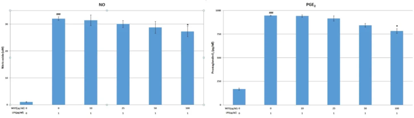
Fig. 2. LPS induced NO and PGE2 production. RAW 264.7 cells were preincubated with 10, 25, 50 and 100 μg/ml of Woogakseungmatang.
The cells were treated with 1 μg/ml of LPS to induce NO and PGE2 production. The NO production was measured by Griess Reagent System, and the PGE2 production was measured by ELISA as described in materials and methods. Data are represented as means±SEM.Significantly different from control (#) or LPS alone (*); ###
p
<0.001, *p
<0.05.Fig. 1. This graph shows the cytotoxic effects of Woogak- seungmatang extract in macrophage.
There is no significant cytoxicity up to 150 μg/ml.
significance (** p <0.01; * p <0.05).
Results
1. Cytotoxicity
The cell viability was 100±0.49, 99.46±0.69, 98.17±0.57, 97.55±1.89, 92.18±2.97, 85.39±1.54, and 76.11±2.81% in the control group and at 10, 25, 50, 100, 150 and 200 μg/ml, respectively. Woogakseungmatang extract showed no cytotoxicity at the 10, 25, 50, and 100 μg/ml concentrations, but decreased by about 15 and 24% at 150 and 200 μg/ml compared with the control; thus, the 10, 25, 50, and 100 μg/ml concentrations were selected in subsequent experiments (Fig. 1).
2. NO production
The means of NO treated with LPS alone is 31.40 μM. The NO production was 100.00±0.65, 98.15±1.95, 93.79±1.28, 88.77±2.18, and 85.13±1.93% in the control group and at 10, 25, 50, and 100 μg/ml, respectively. Woogakseungmatang extract showed approximately 85% NO production at 100 μg/ml compared with the control; a significant difference was observed (Fig. 2).
3. PGE
2production
The means of PGE
2treated with LPS alone is 946.27 pg/ml.
The PGE2 production was 100±0.42, 99.51±1.29, 96.75±2.68, 89.16±1.94, and 82.64±2.54% in the control group and at 10, 25, 50, and 100 μg/ml, respectively.
Woogakseungmatang extract showed a significant decrease in the amount of PGE
2produced at 100 μg/ml compared with the control (Fig. 2).
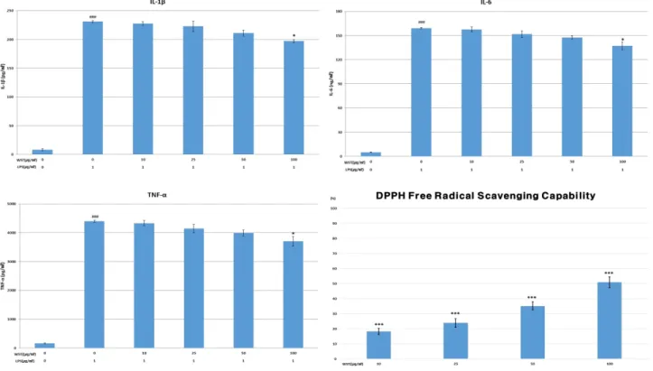
4. Inflammatory cytokines
Woogakseungmatang extracts were treated with cells, and their cytokine production was measured.
The means of IL-1β treated with LPS alone is 231.62 pg/ml. The IL-1β production was 100±0.79, 98.54±1.28, 96.41±3.97, 91.25±2.19, and 85.24±1.18% in the control group and at 10, 25, 50, and 100 μg/ml, respectively.
The IL-1β production was reduced by approximately 9%
at 100 μg/ml compared with the control. There was a
Fig. 3. Inhibition of LPS-induced IL-1β, IL-6, TNF-α and DPPH radical by Woogakseungmatang extract.
RAW 264.7 cells were preincubated with 10, 25, 50 and 100 μg/ml of Woogakseungmatang extract. The cells were treated with 1 μg/ml of LPS. The IL-1β, IL-6 and TNF-α production was measured by ELISA as described in materials and methods. Data are represented as means±SEM.Significantly different from control (#) or LPS alone (*); ###,***
p
<0.001, *p
<0.05.significant decrease at 100 μg/ml.
The means of IL-6 treated with LPS alone is 159.54 ng/ml.
The IL-6 production was 100±0.69, 99.10±1.96, 95.31±
2.84, 92.74±1.48, and 86.18±3.37% in the control group and at 10, 25, 50, and 100 mg/ml, respectively. Woogakseungma- tang extract reduced IL-6 production by approximately 12, 18, and 20% at the 25-, 50-, and 100-μg/ml concentrations, respectively. There was a significant decrease at 100 μg/ml.
The means of TNF-α treated with LPS alone is 4395.59 pg/ml. The TNF-α production was 100±0.78, 98.52±2.19, 94.21±3.46, 90.77±2.43, and 84.20±3.94% in the control group and at 10, 25, 50, and 100 μg/ml, respectively. The TNF-α production was also reduced by approximately 19 and 25% at 50 and 100 μg/ml, respectively. There was a significant decrease at 100 μg/ml (Fig. 3).
5. Antioxidative activity
The DPPH radical scavenging activity was 18.26±2.14,
23.87±2.96, 35.11±3.73, and 50.89±3.59% compared with the control group at 10, 25, 50, and 100 μg/ml, respectively.
The DPPH radical scavenging activity of ascorbic acid and Woogakseungmatang extract was measured. The ascorbic acid-treated group showed more than 100% activity at 50 μg/ml. Woogakseungmatang extract significantly increased the DPPH radical scavenging ability in a concentration- dependent manner. The criteria for the data is that the reagents have not been processed. Woogakseungmatang extract at 100 μg/ml showed the highest DPPH radical scavenging ability (more than 50%) (Fig. 3).
Discussion
Seogak is rhinoceros horn. In Korean medicine, it is said
to have the effect of lowering fever, reducing edema, and
eliminating side effects; however, rhinoceros is an
internationally protected animal whose horns have limited use. Woogak has the same effect as Seogak because it has the effect of lowering and treating fever in Korean medicine.
Seogak and Woogak have the same main components;
therefore, Woogak is the most commonly used substitute for Seogak
3).
Macrophages protect the body from foreign substances, viruses, microorganisms, etc. when they invade. They are robust and have a longer lifespan than neutrophils; in addition, they protect against the treatment of waste products and microorganisms that neutrophils cannot digest; they are also known to produce inflammatory cytokines and antitumor effects and have been used in this study.
As a result of the cytotoxicity test for Woogakseungmatang, approximately 15% of cells were killed at the 200-μg/ml concentration. Since this is a globally significant figure, the experiment proceeded to use 100 μg/ml.
NO is an inflammatory factor and a major cause of various chronic inflammatory diseases. When inflammation occurs, macrophages are activated and produce NO. NO-activated leukocytes and macrophages help regenerate tissues after removing foreign matter; however, excess NO is known to cause cerebral infarction, degenerative neurological diseases, diabetes, and similar issues
8). Thus, Woogakseungmatang extract showed the effect of reducing NO by approximately 15% at 100 μg/ml compared with the control, which is statistically significant.
The main symptoms of inflammation are pain, fever, redness, and swelling
9); PGE
2modulates these symptoms and increases the vascular permeability to spread leukocytes to inflammation sites. It is considered an inflammatory mediator, but induces IgE secretion depending on the target cell and promotes interleukin-4 (IL-4) and interleukin-5 (IL-5) production
10). Woogakseungmatang extract showed the effect of reducing PGE
2significantly by approximately 18% at 100 μg/ml compared with the control.
In general, the inflammatory response of macrophages activated by LPS involves inflammatory mediators, such as pro-inflammatory cytokines, NO, PGE
2, LTB4, iNOS, and COX-2
11). Representative inflammatory cytokines involved in
the development of inflammation are IL-1β, IL-6, and TNF- α. Among these, IL-1β causes an inflammatory reaction, is involved in fever, and activates lymphocytes and neutrophils.
IL-6 promotes the production of acute-phase protein (CRP) and develops chronic stages of inflammatory diseases, including allergic diseases. In addition, TNF-a activates leukocytes by causing an inflammatory reaction. However, excess cytokine causes thrombosis and tissue damage
12). In this study, Woogakseungmatang extract significantly decreased IL-1β production at the 100-μg/ml concentration, IL-6 production was significantly decreased at 10 and 100 μg/ml, and TNF-α was significantly reduced at 100 μg/ml. These results show the potential of Woogakseungmatang extract to be used to prevent and treat various inflammatory diseases through the effective reduction of inflammatory cytokines
13).
Studies on the relationship between the cause of disease and oxidative byproducts by reactive oxygen species are underway
14). Active oxygen is known to be harmful to proteins, biological membranes, and DNA. Therefore, studies on antioxidants have been actively conducted recently
15). The DPPH free radical scavenging capability showed a tendency to increase in a concentration-dependent manner after Woogakseungmatang extract treatment. At the concentration of 100 μg/ml, it showed more than 50% scavenging ability.
Woogakseungmatang extract inhibits the inflammatory process by significantly reducing cytokine IL-6 and TNF-a production. Woogakseungmatang extracts should be studied clinically and experimentally.
Conclusion
This paper investigates the anti-inflammatory effect of Woogakseungmatang extract using RAW 264.7 cells to measure the amounts of MTT, NO, and PGE
2and cytokines IL-1β, IL-6, and TNF-α. Thus, the following conclusions were obtained:
1. There was no significant change in cell survival up to
100 μg/ml for Woogakseungmatang extract; cell viability
was 76% at 200 μg/ml.
2. The amount of NO produced was significantly decreased by 85% at 100 μg/ml compared with the control.
3. The amount of PGE
2produced was significantly reduced by 82% at 100 μg/ml compared with the control.
4. IL-1β, IL-6, and TNF-α were significantly reduced at 100 μg/ml compared with the control group.
5. The DPPH free radical scavenging capability was more than 50% at 100 μg/ml.
According to this study, the Woogakseungmatang showed only a slight anti-inflammatory effect at 100 μg/ml the highest concentration that does not show cytotoxicity. In addition, it was difficult to confirm the concentration- dependent anti-inflammatory effect of the Woogakseungmatang.
Therefore, this study means to confirm the potential anti- inflammatory effects of Woogakseungmatang. A variety of anti-inflammatory studies of Woogakseungmatang are needed.
Acknowledgements
This paper was supported by the Semyung University Research Grant of 2017.
References
1. Vinay Kumar, Abbas, ASTER, Fausto, Robbins &Cotran patho- logic basis of disease. 8th, Seoul: Bummoon education. 2011 : 223-41.
2. Cheon MS, Yoon TS, Choi GY, Kim SJ, Lee AY, Moon BC, et al.
Comparative Study of Extracts from Rhubarb on Inflammatory Activity in RAW 264.7 Cells. Korean J. Medicinal Crop Sci. 2009 ; 17(2) : 109-14.
3. The Korea Society of Korean Herbal medicine textbook compi- lation committee. Herbal medicine. 2nd. Seoul: Younglimsa.
2007 : 229-30
4. Heo J. Donguibogam. 3rd. Seoul: Donguibogam Publishers.
2006 : 1003-5.
5. The Korea Society of acupuncture and moxibution textbook compilation committee. Acupuncture and moxibution textbook.
2nd. Paju: Hanmi medical Publishers. 2017 : 229-30
6. Cho GS, Kim JH, Chung SH, Shin GJ, Lee WC. The clinical ob- servation on I case of patient with trigeminal neuralgia. The J. of internal Korean medicine. 2000 ; 21(3) : 505-10.
7. Lee SI, Lee BJ, Lee TJ. Comparative Experimental Study of Anti-Thrombosis Effect Comparing Cornu Bubali , Cornu Bos tauri , Cornu Rhinoceri. The Korean J. Herbology. 1988 ; 3(1) : 19-48.
8. Kain V, Prabhu SD, Halade GV. Inflammation revisited: in- flammation versus resolution of inflammation following my- ocardial infarction. Basic Res Cardiol. 2014 ; 109(6) : 444.
https://doi.org/10.1007/s00395-014-0444-7
9. The Korea Society of Korean Pathology textbook compilation committee. Korean Pathology. Iljungsa. 2004 : 109-12.
10. Chapter of The Korea Society of Pathlogists. Summarize Pathology. 1st. Seoul : Jungmoongak. 2008 : 81
11. Alanazi AM, El-Azab AS, Al-Suwaidan IA, ElTahir KE, Asiri YA, Abdel-Aziz NI, et al. Structure-based design of phthalimide de- rivatives as potential cyclooxygenase-2 (COX-2) inhibitors: an- ti-inflammatory and analgesic activities. Eur J Med Chem. 2015 ; 92(6) : 115-23. https://doi.org/10.1016/j.ejmech.2014.12.039 12. Fischer R, Maier O. Interrelation of oxidative stress and in-
flammation in neurodegenerative disease: role of TNF. Oxid Med Cell Longev. 2015 ; 2015(1) : 1-18. https://doi.org/10.11 55/2015/610813
13. Schett G, Elewaut D, McInnes IB, Dayer JM, Neurath MF. How cytokine networks fuel inflammation: Toward a cytokine- based disease taxonomy. Nat Med. 2013 ; 19(7) : 822-4.
https://doi.org/10.1038/nm.3260
14. Wiseman H. Dietary inflence on membrane function; im- portwnt in protection against oxdative damage and disease.
7th. London : Nutritional Biochemistry. 1996 : 2-6. https://doi- .org/10.1016/0955-2863(95)00152-2
15. Chang S, Ostric A. National antioxidants from rosmary and sage.
J. Food Sci. 1977 ; 42(1) : 1102-10.
국문초록
목적 :