• 검색 결과가 없습니다.

Anti-inflammatory Effects of Various Mushrooms in LPS-stimulated RAW264.7 Cells

N/A
N/A
Protected

Academic year: 2021

Share "Anti-inflammatory Effects of Various Mushrooms in LPS-stimulated RAW264.7 Cells"

Copied!
11
0
0

로드 중.... (전체 텍스트 보기)

전체 글

(1)

Introduction

Mushrooms have a long history of use as a part of the human diet in many regions worldwide due to their organo- leptic characteristics and nutritional value (Reid et al., 2017).

They have a high protein content with almost all essential amino acids and are rich in diverse minerals and vitamin B, representing a good dietary source of these important nutrients (Robaszkiewicz et al., 2010). In addition to attracting a great deal of interest in many areas of foods, mushrooms are also

useful for preventing diseases, such as hypertension, hyper- cholesterolemia, and cancer (Lull et al., 2005). Medicinal mushrooms have an established history of use in traditional Eastern therapies. Based on historical customs of North Eastern Asia, medicinal mushrooms such as Ganoderma lucidum (Reishi), Lentinus edodes (Shiitake), and Inonotus obliquus (Chaga) have been collected and hot-water-soluble fractions have been extracted (Wasser, 2002). However, appropriate preparation methods for mushrooms in food processing and pharmaceutical production to regulate cellular immune responses are still elusive. Furthermore, the types of mushrooms that have greater biological and pharmaceutical activities for enhancing the immune response have not yet

Anti-inflammatory Effects of Various Mushrooms in LPS-stimulated RAW264.7 Cells

Kyung Hye Seo

1

*, Jeong-Yong Park

2,3

, Hyung-Jun Noh

1

, Ji Yeon Lee

2,3

, Eun Young Lee

2

, Jae-Gu Han

4

, Jin Hyo Kim

5

and Mi Sun Cheong

6

1

Researcher and

2

Assistant, Department of Herbal Crop Research, National Institute of Horticultural & Herbal Science, Eumsung 27709, Korea

3

Student, Department of Food Science and Biotechnology, Chungbuk National University, Cheongju 28644, Korea

4

Researcher, Mushroom Research Division, National Institute of Horticultural & Herbal Science, Eumsung 27709, Korea

5

Professor and

6

Researcher, Institute of Agriculture and Life Science (IALS), Gyeongsang National University, Jinju 52828, Korea

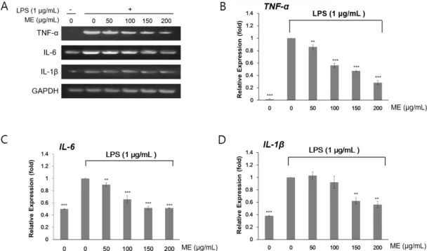
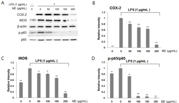
Abstract - Mushrooms have been widely cultivated and consumed as foods and herbal medicines owing to their various biological properties. However, few studies have evaluated the anti-inflammatory effects of mushrooms. Here, we investigated the effects of mushroom extracts (MEs) on lipopolysaccharide (LPS)-induced inflammation in macrophages (RAW264.7 cells). First, we extracted MEs with either water or ethanol. Using LPS-treated RAW264.7 cells, we measured cell proliferation and NO production. Gene expression of tumor necrosis factor-α (TNF-α), interleukin (IL)-6 (IL-6), and IL-1β was assessed by RT-PCR, and protein abundance of inducible NO synthase (iNOS) and cyclooxygenase-2 (COX-2) and phosphorylation of p65 were determined by immunoblotting. MEs prepared using both water and ethanol inhibited LPS-induced inflammation in RAW264.7 cells. Nitric oxide (NO) levels induced by LPS were reduced by treatment with MEs. Isaria japonica Yasuda water extracts and Umbilicaria esculenta (Miyoshi) Minks ethanol extracts significantly decreased the mRNA expression of inflammation-related cytokine genes including TNF-α, IL-6, and IL-1β. Similarly, the protein abundance of iNOS and COX-2 was also decreased. The phosphorylation of p65, a subunit of nuclear factor-κB was at least partly suppressed by MEs. This study suggests that mushrooms could be included in the diet to prevent and treat macrophage-related chronic immune diseases.

Key words – Anti-inflammation, Isaria japonica Yasuda, Macrophage, Medicinal mushrooms, Umbilicaria esculenta (Miyoshi) Minks

*Corresponding author. E-mail : [email protected] Tel. +82-43-871-5785

ⓒ 2018 by The Plant Resources Society of Korea

Original Research Article

(2)

been identified.

Inflammation is a biological defense response of the body to harmful stimuli, including tissue damage or pathogen infection (Park et al., 2018); this response is required for removal of damaging agents. Chronic inflammation can be differentially classified from acute inflammation and is generally associated with the development of diseases, such as atherosclerosis, asthma, rheumatoid arthritis, and cancer (Libby et al., 2002; Du et al., 2015).

During the inflammatory response, inflammatory cells such as macrophages secrete inflammatory mediators including nitric oxide (NO), tumor necrosis factor- α (TNF-α), interleukin (IL)-6, and IL-1 β (Mann et al., 2005). Lipopolysaccharide (LPS), a well-known endotoxin, activates the transcription factor nuclear factor- κB (NF-κB) and induces the expression of inflammatory cytokines, such as inducible NO synthase (iNOS) and cyclooxygenase-2 (COX-2), resulting in the production of inflammatory mediators (Guzik et al., 2003;

Yi et al., 2017). Currently, many anti-inflammatory drugs cause side effects such as gastritis, nephritis, and cardiac disorders. Accordingly, many studies have attempted to identify safer anti-inflammatory substances among commonly consumed dietary foods.

In this study, we aimed to investigate whether mushroom extracts possessed anti-inflammatory effects. Primarily, we considered that mushroom extracts should be easily prepared and ready to eat. For this purpose, the solvents were applied either water or ethanol. To this end, we examined the production of LPS-induced NO using RAW264.7 cells and evaluated the mRNA levels of cytokines and protein accumu- lation of iNOS and COX-2 following modification of NF- κB protein levels. Our data provide insights into the utility of mushroom extracts (MEs) in the regulation of immune responses and anti-inflammatory activities.

Materials and Methods

ME preparation and preparation of extracts

Mushrooms were purchased from Yeoncheon, South Korea in June, 2014. The dried fruiting body at 50 ℃ for 3 days of 22

species of mushroom was placed into a flask and extracted three times with 10 volume of 70% ethanol or water at room temperature for 1 day, and then filtered through a PTEE syringe. The filtrate was condensed by evaporation under reduced pressure at 50℃ and freeze-dried, and the weight was measured.

Cell lines

RAW264.7 macrophages were obtained from the Korean Cell Line Bank (KCLB NO. 40071, Seoul, Korea). The cells were grown in Dulbecco’s modified Eagle’s medium (Gibco Life Technologies, Carlsbad, CA, USA) containing 10% fetal bovine serum, 50 ㎍/㎖ penicillin, and 50 ㎍/㎖ streptomycin in a 5% CO chamber at 37℃.

Cell viability assay

RAW264.7 cells were seeded in 96-well plates at a density of 1 × 10

4

cells/well. The cultured cells were treated with 1 ㎍/㎖

LPS for 4 h and MEs were then added in dimethylsulfoxide (DMSO) at various concentrations. Solvent control cells were treated with LPS alone. After 24 h of culture with or without samples, MTS working solution (Promega, CA, USA) was added into the culture plates for 4 h at 37℃. The absorbance of individual wells was measured at 540 nm using a microplate reader (Molecular Devices, CA, USA), and cell viability was calculated as the percent absorbance relative to LPS-treated control cells.

NO assay

The level of NO production in RAW264.7 cell supernatants was determined using Griess reagent (Promega), according to the manufacturer’s instructions. Briefly, the extracts were treated as described for the cell viability assays, and the cell culture supernatants were then collected in 96-well plates for nitrite assays. The absorbance was measured at 540 nm using a microplate reader (Molecular Devices, CA, USA).

The concentration of nitrite was calculated from a standard

curve.

(3)

Total RNA extraction, cDNA synthesis, and reverse transcription polymerase chain reaction (RT-PCR) analysis

Total RNA was isolated from RAW264.7 cells using TRIzol Reagent (Invitrogen, NY, USA), and 1 ㎍ of RNA was used for cDNA synthesis. RT-PCR was performed where each cDNA was amplified using gene-specific primers (Table 1) and a Maxime PCR RedMix kit (iNtRON Biotechnology, Seongnam, Korea). GADPH expression was used to normalize the expression value in each sample and relative expression values were determined against the average value of LPS- treated samples without MEs using ImageJ software.

Immunoblotting analysis

RAW264.7 cells were collected and lysed in ice-cold RIPA buffer (20 mM Tris-HCl, pH 7.5, 150 mM NaCl, 1 mM Na

2

-ethylenediamintetraacetic acid, 1 mM ethylene glycol-bis [ β-aminoethyl ether]-N,N,N',N'-tetraacetic acid, 1% NP-40, 1% sodium deoxycholate, 2.5 mM sodium pyrophosphate, 1 mM β-glycerophosphate, 1 mM Na

3

VO

4

, and 1 ㎍/㎖ leupeptin) for 30 min, and protein levels were measured using the BCA assay (Thermo Scientific, Rockford, IL, USA). The extracted protein was separated using 12% sodium dodecyl sulfate- polyacrylamide gel electrophoresis and then transferred onto nitrocellulose membranes (GE Healthcare, Chalfont St. Giles, UK). After blocking with Tris-buffered saline containing 5% nonfat dry milk and 0.1% (w/v) Tween 20, the mem- branes were immunoblotted with specific antibodies and then subjected to enhanced chemiluminescence (ECL kit;

GE Healthcare). The relative band intensities were determined

using chemiluminescence (Davinch-chemi Chemiluminescence Imaging System; CAS-400SM; Seoul, Korea). The relative protein intensity of each sample (200 ㎍/㎖) was calculated by normalizing against the intensity of β-actin as a loading control.

Statistical analysis

Unless otherwise indicated, values represent the mean ± standard deviation (SD) of data obtained from triplicate experiments. Statistical analysis was performed using Student’s t-tests.

Results

Identification of mushroom extracts (MEs)

To obtain MEs, either water or 70% ethanol, herein referred to simply as ethanol, was used as the extraction solvent based on the consideration that MEs should be directly edible. We chose the dried fruiting body of the mushrooms, and ex- traction was performed at room temperature for 1 day. The MEs are listed in Table 2.

Effects of MEs on LPS-induced cell proliferation in RAW264.7 cells

To examine the effects of cell viability in the presence of MEs, we performed MTS assays using RAW264.7 cells, which were derived from mouse leukemic monocyte macrophages.

The MTS assay is commonly used for analyze cell cytotoxicity and its result is correlated with cell proliferation. Cell pro- Table 1. The pro-inflammatory gene primer used for RT-PCR

Gene Sense and antisense sequences Expected product size (bp)

IL-6 5 ′-GTACTCCAGAAGACCAGAGG-3′ 308  

5 ′-TGCTGGTGACAACCACGGCC-3′

TNF- α 5 ′-TTGACCTCAGCGCTGAGTTG-3′ 364

5 ′-CCTGTAGCCCACGTCGTAGC-3′

IL-1 β 5 ′-CAGGATGAGGACATGAGCACC-3′ 447

5 ′-CTCTGCAGACTCAAACTCCAC-3′

GAPDH 5 ′-CGGAGTCAACGGATTTGGTCGTAT-3′ 306

5 ′-AGCCTTCTCCATGGTGGTGAAGAC-3′

(4)

liferation was not changed by LPS (Table 3) or ME treatment in the absence of LPS (data not shown). LPS-induced cell proliferation was not altered by around 36 different spices

of MEs (Table 2, Table 3). However, some MEs caused reduced cell proliferation under the LPS treatment. For example, Isaria japonica Yasuda (I. japonica) and Inonotus Table 2. Relative effects of mushroom extracts on LPS-treated RAW264.7 cells

Extracted

Solvent Mushroom The rate of

cell growth change

NO contents (µM)

The rate of NO change

Water

Pleurotus eryngii (DC.) Quél. 0.928 26.45±1.43 0.926

Tricholoma giganteum Massee 1.018 26.25±0.52 0.988

Tricholoma matsutake 0.902 26.26±1.70 0.879

Sparassis crispa (Wulfen) Fr. 0.951 28.95±2.36 1.052

Agaricus bisporus (J.E. Lange) Imbach 1.051 26.21±1.20 0.993

Pleurotus cornucopiae (paulet) Rolland var,

citrinopileatus (Sing.) Ohira 0.942 3.75±0.69 0.134

Pleurotus ostreatus (Jacq.: Fr.) P. Kumm 1.096 27.86±0.15 0.985

Pleurotus salmoneostramineus Lj.N. Vassiljeva 1.206 25.48±1.96 0.975

Hypsizygus marmoreus (Peck) H.E. Bigelow 1.064 29.12±3.44 1.023

Isaria japonica Yasuda 0.849 4.25±0.74 0.258

Inonotus obliquus (Fr.) Pilat 1.023 26.68±0.76 0.930

Lentinula edodes (Berk.) Pegler 0.989 9.92±0.70 0.338

Ganoderma lucidum (Curtis) P. Karst. 0.953 0.17±0.36 0.006

Antler-Ganoderma lucidum (Curtis) P. Karst. 1.104 26.47±1.17 0.940 Flat Ganoderma lucidum (Curtis) P. Karst. 1.208 28.20±1.19 1.001

Grifola frondosa (Dicks.) Gray 1.017 16.74±0.65 0.596

Flammulina velutipes (Curtis) Singer 1.285 26.35±2.45 0.880

Hypsizygus marmoreus (Peck) H.E. Bigelow 0.941 21.02±1.89 0.818

Gold-Hypsizygus marmoreus (Peck) H.E. Bigelow 0.956 26.17±2.22 0.976

Ethanol

Pleurotus eryngii (DC.) Quél. 1.082 17.68±2.50 0.635

Tricholoma giganteum Massee 0.928 18.40±2.63 0.652

Tricholoma matsutake 1.370 17.28±1.18 0.658

Sparassis crispa (Wulfen) Fr. 1.384 15.21±0.99 0.597

Agaricus bisporus (J.E. Lange) Imbach 0.897 17.22±1.17 0.642

Pleurotus ostreatus (Jacq.: Fr.) P. Kumm 1.307 17.31±1.03 0.657

Pleurotus salmoneostramineus L. Vass 0.900 14.18±1.04 0.540

Hypsizygus marmoreus (Peck) H.E. Bigelow 1.379 13.77±1.55 0.554

Isaria japonica Yasuda 0.979 0.035±0.12 0.008

Inonotus obliquus (Fr.) Pilat 0.998 2.17±0.35 0.219

Lentinula edodes (Berk.) Pegler 1.452 15.68±1.79 0.593

Ganoderma lucidum (Curtis) P. Karst. 0.997 4.96±1.22 0.201

Antler-Ganoderma lucidum (Curtis) P. Karst. 1.114 17.30±0.88 0.648

Grifola frondosa (Dicks.) Gray 0.737 15.40±1.19 0.582

Phellinus linteus (Berk. Et Curt.) Teng 1.211 0.05±0.14 0.013

Umbilicaria esculenta (Miyoshi) Minks 0.952 2.00±1.10 0.119

Cordyceps militaris 1.246 18.77±1.00 0.716

DMSO 1.000 30.68±2.17 1.000

(5)

Table 3. The mushroom extracts effect of cell proliferation and NO release content

Exracted

Solvent Mushroom

Cell proliferation (% of LPS-treated control)

NO contents (µM)

100 µg/ml 200 µg/ml 100 µg/ml 200 µg/ml

Water

Pleurotus eryngii (DC.) Quél. 103.3 ±10.29 95.90±12.76 28.56±0.28 26.45±1.43 Tricholoma giganteum Massee 106.88±13.17 108.84±3.68 26.56±1.37 26.25±0.52

Tricholoma matsutake 105.69±13.61 95.37±8.22 29.88±1.29 26.26±1.70

Sparassis crispa (Wulfen) Fr. 91.65±3.47 87.12±10.32 27.51±1.05 28.95±2.36 Agaricus bisporus (J.E. Lange) Imbach 89.74±8.92 94.33±17.90 26.39±1.03 26.21±1.20 Pleurotus cornucopiae (paulet) Rolland var,

citrinopileatus (Sing.) Ohira 80.68±3.20 76.04±4.00 28.08±1.35 3.75±0.69 Pleurotus ostreatus (Jacq.: Fr.) P. Kumm 88.94±12.90 97.45±7.43 28.28±3.37 27.86±0.15 Pleurotus salmoneostramineus Lj.N. Vassiljeva 93.77±11.56 113.09±7.51 26.12±0.35 25.48±1.96 Hypsizygus marmoreus (Peck) H.E. Bigelow 89.57±3.93 95.27±8.60 28.46±0.27 29.12±3.44

Isaria japonica Yasuda 82.01±7.81 81.13±9.34 16.45±3.15 4.25±0.74

Inontus obliquus 79.61±10.35 81.46±5.09 28.69±1.55 26.68±0.76

Lentinula edodes (Berk.) Pegler 87.54±10.52 86.59±5.57 29.32±0.93 9.92±0.70 Ganoderma lucidum (Curtis) P. Karst. 91.74±12.99 87.39±3.95 27.12±1.48 0.17±0.36 Antler-Ganoderma lucidum (Curtis) P. Karst. 107.82±17.47 118.98±7.90 28.16±0.77 26.47±1.17 Flat Ganoderma lucidum (Curtis) P. Karst. 94.81±8.64 114.57±10.39 28.18±1.14 28.20±1.19 Grifola frondosa (Dicks.) Gray 105.27±9.67 107.03±5.81 28.10±1.20 16.74±0.65 Flammulina velutipes (Curtis) Singer 90.78±15.24 116.67±10.10 29.93±0.74 26.35±2.45 Hypsizygus marmoreus (Peck) H.E. Bigelow 88.41±6.44 83.16±13.42 25.70±1.00 21.02±1.89 Gold-Hypsizygus marmoreus (Peck) H.E. Bigelow 93.31±5.51 89.21±11.70 26.82±0.81 26.17±2.22

Ethanol

Pleurotus eryngii (DC.) Quél. 91.74±3.43 99.29±10.72 27.86±0.15 17.68±2.50 Tricholoma giganteum Massee 124.06±18.08 115.18±12.33 28.20±1.19 18.40±2.63

Tricholoma matsutake 97.02±8.68 132.92±8.83 26.28±0.86 17.28±1.18

Sparassis crispa (Wulfen) Fr. 95.45±27.14 132.11±8.21 25.48±1.96 15.21±0.99 Agaricus bisporus (J.E. Lange) Imbach 106.27±6.68 95.37±8.22 26.84±0.81 17.22±1.17 Pleurotus ostreatus (Jacq.: Fr.) P. Kumm 101.21±2.53 132.33±9.11 26.35±2.45 17.31±1.03 Pleurotus salmoneostramineus L. Vass 130.85±11.59 117.76±20.26 26.26±1.70 14.18±1.04 Hypsizygus marmoreus (Peck) H.E. Bigelow 97.59±5.22 134.57±10.27 24.86±1.32 13.77±1.55

Isaria japonica Yasuda 54.81±6.77 53.64±10.50 4.25±0.74 0.035±0.12

Inontus obliquus 58.77±5.35 58.66±3.19 9.92±0.70 2.17±0.35

Lentinula edodes (Berk.) Pegler 94.09±10.98 136.6±11.17 26.45±1.43 15.68±1.79 Ganoderma lucidum (Curtis) P. Karst. 85.08±11.09 84.80±10.38 24.65±0.21 4.96±1.22 Antler-Ganoderma lucidum (Curtis) P. Karst. 92.71±6.47 103.26±13.37 26.68±0.76 17.30±0.88 Grifola frondosa (Dicks.) Gray 113.86±10.39 83.90±3.03 26.47±1.17 15.40±1.19 Phellinus linteus (Berk. Et Curt.) Teng 54.30±4.81 65.78±9.88 3.75±0.69 0.05±0.14 Umbilicaria esculenta (Miyoshi) Minks 102.38±10.28 97.48±5.19 16.74±0.65 2.00±1.10

Cordyceps militaris 101.65±8.48 126.62±16.75 26.21±1.20 18.77±1.00

  -LPS 103.01±2.49 2.17±0.02

  1 µg/ml LPS 100±00 30.68±2.17

(6)

obliquus (Fr.) Pilát (I. obliquus) ethanol extracts showed only 53.64% ± 9.10% and 58.66% ± 2.76% cell proliferation, respectively, relative to the control, indicating that these MEs may exhibit cytotoxicity in RAW264.7 cells (Table 3).

Indeed, even when using a relatively low concentration of 100 ㎍/㎖ I japonica and I. obliquus MEs, cell growth reached only 54.81% ± 6.77% and 58.77% ± 5.35%, respectively (Table 3). Thus, although some MEs affected cell proliferation, most MEs yielded 100% or higher viability, indicating that these MEs did not exert cytotoxic effects in RAW264.7 cells.

Effects of MEs on LPS-stimulated NO production in RAW264.7 cells

To observe LPS-induced NO production in RAW264.7 cells in the presence of MEs, cells were treated with LPS (1

㎍/㎖) for 4 h and then treated with DMSO or various MEs for 24 h. After 24 h of incubation, the NO levels in the cellular supernatants were assessed by Griess assays. Treatment with LPS increased NO production (30.68 ± 2.17 µM) compared with that in the absence of LPS (2.17 ± 0.02 µM; Table 2, Table 3). In contrast, post-treatment with 200 ㎍/㎖ MEs resulted in reduced LPS-induced NO generation (Table 3).

Specifically, MEs prepared using Pleurotus cornucopiae (paulet) Rolland var. citrinopileatus (Sing.) Ohira, and I japonica extracts with water as well as those prepared using I. obliquus t and U. esculenta extracts with ethanol reduced NO contents to 3.75 ± 0.69 µM, 4.25 ± 0.74 µM, and 2.17 ± 0.35 µM, 2.00 ± 1.10 µM, respectively, similar to that without LPS (2.17 ± 0.02 µM; Table 3). We further tested NO production in the presence of 100 ㎍/㎖ MEs to investigate the effects of a lower concentration of MEs. Notably, 100 ㎍/

㎖ MEs yielded results similar to those above, except that the NO concentration was higher than that induced by 200 ㎍/㎖

MEs (Table 3), indicating that LPS-induced NO contents were reduced in an ME concentration-dependent manner.

Statistically significant differences were calculated by Student’s t-test, p < 0.05 (*), p < 0.01(**), and p < 0.001 (***). As NO is a signaling molecule involved in acute and chronic inflammatory diseases, NO levels are used to be representing

inflammation. These results suggest that extracts from several mushrooms including I japonica, I. obliquus, U. esculenta, and P. linteus exerted anti-inflammatory effects via reduction of NO in LPS-stimulated RAW264.7 cells.

Before further investigating the anti-inflammatory effects of MEs, we summarized the above results for cell proliferation and NO reduction. The values were obtained by comparing the changes in cell proliferation and NO contents between 200 ㎍/㎖ MEs and 100 ㎍/㎖ MEs (Table 2). Thus, we chose two ME candidates: I. japonica water extract and U. esculenta ethanol extract due to that the change rate of cell growth was close to 1 for no change (i.e less cytotoxic), and the change rate of NO content was around 0.2 for 80% reduction on the treatment of 200 ㎍/㎖ MEs (i.e effective for NO reduction) (Table 2).

Effects of MEs on the LPS-induced increase in TNF- α, IL-6, and IL-1 β mRNAs in RAW264.7 cells

To evaluate the anti-inflammatory effects of these two MEs, we analyzed the expression of inflammatory marker genes by reverse transcription PCR. Because murine macro- phages produce pro-inflammatory cytokines, such as TNF- α, IL-6, and IL-1β, during the inflammatory response, LPS treatment increases the these transcripts (Mann et al., 2005).

Given the screening results for the chosen two MEs, RAW264.7 cells were pre-incubated for 4 h with 1 ㎍/㎖ LPS and cultured 24 h more with the indicated concentrations (50 –200 ㎍/㎖) of MEs. LPS treatment induced TNF-α, IL-6, and IL-1 β gene expression. However, LPS-induced accumulation of TNF- α, IL-6, and IL-1β mRNA was significantly reduced after treatment with both I. japonica extracts (Fig. 1) and U.

esculenta extracts (Fig. 2). In addition, reduction of the these

transcripts was much more apparent following treatment

with the high concentration (200 ㎍/㎖) of MEs compared

with that after treatment with a low concentration (50 ㎍/㎖)

of MEs (Fig. 3B –D and Fig. 4B–D). Statistically, the values

revealed that the difference were significant with p < 0.05

(*), p < 0.01(**), and p < 0.001(***) by Student’s t-test. This

result indicated that both I. japonica and U. esculenta extracts

suppressed LPS-mediated transcription of pro-inflammatory

(7)

Fig. 1. Effects of water extracts of I. japonica on pro-inflammatory gene expression in RAW264.7 cells. (A) The expression levels of TNF- α, IL-6, and IL-1β mRNAs were determined by RT-PCR analysis. Cells were treated with 1 μg/mL LPS and then I. japonica extracts at various concentrations for 24 h. GADPH was used as an internal control. The expression levels of TNF- α (B), IL-6 (C), and IL-1 β (D) were determined by densitometry analysis. Values are expressed as the mean ± SD of four determinations.

Significance was determined using Student’s t-tests: *p < 0.05, **p < 0.01, and ***p < 0.001 versus cells treated with LPS alone.

Fig. 2. Effects of ethanol extract of U. esculenta on pro-inflammatory gene expression in RAW264.7 cells. (A) The expression levels of TNF- α, IL-6, and IL-1β mRNA was determined by RT-PCR analysis. Cells were treated with 1 ㎍/㎖ LPS and then U. esculenta extracts at various concentrations for 24 h. GADPH was used as an internal control. The expression levels of TNF- α (B), IL-6 (C), and IL-1 β (D) were determined by densitometry analysis. Values are represented as the mean ± SD of four determinations.

Significance was determined using Student’s t-tests: *p < 0.05, **p < 0.01, and ***p < 0.001 versus cells treated with LPS alone.

(8)

Fig. 3. Effects of water extracts of I. japonica on the NF- κB pathway in RAW264.7 cells. (A) COX-2, iNOS, and p65 protein abundance was determined by a western blotting. Relative protein abundance of IL-6 (B) and IL-1 β (C) and the level of Phosphorylated p65 (D), and were determined by densitometry analysis. Values are expressed the mean ± SD of four determinations.

Significance was determined using Student’s t-tests: *p < 0.05, **p < 0.01, and ***p < 0.001 versus cells treated with LPS alone.

Fig. 4. Effects of ethanol extracts of U. esculenta on the NF- κB pathway in RAW264.7 cells. (A) COX-2, iNOS, and p65 protein levels were determined by western blot analysis. IL-6 (B) and IL-1 β (C) protein levels were determined by densitometry analysis.

Phosphorylated p65 levels were determined (D). Values are expressed as the mean ± SD of four determinations. Significance was

determined using one-factor t-tests: *p < 0.05, **p < 0.01, and ***p < 0.001 versus cells treated with LPS alone.

(9)

cytokines.

Effects of MEs on LPS-induced iNOS, COX-2, and p65 expression in RAW264.7 cells

Next, we examined whether I. japonica and U. esculenta extracts suppressed the expression of transcription factors related to the inflammatory process, e.g., iNOS and COX-2.

iNOS and COX-2 are key pro-inflammatory proteins, and the expression of these proteins is controlled by signaling downstream of NF- κB (T and A, 2006). To test this hypo- thesis, we investigated iNOS and COX-2 protein expression and p65 protein modification by western blotting following treatment with MEs. p65, a subunit of NF- κB, undergoes phosphorylation (p-P65) to activate gene expression, including COX-2 and iNOS genes (T and A, 2006).

LPS treatment increased iNOS and COX-2 expression by inducing the inflammatory response in RAW264.7 cells. As expected, immunoblotting analyses indicated that iNOS and COX-2 expression and post-translation modification of p65 by LPS pretreatment were inhibited after incubation with I.

japonica (Fig. 3) and U. esculenta (Fig. 4) MEs. In addition, p65 protein abundance was slightly increased by LPS, whereas p65 protein phosphorylation was dramatically decreased (Fig. 3D and Fig. 4D). These results demonstrated that both I. japonica and U. esculenta extracts suppress LPS-stimulated p65 phosphorylation and affected COX-2 and iNOS protein abundance. Because the pro-inflammatory enzyme iNOS is responsible for NO production, inhibition of iNOS protein accumulation resulted in reduced NO content following treatment with I. japonica and U. esculenta extracts.

Discussion

Macrophages play important roles in inflammation through the production of several pro-inflammatory molecules, in- cluding NO. Uncontrolled, excessive NO has been shown to be associated with a range of inflammatory diseases, including arteriosclerosis, ischemic reperfusion, hypertension, and septic shock (Alvarez and Evelson, 2007; Pacher et al., 2007). Recent studies have demonstrated that plant foods,

including fruits, vegetables, and medicinal herbs, are excellent sources of antioxidant molecules that effectively inhibit the inflammatory process by influencing different molecular targets (Tsai et al., 2007).

Edible mushrooms are used as functional foods due to their high content of bioactive compounds. However, their anti-inflammatory properties and the effects of processing steps on their bioactivity have not been systemically investi- gated. The anti-inflammatory effect of mushrooms of water extracts were reported using Sparassis cispa (Wulfen) Fr.

(Kim et al., 2012), Ganoderma lucidum (Curtis) P. Karst (Kuo et al., 2006) and Grifola frondosa (Dicks.) Gray (Radic et al., 2010). The anti-inflammatory effect of mushrooms of ethanol extracts were reported Pleurotus eryngii (DC.) Quél (Lin et al., 2014), Agaricus bisporus (J.E. Lange) Imbach (Gunawardena et al., 2014; Taofiq et al., 2015), Pleurotus ostreatus (Jacq.: Fr.) P. Kumm (Taofiq et al., 2015), Hypsizygus marmoreus (Peck) H.E. Bigelow (Chien et al., 2016), Inonotus obliquus (Fr.) Pilat (Park et al., 2005; Van et al., 2009; Ma et al., 2013), Lentinula edodes (Berk.) Pegler (Gunawardena et al., 2014), Ganoderma lucidum (Curtis) P. Karst (Yoon et al., 2013). Accordingly, in this study, we found that MEs effectively suppressed LPS-induced inflammation. In addition, we showed that water extracts of I. japonica and ethanol extracts of U. esculenta inhibited the LPS-induced increase in NO production and pro-inflammatory cytokine expression in RAW264.7 cells.

LPS, a component of the outer membrane of gram-negative

bacteria, activates macrophages and promotes the synthesis

and release of molecules involved in the inflammatory

response, such as cytokines, NO, and pro-inflammatory

enzymes (O'Connell et al., 1998). We observed that MEs

prepared using water or ethanol effectively reduced LPS-

induced NO accumulation in RAW264.7 macrophages,

suggesting that the MEs mitigated the pro-inflammatory

reaction. Accumulating evidence has shown that NO is

directly involved in the development of inflammation (Aktan,

2004; Pacher et al., 2007). In particular, high concentrations

of NO, synthesized by iNOS, can mediate inflammation and

cause cell death by triggering apoptosis (Wink and Mitchell,

(10)

1998).

Interestingly, both water extracts of I. japonica and ethanol extracts of U.esculenta elicited changes in TNF- α, IL-1β, and IL-6 mRNA expression and in accumulation of iNOS and COX-2 enzymes, and they modulated p65 phosphorylation, suggesting that these MEs could inhibit the expression of the pro-inflammatory cytokines and modulate NF- κB signaling to control NO. Therefore, the findings of our study reveal that water extracts of I. japonica and ethanol extracts of U. esculenta possess a protective effect in LPS-induced RAW264.7 cells. In addition, these results support that mushrooms are healthy foods and sources of medicinal components. One benefit of these MEs is that they can be prepared easily, and this benefit might contribute to developing healthy beverages or drugs to prevent or treat inflammatory diseases.

Acknowledgments

This study was performed with the support of the Coo- perative Research Program for Agriculture Science and Technology Development (project no. PJ01134401), the Rural Development Administration, Republic of Korea.

References

Aktan, F. 2004. iNOS-mediated nitric oxide production and its regulation. Life Sci. 75:639-653.

Alvarez, S. and P.A. Evelson. 2007. Nitric oxide and oxygen metabolism in inflammatory conditions: sepsis and exposition to polluted ambients. Front Biosci. 12:964-974.

Chien, R.C., Y.C. Yang, E.I. Lai and J.L. Mau. 2016. Anti- inflammatory effects of extracts from the medicinal mushrooms Hypsizygus marmoreus and Pleurotus eryngii (Agaricomycetes).

International Journal of Medicinal Mushrooms 18:477-487.

Du, C., M. Bhatia, S.C. Tang, M. Zhang and T. Steiner. 2015.

Mediators of inflammation: Inflammation in cancer, chronic diseases, and wound healing. Mediators Inflamm. 2015:570653.

Gunawardena, D., L. Bennett, K. Shanmugam, K. King, R.

Williams, D. Zabaras, R. Head, L. Ooi, E. Gyengesi and G.

Münch. 2014. Anti-inflammatory effects of five commercially

available mushroom species determined in lipopolysaccharide and interferon-γ activated murine macrophages. Food Chemistry 148:92-96.

Guzik, T.J., R. Korbut and T. Adamek-Guzik. 2003. Nitric oxide and superoxide in inflammation and immune regulation. J Physiol Pharmacol. 54:469-487.

Kim, H.H., S.Y. Lee, T.S. Singh, J.K. Choi, T.Y. Shin and S.H.

Kim. 2012. Sparassis crispa suppresses mast cell-mediated allergic inflammation: Role of calcium, mitogen-activated protein kinase and nuclear factor- κB. International Journal of Molecular Medicine 30:344-350.

Kuo, M.C., C.Y. Weng, C.L. Ha and M.J. Wu. 2006. Ganoderma lucidum mycelia enhance innate immunity by activating NF- κB. Journal of Ethnopharmacology 103:217-222.

Libby, P., P.M. Ridker and A. Maseri. 2002. Inflammation and atherosclerosis. Circulation 105:1135-1143.

Lin, J.T., C.W. Liu, Y.C. Chen, C.C. Hu, L.D. Juang, C.C. Shiesh and D.J. Yang. 2014. Chemical composition, antioxidant and anti-inflammatory properties for ethanolic extracts from Pleurotus eryngii fruiting bodies harvested at different time.

LWT-Food Science and Technology 55:374-382.

Lull, C., H.J. Wichers and H.F. Savelkoul. 2005. Antiinflammatory and immunomodulating properties of fungal metabolites.

Mediators Inflamm. 2005:63-80.

Ma, L., H. Chen, P. Dong and X. Lu. 2013. Anti-inflammatory and anticancer activities of extracts and compounds from the mushroom Inonotus obliquus. Food Chemistry 139:503-508.

Mann, J.R., M.G. Backlund and R.N. DuBois. 2005. Mechanisms of disease: Inflammatory mediators and cancer prevention.

Nat Clin Pract Oncol. 2:202-210.

O'Connell, M.A., B.L. Bennett, F. Mercurio, A.M. Manning and N.

Mackman. 1998. Role of IKK1 and IKK2 in lipopolysaccharide signaling in human monocytic cells. J Biol Chem. 273:30410- 30414.

Pacher, P., J.S. Beckman and L. Liaudet. 2007. Nitric oxide and peroxynitrite in health and disease. Physiol Rev. 87:315-424.

Park, S.B., H.M. Song, H.N. Kim, G.H. Park, H.J. Son, Y. Um, J.A. Park and J.B. Jeong. 2018. Anti-inflammatory effect of Biji (Soybean curd residue) on LPS-stimulated RAW264.7 Cells. Korean J. Plant Res. 31:117-123.

Park, Y.M., J.H. Won, Y.H. Kim, J.W. Choi, H.J. Park and K.T.

Lee. 2005. In vivo and in vitro anti-inflammatory and anti- nociceptive effects of the methanol extract of Inonotus obliquus. Journal of Ethnopharmacology 101:120-128.

Radic, N., Z. Jevnikar, N. Obermajer, J. Kristl, J. Kos, F.

(11)

Pohleven and B. Strukelj. 2010. Infuence of culinary- medicinal Maitake mushroom, Grifola frondosa (Dicks.: Fr.) SF Gray (Aphyllophoromycetideae) polysaccharides on gene expression in Jurkat T lymphocytes. International Journal of Medicinal Mushrooms 12:245-255.

Reid, T., M. Munyanyi and T. Mduluza. 2017. Effect of cooking and preservation on nutritional and phytochemical composition of the mushroom Amanita zambiana. Food Sci Nutr. 5:538- 544.

Robaszkiewicz, A., G. Bartosz, M. Lawrynowicz and M.

Soszynski. 2010. The role of polyphenols, beta-carotene, and lycopene in the antioxidative action of the extracts of dried, edible mushrooms. J Nutr Metab. 2010:173274.

Trianthi, P. and A. Aggarwal. 2006. NF-kB transcription factor:

a key player in the generation of immune response. Current Science 90:519-531.

Taofiq, O., R.C. Calhelha, S. Heleno, L. Barros, A. Martins, C.

Santos-Buelga, M.J.R. Queiroz and I.C. Ferreira. 2015. The contribution of phenolic acids to the anti-inflammatory activity of mushrooms: Screening in phenolic extracts, individual parent molecules and synthesized glucuronated and methylated derivatives. Food Research International. 76:821-827.

Tsai, P.J., T.H. Tsai, C.H. Yu and S.C. Ho. 2007. Evaluation of NO-suppressing activity of several Mediterranean culinary spices. Food Chem Toxicol. 45:440-447.

Van, Q., B. Nayak, M. Reimer, P. Jones, R. Fulcher and C.

Rempel. 2009. Anti-inflammatory effect of Inonotus obliquus, Polygala senega L., and Viburnum trilobum in a cell screening assay. Journal of Ethnopharmacology 125:487-493.

Wasser, S.P. 2002. Medicinal mushrooms as a source of antitumor and immunomodulating polysaccharides. Appl Microbiol Biotechnol. 60:258-274.

Wink, D.A. and J.B. Mitchell. 1998. Chemical biology of nitric oxide: Insights into regulatory, cytotoxic, and cytoprotective mechanisms of nitric oxide. Free Radic Biol Med. 25:434-456.

Yi, M., C. Kang and H. Bu. 2017. Anti-inflammatory and tyrosinase inhibition effects of amaranth (Amaranthus spp. L.) seed extract. Korean J. Plant Res. 30:144-151.

Yoon, H.M., K.J. Jang, M.S. Han, J.W. Jeong, G.Y. Kim, J.H.

Lee and Y.H. Choi. 2013. Ganoderma lucidum ethanol extract inhibits the inflammatory response by suppressing the NF- κB and toll-like receptor pathways in lipopolysaccharide- stimulated BV2 microglial cells. Experimental and Therapeutic Medicine 5:957-963.

(Received 18 May 2018 ; Revised 24 July 2018 ; Accepted 24 July 2018)

수치

Table 3. The mushroom extracts effect of cell proliferation and NO release content Exracted  Solvent Mushroom  Cell proliferation (% of LPS-treated control)  NO contents(µM) 100 µg/ml 200 µg/ml 100 µg/ml 200 µg/ml Water
Fig. 1. Effects of water extracts of I. japonica on pro-inflammatory gene expression in RAW264.7 cells
Fig. 3. Effects of water extracts of I. japonica on the NF- κB pathway in RAW264.7 cells

참조

관련 문서

4 &gt; Effects of Taro on COX-2 expression and iNOS expression(hot water) in human thyroid cancer cells. The cells were pretreated for 48hours with either

And Western Blotting was used to see the effects of Cnidium officinale MAKINO extracts on inducible Nitric Oxide Synthase (iNOS) and cyclooxygenase-2 (COX-2) expression..

• Theory can extent to molten polymers and concentrated solutions.. The single-molecule bead spring models.. a)

Inhibitory effects of the concentration of methanol extracts from Plantago Asiatica against the COX-2 protein expression and iNOS expression of the MDA-MB-231 human

포에서 We s t e r nbl ot 을 통해 HJ ME가 i NOS의 단백질 발현을 농도의존적으로 저해 하는 것을 확인하였다( Fi g.6) .그러므로 환삼덩굴은 i NOS의 발현을

Inhibitory effects of classified methanol extracts of Gastrodia elata Blume on the COX-2 and iNOS expression of human colorectal cancer cell lines...

In conclusion, the data acquired in this study demonstrate that pinosylvin can inhibit LPS-induced expression of pro-inflammatory mediators, and the

This study supports that flavonoids present in PCE has anti-adipogenic and anti- diabetic effects by activating AMPK and normalizing the expression of