J Korean Soc Food Sci Nutr 한국식품영양과학회지
41(11), 1645~1648(2012) http://dx.doi.org/10.3746/jkfn.2012.41.11.1645
삼채 뿌리 메탄올 추출물이 LPS가 유도된 RAW264.7 세포에 대한 항염증 효과
⁃
연구노트⁃
김창현․이미애․김태운․장자영․김현주
† 세계김치연구소
Anti-inflammatory Effect of Allium hookeri Root Methanol Extract in LPS-induced RAW264.7 Cells
Chang-Hyun Kim, Mi-Ai Lee, Tae-Woon Kim, Ja Young Jang, and Hyun Ju Kim
†
World Institute of Kimchi, Gwangju 463-746, KoreaAbstract
Allium hookeri, a member of the onion family, has long been mainly cultivated for food and medicinal use in Southeast Asia countries owing to its various biological properties. However, no studies of the anti-in- flammatory effects of A. hookeri extracts have been conducted to date. Therefore, this study was investigated the potential of the methanol extract of A. hookeri to suppress the inflammation in lipopolysaccharide (LPS)- induced mouse macrophage RAW264.7 cells. This study was performed on macrophage cells that were pretreated with 0~500 μg/mL of methanol extract of A. hookeri root prior to LPS treatment. Treatment with methanol extract of A. hookeri root significantly inhibited LPS-induced nitric oxide formation in dose-dependent manner.
Treatment of A. hookeri root also significantly decreased LPS-induced TNF-α and IL-6 production. The results of this study provide new evidence of the anti-inflammatory properties of A. hookeri and indicate that it may have a potential therapeutic use for the prevention and treatment of macrophage derived chronic immune diseases.
Key words: Allium hookeri, macrophage, nitric oxide, pro-inflammatory cytokines
†
Corresponding author. E-mail: [email protected]
†
Phone: 82-62-610-1725, Fax: 82-62-610-1850
서 론
최근 국내에서 재배되고 있는 삼채(
Allium hookeri)는 파 속(屬) 식물로 히말라야 산맥 해발 1,400~4,200미터 이상의 초원지대에서 자생하며, 중국, 인도, 부탄, 스리랑카, 미얀마 등에 분포한다(1). 고대 중국인들은 3000년 전부터 식용과 약용으로 사용해 왔으며 뿌리, 잎, 순 모두가 식용으로 가능 하고 식이 유황화합물이 마늘보다 6배 많다고 알려져 있다.
또한 삼채는 단백질, 당, 섬유소, ascorbic acid, phytosterol, total phenol 등이 양파보다 많이 함유되어 있어 양파 대체양 념으로 널리 사용되어지는 의학식품(medicinal food)이다 (1). 유황화합물을 많이 포함하는 파, 마늘, 양파 등
Allium속 식물은 항산화, 항암, 항 혈액응고, 항 콜레스테롤, 항균작용 및 혈당 강하 작용 등 다양한 생리활성을 가지고 있음이 보 고되었으나(2-7), 삼채의 생리활성에 관한 연구는 미비한 실 정이다.
염증 반응은 외부 자극에 대한 생체의 정상적인 방어 기작 이며, 지속적인 염증반응은 조직의 손상을 유발하여 결과적 으로 관절염, 당뇨병, 동맥경화 및 암 등의 여러 가지 질환의 원인이 된다(8,9). 염증반응이 일어나면 대식세포와 같은 염
증세포들은 nitric oxide(NO), prostagladin E2(PGE2), tu- mor necrosis factor-α(TNF-α), interleukin-1β(IL-1β) 등 염증 매개물질을 분비한다(10). 내독소로 알려진 lipopoly- saccharide(LPS)는 그람 음성균의 세포 외막에 존재하여, 대식세포 또는 단핵세포에서 세포내 전사요소인 nuclear factor-κB(NF-κB)의 활성화를 유도하여 염증성 cytokine, inducible nitric oxide synthase(iNOS), cyclooxygenase-2 (COX-2)의 유전자 발현을 유도하며, 염증 매개물질을 생성 하므로 이들 효소의 발현을 저해하거나, 이들 유전자의 발현 에 있어 주요 신호전달 분자인 전사인자들의 활성을 조절하 는 물질은 항염증제로서 개발 가능성이 높다(11). 지금까지 개발된 항염증제는 위염, 신장염 및 심장질환 등을 초래함으 로써 인체 안전성면에서 문제점을 안고 있어 그 사용이 일부 제한되고 있기에 현재 천연물로부터 보다 안전한 항염증 물 질을 검색하는 연구가 활발하게 진행되고 있을 뿐 아니라 일상 식생활에서 섭취하는 식품 중 항염증제로 이용하기 위 한 물질이 계속 탐색되고 있다.
따라서 본 연구에서는 마우스 대식세포 RAW264.7에서
LPS를 이용하여 인위적인 활성화 및 염증 반응을 유도한
후, 삼채
뿌리메탄올 추출물의 항염증 효과를 알아보고자
1646 김창현․이미애․김태운․장자영․김현주
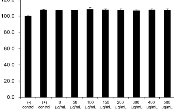
0.0 20.0 40.0 60.0 80.0 100.0 120.0
C e ll v ia b ili ty ( % o f u n tr e a te d c o n tr o l) .
(-) (+) 0 50 100 150 200 300 400 500 control control μg/mL μg/mL μg/mL μg/mL μg/mL μg/mL μg/mL μg/mL
Fig. 1. Effects of A. Hookeri root methanol extracts and LPS on the cell viability in RAW264.7 macrophage cells. The cells were pre-treated with the various concentrations of Allium hook- eri extracts (methanol extract from the root of A. hookeri) for 2 hr and then treated with LPS (2 μg/mL) for 20 hr. The concen- trations of A. hookeri extracts are 0, 50, 100, 150, 200, 300, 400 and 500 μg/mL. The rates of cell viability were measured by CCK-8 assay. The data shown are means±SD of three in- dependent experiments.
하였다.
재료 및 방법
시료
본 실험에 사용한 삼채는 경남 고성에서 구입하여 뿌리부 분을 동결 건조하여 분쇄한 후 분말은 메탄올과 1:9 비율로 취하여 상온에서 24시간씩 3회 추출하였다. 추출액은 회전 식 진공농축기(EYELA, Tokyo, Japan)를 이용하여 농축한 후 세포실험에 사용하였다. 농축된 샘플은 DMSO에 녹여서 각 농도별 세포에 처리하였다.
세포 배양
실험에 사용된 마우스 대식 세포 RAW264.7은 한국세포 주은행(Seoul)에서 구입하였다. 세포는 습윤한 5% CO
2, 37
oC 배양기(SANYO, San Diego, CA, USA)에서 배양하였 으며 배양액은 10% fetal bovine serum(FBS), 1% pen- icillin-streptomycin을 함유한 DMEM 배지를 사용하였다.
FBS, penicillin-streptomycin 및 배지는 Gibco(Invitrogen, Grand Island, NY, USA)에서 구입하였다. 배지는 2~3일마 다 교환하였으며 세포가 80% 이상 자랐을 때 phosphate buffered saline solution(PBS)로 세척한 후 cell scraper를 사 용하여 계대배양 하였다.
세포독성 측정
삼채 뿌리 메탄올 추출물의 농도에 따른 RAW264.7 세포 의 생존율을 측정하기 위해 cell viability assay를 실시하였 다. 100 μL(10,000 cells/well)의 세포 부유액을 96 well plate 에 분주 후, CO
2배양기 안에서 4시간 동안 전 배양(pre-in- cubation)을 한 후, 0(0.1% DMSO), 50, 100, 150, 200, 300, 400, 500 μg/mL 농도의 삼채
뿌리메탄올 추출액, 2 μg/mL 농도의 LPS를 10% FBS가 함유된 DMEM 배지와 함께 20 시간 배양했다. 배양 후 각 well에 10 μL의 CCK-8 용액 (Dojindo, Tokyo, Japan)을 첨가했다. 2시간 동안 CO
2배양 기 안에서 반응을 시킨 뒤 microplate reader를 사용하여 450 nm 파장에서 흡광도를 측정했다.
Nitric oxide 측정
삼채 뿌리 메탄올 추출물이 LPS가 유도된 RAW264.7 세 포에서 농도별 항염증 효과를 측정하기 위해 삼채
뿌리메탄 올 추출물을 처리 후, NO assay를 시행하였다. 1 mL(500,000 cells/ well)의 세포 부유액을 24 well plate에 분주 후, CO
2배양기 안에서 4시간 동안 전 배양(pre-incubation)을 한 후, 0(0.1% DMSO), 50, 100, 150, 200, 300, 400, 500 μg/mL 농도 의 삼채
뿌리메탄올 추출물과 positive control인 allyl di- sulfide(ADS) 1 ng/mL를 처리한 후, 2 μg/mL 농도의 LPS 를 10% FBS가 함유된 DMEM 배지(without phenol red)와 함께 20시간 반응했다. 20시간 후, 상층액 50 μL를 회수하여 Griees reagent(Promega, Madison, WI, USA)를 이용한
NO assay를 수행하였다.
Cytokine 측정
NO 측정과 같은 방법으로 세포를 배양한 후, 각 well에서 상층액을 회수하였다. 상층액 내 TNF-α와 IL-6의 양은 각 각 ENZO(ENZO bioscience, Farmingdale, NY, USA)에서 구입한 Mouse IL-6 ELISA kit와 Mouse TNF-α ELISA kit를 사용하여 제시된 방법에 따라 처리한 다음, ELISA reader로 흡광도를 측정한 후 standard curve를 바탕으로 TNF-α와 IL-6의 생성량을 계산하였다.
통계학적 검정
각 실험은 3회 이상 반복 실험을 통하여 결과를 얻어 각각 의 시료 농도에 대해 평균±표준편차로 나타내었다. 각 시료 농도군에 대한 유의 검정은 대조군과 비교하여 Student's t-test 한 후, p<0.05 값을 통계적으로 유의성 있는 결과로 간주하였다.
결과 및 고찰
삼채
뿌리메탄올 추출물의 세포 독성효과
삼채
뿌리메탄올 추출물이 마우스 대식세포 RAW264.7 에 미치는 영향을 측정하기 위하여 세포독성 실험을 하였다.
다양한 농도의 삼채
뿌리메탄올 추출물 0, 50, 150, 100, 200, 300, 400, 500 μg/mL를 처리한 후, 세포 생존률을 CCK-8 kit로 측정하였을 때, 각 농도별 대조군과 비교하여 모든 농 도에서 100% 이상의 생존률을 보여 어떠한 독성 효과도 없 는 것으로 확인되었다(Fig. 1).
삼채
뿌리메탄올 추출물의 NO 생성 억제 효과
삼채
뿌리메탄올 추출물을 이용한 NO assay는 Griess's
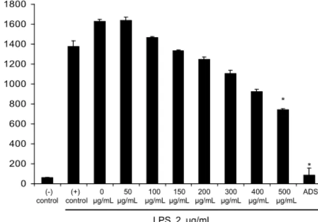
삼채 뿌리 메탄올 추출물이 LPS가 유도된 RAW264.7 세포에 대한 항염증 효과 1647
* *
*
*
0 20 40 60 80 100 120
N O p ro d u c tio n ( % o f L P S s tim u la te d c o n tr o l) .
(-) (+) 0 50 100 150 200 300 400 500 ADS control control μg/mL μg/mL μg/mL μg/mL μg/mL μg/mL μg/mL μg/mL
LPS 2 μg/mL
Fig. 2. Effects of A. Hookeri root methanol extracts on LPS-induced NO generation in RAW264.7 macrophage cells.
The cells were pre-treated with the various concentrations of A.
Hookeri extracts (methanol extract from the A. hookeri root) for 2 hr and then treated with LPS (2 μg/mL) for 20 hr. The amounts of NO were determined by Griess assay. The data shown are means±SD of three independent experiments. Allyl disulfide (ADS) was used as a posive control (1 ng/mL).
*p<0.05 signifi- cantly different from the value in cells treated with LPS in the absence of A. Hookeri extracts, respectively.
*
*
0 200 400 600 800 1000 1200 1400 1600 1800
T N F -α c o n c . (p g /m L ) .
(-) (+) 0 50 100 150 200 300 400 500 ADS control control μg/mL μg/mL μg/mL μg/mL μg/mL μg/mL μg/mL μg/mL
LPS 2 μg/mL
Fig. 3. Effects of A. Hookeri root methanol extracts on TNF- α generation in LPS-stimulated RAW264.7 macrophage cells.
The cells were pre-treated with the various concentrations of A.
Hookeri root methanol extracts for 2 hr and then treated with LPS (2 μg/mL) for 20 hr. The amounts of TNF-α was determined according to the manufactural's instruction of mouse TNF-α kit.
The data shown are means±SD of three independent experiments.
Allyl disulfide (ADS) was used as a posive control (1 ng/mL).
*
p<0.05 is significantly different from the value in cells treated with LPS in the absence of A. Hookeri extracts.
*
*
*
*
*
0 200 400 600 800 1000 1200 1400 1600 1800
IL -6 c o n c . (p g /m L ) .
(-) (+) 0 50 100 150 200 300 400 500 ADS control control μg/mL μg/mL μg/mL μg/mL μg/mL μg/mL μg/mL μg/mL
LPS 2 μg/mL
Fig. 4. Effects of A. Hookeri root methanol extracts on IL-6 generation in LPS-stimulated RAW264.7 macrophage cells.
The cells were pre-treated with the various concentrations of A.
Hookeri root methanol extracts for 2 hr and then treated with LPS (2 μg/mL) for 20 hr. The amounts of IL-6 was determined according to the manufactural's instruction of Mouse IL-6 kit.
The data shown are means±SD of three independent experiments.
Allyl disulfide (ADS) was used as a posive control (1 ng/mL).
*
p<0.05 is significantly different from the value in cells treated with LPS in the absence of A. Hookeri extracts.
method(11)를 이용하여 측정하였다. LPS의 처리에 의한 RAW 264.7 세포에서 NO 생성의 증가는 삼채
뿌리메탄올 추출물 처리군에서 농도의존적으로 감소되는 것으로 확인 되었다(Fig. 2). 특히 삼채
뿌리메탄올 추출물 300 μg/mL 처리군에서는 LPS만을 처리한 군과 비교했을 때, 50% 수준 까지 NO 생성이 감소되는 것으로 확인되었으며, 특히 500 μ g/mL 처리군에서는 거의 대조군 수준까지 감소되는 것으 로 확인되었다.
NO는 혈액응고, 혈압 및 신경전달 기능의 조절 등 생리적 역할을 하지만 고농도의 NO 생성은 peroxynitrite, nitrogen dioxide와 같은 유해물질을 생성하여 암의 형성과 진행에 중요한 역할을 하고 세포내 유해한 산화물질의 축적, DNA 손상을 일으키며 mitochondria에 감지되어 cytochrome C, apoptosis inducing factor를 방출하게 하여 세포자연사를 초래하는 것으로 알려져 있다(12,13).
황 함유 채소는 우리나라 전통 음식인 김치의 양념채소로 쓰이고 있을 뿐 아니라 황 함유 화합물은 생체의 대사 과정 중에 산화, 환원에 깊이 관여하여 radical에 의한 세포 손상 을 막을 수 있다(4). 마늘과 양파 추출물을 대사증후군을 가 진 동물모델에 투여하였을 때, 산화 스트레스와 내피세포 염증인자의 발현을 감소시키고 eNOS 활성을 증가시켜 항 산화 및 항염증효과를 나타낸다고 보고하였다(5). 마늘의 주 요 성분인
S-allylcysteine은 세포 내 glutathione의 고갈 방 지와 peroxide 제거에 의해 산화된 LDL로부터 동맥내피세 포를 보호하고 TNF-α와 hydrogen peroxide에 의해 활성화 된 NF-κB를 억제하는 것으로 보고되었다(14). 또한 최근에 는 양파에 존재하는 주요 flavonoid인 quercetin의 항염증효 과도 보고되었다(15,16).
삼채
뿌리메탄올 추출물의 cytokine 생성 억제 효과
다음은 RAW264.7 세포에서 LPS에 의해 유도된 대표적
인 염증성 cytokine인 TNF-α와 IL-6의 생성양에 미치는
삼채
뿌리메탄올 추출물의 영향을 조사하였다. LPS만을
처리한 군에서 TNF-α는 1,378 pg/mL로 높은 증가를 보였
으나, 삼채
뿌리메탄올 추출물 0, 50, 150, 100, 200, 300,
400, 500 μg/mL 처리군에서는 농도의존적으로 TNF-α의 생
성을 억제하는 것으로 확인되었다(Fig. 3). IL-6의 경우는
LPS만을 처리한 군에서 1,442 pg/mL로 높은 증가를 보였으
1648 김창현․이미애․김태운․장자영․김현주
나, 삼채
뿌리메탄올 추출물 150 μg/mL 처리군에서 1,259 pg/mL로 13% 감소되는 것을 확인하였다. 특히 300, 400, 500 μg/mL 처리군에서 각각 1,023 pg/mL, 886 pg/mL, 721 pg/mL로 30%, 39%, 50% 감소효과를 보였다(Fig 4.).
혈관관련 염증 반응은 염증 세포들(neutrophils, lympho- cytes, monocytes, macrophages), 내피세포, 혈관 평활근 세 포와의 복잡한 상호 작용과 관련되고, 대식세포, T-세포, 단 핵구, 혈소판, 내피세포에 의해 생성된 cytokine은 염증성 혈관 질환의 발병에 중요한 역할을 한다(17). 마늘 추출물은 동맥내피세포에서 ROS 생성, VCAM-1 발현, NF-κB 활성 화를 억제시켜 염증성 관련질환의 예방에 효과적인 것으로 보고되었다(18,19). 또한 LPS로 유도된 BV2 세포에서 다양 한 파 추출물은 염증성 cytokine 발현 및 생성, iNOS, COX- 2의 발현을 조절함으로써 염증반응을 효과적으로 억제하는 것으로 보고되었다(20).
요 약
본 연구에서는 삼채 뿌리 메탄올 추출물의 항염증 효과를 확인하기 위하여 마우스 대식세포인 RAW264.7 세포에 LPS를 처리한 결과 세포 생존율은 다양한 농도의 삼채
뿌리메탄올 추출물에서 세포독성이 없는 것으로 나타났다. 삼채 뿌리 메탄올 추출물은 다양한 농도에서 LPS만을 처리한 대 조군과 비교하였을 때, NO 생성을 농도 의존적으로 감소시 키는 것으로 확인되었다. 또한 염증성 사이토카인인 TNF-α 와 IL-6의 생성양 역시 LPS만을 처리한 군과 비교했을 때, 각각의 농도에서 농도의존적으로 감소하는 경향을 보였다.
본 연구에 사용된 삼채 뿌리 메탄올 추출물의 염증 억제 기 작에 관한 추가적인 연구의 수행과 활성물질의 동정에 관한 연구가 추가로 이루어져야겠지만, 본 연구의 결과는 삼채
뿌리메탄올 추출물은 NO 및 염증성 사이토카인의 생성을 조절함으로써 대식세포 유래의 염증 반응을 효과적으로 억 제하고, 염증성 매개질환에 탁월한 효능이 있을 것으로 사료 되며, 예방물질로서 활용될 수 있을 것으로 기대된다.
감사의 글
본 논문은 세계김치연구소 주요사업과제(KE1201-3)의 지원에 의하여 이루어진 결과의 일부입니다.
문 헌