Vol.31(2): 97~104 (2004)
한우 myostatin 유전자의 SNP 및 발현분석
유성란 ․ 정기철 ․ 상병찬 ․ 이준헌*
SNP and Expression Analyses of Myostatin Gene in Korean Cattle (Hanwoo)
Yu, Seong-Lan ․ Jung, Kie-Chul ․ Sang, Byung-Chan ․ Lee, Jun-Heon
ABSTRACT
Myostatin is a transforming growth and differentiation factor-β family member that acts as a negative regulator of muscle growth. Previously, mutations in the myostatin gene were known to be related to double muscling phenotypes in cattle. Because myostatin gene is highly related to muscle mass, also meat quality, in cattle, we sequenced whole myostatin mRNA and investigated the SNPs (Single Nucleotide Polymorphisms) in Korean cattle (Hanwoo). The results indicated that Hanwoo had an SNP in nt2385 and this mutation can be a useful marker with further verifications. We also investigated expression patterns of the myostatin gene from various muscle tissues and organs. Northern blotting results indicated that myostatin expression was restricted in muscles with variable expression levels. The results presented here can be used as a valuable information for meat quality related traits and muscle mass in cattle.
Keywords : Hanwoo, meat quality, myostatin, SNP
1
이 논문은 바이오그린21사업 지원에 의해 이루어진 연구결과이다.
충남대학교 농업생명과학대학 동물자원과학부 (Division of Animal Science and Resources, College of Agriculture and Life Sciences, Chungnam National University, Daejeon 305-764, Korea)
*교신저자 : 이준헌(E-mail : [email protected], Tel : 042-821-5779)
I. 서 론
Myostatin은 15kDa의 단백질로서 활성을 가진 376개의 amino acid propeptide로 구성되어 있으 며 주로 골격근에서 합성된다. 이 단백질은 배아 의 발달 및 조절과 성체의 조직 항상성 유지에 중 요한 역할을 하는 성장․분화와 관련된 TGF-β superfamily에 속하며 Growth/Differentiation factor 8(GDF8)이라고 불려지기도 한다(McPherron 등, 1997). 이 유전자는 생쥐와 소에서 배아 초기 형 성단계 동안 배아의 근판(myotome)에서 주로 발현되고(McPherron 등, 1997), 성체가 된 후에 는 골격근에서 계속 발현하는데, 이 발현은 근육 의 결합조직에 존재하는 것이 아니라 근섬유의 세포질에서 발현된다고 알려져 있다(Krik 등, 2000).
Gonzalez-Cadavid 등(1998)이 밝힌 Myostatin 유전자의 구조는 사람에서 3개의 exon으로 구성 되고 있으며, 3개의 전사개시 부위가 있는 것으로 추정이 되고 있다. 소에서도 사람과 마찬가지로 이 유전자는 3개의 exon으로 구성되며 염색체상의 위치는 2q14-q15으로 알려져 있다(Masabanda 등, 1998). 그 후 Ferrell 등(1999)이 인간의 myostatin 유전자 염기서열을 밝혀냈으며, 5개의 mutation site(A55T, K153R, E164K, P198A, 1225T)를 찾 아냈다. 특히 이 유전자의 돌연변이는 정상의 개 체보다 2배 이상 근육이 발달하는 muscular hypertrophy증상을 보여서 많은 연구자들이 이 유 전자를 연구하는 계기가 되었다(Grobet 등, 1997).
그 후 이 유전자의 3번째 exon에서 염기 G가 T로 변환되는 것은 double-muscle과 관련이 있다는 보 고가 있었다(Marchitelli 등, 2003). 또한 Myostatin knock-out mice에서는 대조구보다 2배 이상 근 육이 발달되는 특징과 근육내 지방함량이 적은
표현형을 보였는데 이는 myostatin 유전자가 골격 근의 발달에 있어 negative regulator 기능을 하고 (McPherron 등, 1997), 근육내의 preadipocyte differentiation을 억제하기 때문에 나타나는 표현 형이라고 추정하였다(Kim 등, 2001). 즉 myostatin 유전자는 근육형성단계(myogenesis)와 지방형성 단계(adipogenesis)를 조절하는 switch 역할을 한 다고 알려져 왔다.
최근까지의 연구 결과를 기초하여 볼 때, 근육의 성장 조절에 대한 기전은 아직까지 확실히 밝혀지 지 않았지만 myostatin 유전자의 발현을 억제하는 follistatin과 activin IIB receptor 단백질들이 발견 되었으며 이들 유전자들이 생쥐에서 myostatin 유 전자를 knock-out 했을 때와 같은 double-muscling phenotype을 보였다(Lee and McPherron, 2001).
이와 같이 myostatin 유전자는 근육의 발달을 조 절하는데 중요한 역할을 하고 있으며 본 연구는 한국 재래종인 한우에서 myostatin의 돌연변이와 발현양상을 알아냄으로서 한우의 성장을 증진시 키는 기초 자료를 얻기 위하여 본 연구를 수행하 였다.
Ⅱ. 재료 및 방법
2.1 공시재료
이 연구에서 SNP 분석을 위해 사용된 공시재
료는 축산연구소에서 사육된 근연관계가 먼 한우
3개체가 이용이 되었으며 발현분석에 이용된 공
시재료는 축산연구소에서 도축된 12개월령 한우
의 9개 근육 부위(안심, 갈비, 등심, 목심, 채끝,
우둔, 설도, 사태, 양지) 및 7개의 장기 조직(좌
심방, 우심방, 간, 비장, 신장, 우심실, 좌심실)을
이용하였다. RT-PCR 및 Northern blotting을 위
한 시료는 mRNA의 degradation을 막기 위하여 도축 후 30분 이내에 모든 조직이 채취되었으며, 채취된 조직은 바로 액체 질소에 넣어 실험실로 옮겨졌다.
2.2 Total RNA 추출
한우의 각 조직 1.5g을 15 ㎖의 Tri Reagent (Molecular Reseach Center, INC.) 용액으로 분쇄 한 다음 chloroform 7.5 ㎖을 첨가하여 혼합한 후 4℃에서 12,000 rpm으로 원심분리하여 RNA층을 얻었다. RNA층을 새로운 튜브에 옮긴 후 0.7 volume의 isopropanol로 RNA를 완전히 침전시킨 다음 75% ethanol-DEPC (diethylpyrocarbonate) 로 씻어 낸 후 DEPC처리된 RNase-free H
2O에 RNA를 용해시켰다. 추출된 RNA의 농도는 Spectrophotometer (Ultrospec2000, Pharmacia, U.S.A.)를 이용하여 260 nm 파장에서 측정한 후 -70℃에 저장하여 실험에 사용하였다.
2.3 PCR, RT-PCR 및 Cloning
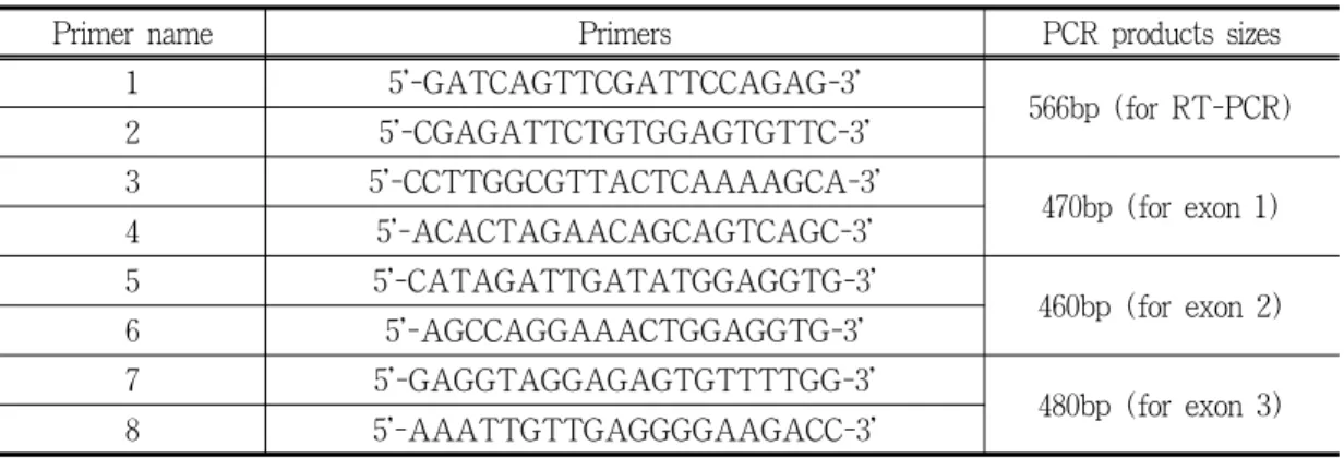
소에서 이미 밝혀진 myostatin 유전자의 염기 서열(GenBank accession number: AF019620)을 기초를 하여 각 exon을 모두 증폭하여 sequence
Table 1. Primers used for amplifying bovine myostatin gene in this study
Primer name Primers PCR products sizes
1 5'-GATCAGTTCGATTCCAGAG-3'
566bp (for RT-PCR)
2 5'-CGAGATTCTGTGGAGTGTTC-3'
3 5'-CCTTGGCGTTACTCAAAAGCA-3'
470bp (for exon 1)
4 5'-ACACTAGAACAGCAGTCAGC-3'
5 5'-CATAGATTGATATGGAGGTG-3'
460bp (for exon 2)
6 5'-AGCCAGGAAACTGGAGGTG-3'
7 5'-GAGGTAGGAGAGTGTTTTGG-3'
480bp (for exon 3)
8 5'-AAATTGTTGAGGGGAAGACC-3'
분석을 할 수 있는 primer를 표 1과 같이 제작을 하였고, PCR의 반응조건은 94℃에서 5분으로 1 회 실시한 후, 94℃에서 30초, 58℃에서 30초, 7 2℃에서 30초씩 총 35회를 증폭시켰고, 마지막으 로 72℃에서 10분간 더 반응시켰다. PCR이 끝난 후 5㎕의 증폭산물을 1.2%의 agarose gel에 전기 영동하여 증폭여부 및 크기를 확인하였다. 이 증폭 산물들은 pGEM-T easy vector(Promega, U.S.A.) 에 ligation시켜 cloning한 후 제한 효소인 EcoRI 의 처리로 insert의 유무 및 정확한 insert size를 확인하였다.
Reverse transcription (RT)은 total RNA 1㎍
을 이용하여 실시하였다. RNA 1㎍에 1×enzyme buffer, 50ng oligo dT, 0.25 mM dNTPs, 1 Unit reverse transcriptase(Promega, U.S.A.), 0.5 Unit Recombinant RNasin ribonuclease inhibitor (Promega, U.S.A.) 조성으로 42℃에서 1시간 반 응한 결과 생성된 1st cDNA를 이용하여 PTC- 200 (MJ Research, U.S.A.)에서 PCR을 실시하 여 각 조직의 myostatin 유전자를 증폭하였다.
PCR 혼합액에는 1st cDNA, 0.2 mM dNTPs,
primer(10 pmol), 2.5 Unit Taq polymerase(Bioneer,
KOREA), 1×buffer조성으로 증폭시켰다.
2.4 myostatin 유전자의 염기서열 결정 및 분석 각 clone들의 염기서열을 결정하기 위하여 재조 합된 Plasmid DNA 200ng을 Bigdye terminator (PE Applied Biosystems, U.S.A)를 사용하여 cycle sequencing reaction을 실시하였다. PCR 반 응은 PE 9600(PE Applied Biosystems, USA)을 이용하여 96℃에서 1초, 50℃에서 5초, 60℃에서 4분간의 조건으로 25회 시켰고, 반응이 끝난 PCR 산물은 ethanol로 정제한 후, ABI 377 자동염기 서열 분석장치(PE Applied Biosystems, U.S.A) 를 사용하여 염기서열을 결정하고 분석하였다.
2.5 Northern blot analysis
각 조직에서 추출한 total RNA 40㎍을 3.3 volume의 sample buffer(1.3×MOPS [3-(N- morpholino)-propane sulfonic acid] running buffer, 8.1% formaldehyde, 65% formamide)에 넣고 65℃에서 5분간 반응시켜 denaturation후, 0.7 volume의 loading dye(50% glycerol, 0.4%
Bromophenol Blue, 0.1% Xylene Cyanol)와 섞어 formaldehyde-agarose gel(1% agarose, 1×MOPS, 6.2% formaldehyde)에서 80 volt로 3시간동안 전 기영동한 후 ethidium bromide로 염색하였다. 추 출된 mRNA의 상태는 18s, 28s, ribosomal RNA 의 순도를 보고 결정을 하였다.
이 gel로부터 RNA를 20× SSC를 이용하여 Nylon-membrane에 삼투압작용으로 20시간 transfer 한 다음 U. V. crosslinker(Upland, U.S.A.)로 RNA 를 membrane에 완전히 고정시켰다. Membrane 은 42℃에서 pre-hybridiztion solution(5×SSC, 5×
Denhart's solution, 100㎍/㎖ Herring sperm DNA, 50% formamide)에 넣고 1시간 동안 반응시킨 후, Redi Prome II kit(Amersharm, U.S.A)를 이 용해 실온에서 1시간동안 [α-
32P]dCTP 방사성
동위원소로 labelling한 myostatin cDNA중 566 bp 를 probe로 이용하여 22시간 정도 hybridization을 실시하였다. Hybridization한 것을 2×SSC, 0.1%
SDS용액으로 한번 세정후, 같은 용액으로 42℃
에서 10분간 세정후에 1×SSC, 0.1% SDS용액으 로 42℃에서 10분간 세정하여 원하는 정도의 signal이 나타내면 X-ray film을 넣고 -70℃에서 3일간 감광시킨 후 현상하였다.
Ⅲ. 결과 및 고찰
3.1 myostatin 유전자의 mRNA 염기서열 및 SNP 분석
Myostatin 유전자는 사람의 경우 mRNA가 2823bp의 길이를 가지고 3개의 exon으로 구성되 어 있는 것으로 보고되었으나(Gonzalez-Cadavid 등, 1998), 소의 myostatin 유전자의 mRNA는 1128bp만이 밝혀져 있었다(McPherron 등, 1997).
이 mRNA sequence를 기초로 하여 제작한 primer 들은 한우에서 각각의 exon을 성공적으로 증폭하 였으며 증폭된 PCR product로부터 염기서열이 분 석되었다. 이미 밝혀진 다른 종과의 mRNA 염기 서열을 비교한 결과 사람, 돼지, 생쥐 및 소의 4 개 종간에 높은 상동성을 확인 할 수 있었으며 (Tabel 2), 특히 돼지와의 상동성이 94%로 가장 높음을 알 수 있었다.
Table 2. Sequence homology table of the myostatin mRNAs among species
(unit: %)
Species Pig Human mouse
Hanwoo 94 91 89
Pig - 91 89
Human - - 89
이 연구를 통하여 밝혀진 한우의 Myostatin 유 전자의 sequence는 다른 품종에서 이미 알려진 SNPs와 비교를 하였으며 혈연관계가 없는 한우 세 마리의 개체를 모두 sequencing한 결과 exon 2 의 nt2385 부위에서 C/T SNP를 확인할 수 있었 다(Table 3). 이 부위는 기존에 밝혀진 SNP와 같 은 것으로서 한우에도 이 SNP가 존재하는 것이
Table 3. Comparison of Myostatin nucleotide and amino acids sequences in Korean cattle (Hanwoo) with known mutations showing double muscling phenotype in other breeds. Nucleotide positions are based on the sequence for the GenBank accession number AB076403.
Exons Mutation sites Animals Sequences
1Exon 1 nt415
Korean Cattle ATTGATCAGTTCGATGTC
I D Q F D V
Known Mutation ATTGATCAGTTAGATGTC
I D Q L D V
Exon 2
nt2385
Korean Cattle (SNP exists)
CCCAAATGTTGCTTCTTT P K C C F F
Known Mutation CCCAAATGTTGTTTCTTT
P K C C F F
nt2390
Korean Cattle TGCTTCTTTAAATTTAGC
C F F K F S Known Mutation TGCTTCT---TAGC
C F *
nt2581
Korean Cattle GGTATTTGGCAGAGCATT
G I W Q S I
Known Mutation GGTATTTGGTAGAGCATT
G I W *
nt2647
Korean Cattle TTAGGCATTGAAATCAAA
L G I E I K
Known Mutation TTAGGCATTTAAATCAAA
L G I *
Exon 3
nt4822
Korean Cattle TGTGATGAACACTCCACA
C D E H S T Known Mutation TGTG---ACA
C R
nt4942
Korean Cattle TCTGGAGAATGTGAATTT
S G E C E F
Known Mutation TCTGGAGAATATGAATTT
S G E Y E F
1