저작자표시-비영리-변경금지 2.0 대한민국 이용자는 아래의 조건을 따르는 경우에 한하여 자유롭게 l 이 저작물을 복제, 배포, 전송, 전시, 공연 및 방송할 수 있습니다. 다음과 같은 조건을 따라야 합니다: l 귀하는, 이 저작물의 재이용이나 배포의 경우, 이 저작물에 적용된 이용허락조건 을 명확하게 나타내어야 합니다. l 저작권자로부터 별도의 허가를 받으면 이러한 조건들은 적용되지 않습니다. 저작권법에 따른 이용자의 권리는 위의 내용에 의하여 영향을 받지 않습니다. 이것은 이용허락규약(Legal Code)을 이해하기 쉽게 요약한 것입니다. Disclaimer 저작자표시. 귀하는 원저작자를 표시하여야 합니다. 비영리. 귀하는 이 저작물을 영리 목적으로 이용할 수 없습니다. 변경금지. 귀하는 이 저작물을 개작, 변형 또는 가공할 수 없습니다.
A Thesis
For the Degree of Master of Science
Effect of castration on leukocyte cytokine expression
and indicators of stress and inflammation in Korean
cattle bull calves
거세가 한우 수송아지의 백혈구 사이토카인
발현과 스트레스와 염증 지표에 미치는 영향
August 2019
By
Seon Pil Yoo
College of Agriculture and Life Sciences
Seoul National University
Effect of castration on leukocyte cytokine expression
and indicators of stress and inflammation in Korean
cattle bull calves
거세가 한우 수송아지의 백혈구 사이토카인
발현과 스트레스와 염증 지표에 미치는 영향
지도교수
백 명 기
이 논문을 농학석사 학위논문으로 제출함
2019 년 8 월
서울대학교 대학원 농생명공학부
유 선 필
유선필의 농학석사 학위논문을 인준함
2019년 8 월
위 원 장 (인)
부위원장 (인)
위 원 (인)
i
This study will be published in elsewhere as a fulfillment of SP Yoo's Master program.
Abstract
This study investigated behavioral, physiological, and inflammatory responses, as well as leukocyte cytokine gene expression, of Korean cattle bull calves following surgical castration. Nineteen Korean cattle bull calves (average body weight, 254.5 kg; average age, 8.2 months) were assigned to one of two treatment groups: control (n = 9) and surgical castration (n = 10). Castration was performed surgically using Newberry knives and a Henderson castrating tool. Blood was collected immediately before castration and at 0.5 h, 6 h, 1 d, 3 d, 7 d, and 14 d after castration, and analyzed cortisol, substance P, and haptoglobin concentrations and leukocyte cytokine gene expression by quantitative real-time PCR. Behaviors were observed for 3 h, from 0.5 to 3.5 h after castration. Feed intake was recorded daily, and body weight was measured 1 d prior to the experiment and 14 d after castration. Castration decreased average daily gain (P < 0.05) and gain-to-feed ratio (P < 0.05). Castration reduced the time spent eating (P < 0.001) and the frequency of eating (P < 0.05) and increased (P < 0.001) the frequency of lying during the 3 h after castration. Castration temporarily increased circulating plasma cortisol (P < 0.001) and salivary cortisol concentrations (P = 0.03) at 0.5 h after castration. Plasma cortisol and salivary cortisol was positively correlated (r = 0.69, P <0.001). Castration temporarily increased (P < 0.05) plasma substance P concentration at 1 d after castration. Castration
ii
increased plasma haptoglobin concentration at 1 d and 3 d after castration. With regard to leukocytes, castration increased (P < 0.05) mRNA levels of interleukin-1-alpha (IL-1A), interleukin-1-beta (IL-1B), interleukin-1 receptor antagonist (IL-1RN), and interleukin-6 (IL-6) at 6 h after castration, and increased (p < 0.05) IL1RN, IL-1A, and IL-6 mRNA levels at 1 d after castration. mRNA level of IL-1A and IL-1B showed a positive correlation with IL-6 (r=0.96, P < 0.001; r = 0.55, P < 0.001). mRNA level of IL-1RN was highly related to the leukocyte mRNA levels of IL-6 and IL-1B (r=0.67, P < 0.001; r=0.83. P < 0.001). In conclusion, castration of Korean cattle bull calves temporarily induced stress, retarded growth, and affected behaviors and inflammatory cytokine gene expression.
Keyword: behavior, castration stress, cytokine gene expression, Korean cattle
iii
Table of Contents
Abstract ... ⅰ Table of Contents ... ⅲ List of tables ... ⅴ List of figures ... ⅶ List of abbreviations ... ⅷ Units and marks ... ⅹⅠ. Introduction ... 1 Ⅱ. Literature review ... 3 1. Animal welfare ... 3 1.1. Stress ... 4 1.2. Castration ... 4 2. Stress indicators ... 7 2.1. Cortisol ... 7 2.2. Substance P ... 8
2.3. Acute phase protein ... 11
2.4. Cytokines ... 12 2.4.1 IL-1β ... 14 2.4.2 IL-1α ... 14 2.4.3 IL-6 ... 15 2.4.4 IL-1ra ... 15 3. Literature cited ... 17
Ⅲ. Materials and methods ... 22
1. Ethical statement ... 22
2. Animals, treatments, and diets ... 22
3. Behavioral parameters ... 25
4. Physiological parameters ... 25
5. Leukocyte isolation ... 27
6. Real-time polymerase chain reaction ... 27
iv
Ⅳ. Results and discussion ... 31
1. Growth performance ... 31
2. Behavior observation ... 32
3. Saliva and plasma cortisol ... 36
4. Plasma substance P ... 41
5. Plasma haptoglobin and leukocyte cytokine gene expression ... 42
Ⅴ. Literature cited ... 51
Ⅵ. Summary in Korean ... 63
v
List of tables
Table 1. The Five Freedoms and Five Provisions for promoting farm animal welfare
Table 2. Acute-phase Proteins (Ceciliani et al., 2002)
Table 3. Ingredients and chemical composition of diets for Korean cattle calves
Table 4. Ethogram of behaviors recorded after castration modified from Marti et al. (2017)
Table 5. Primer sequences and amplicon information for qRT-PCR
Table 6. Effects of castration on growth performance in Korean cattle calves
Table 7. Effect of castration on behaviors from 0.5 h to 3.5 h after castration
Table 8. Effect of castration on saliva cortisol, plasma cortisol, plasma substance P, and plasma haptoglobin during the 14-d postcastration
vi expression during the 14-d postcastrattion
Table 10. Pearson correlation coeffecients and P-value for blood parameters, saliva cortisol and blood leukocyte cytokine transcripts in Korean cattle calves
vii
List of figures
Figure 1. Regulation of Cortisol Secretion (Gardner et al., 2007)
Figure 2. Schematic representation of the biosynthesis of substance P and related peptides
Figure 3. Effect of castration on plasma concentrations of cortisol, substance P, and haptoglobin and saliva cortisol concentrations in Korean cattle calves
Figure 4. Effect of castration on blood leukocyte cytokine gene expression in Korean cattle calves
viii
List of abbreviation
rRNA: ribosomal ribonucleic acid DNA: Deoxyribonucleic acid PCR: Polymerase chain reaction
EDTA: Ethylenediaminetetraacetic acid ELISA: Enzyme-linked immunosorbent assay PC: Plasma cortisol
SC: Saliva cortisol
ACTH: Adrenocorticotropic hormone UK: United Kingdom
AVMA: American Veterinary Medical Association GnRH: Gonadotropin-releasing hormone
CRH: Corticotrophin-releasing hormone NF-κB: Nuclear factor kappa B
NPγ: Neuropeptide γ NPK: Neuropeptide K APP: Acute phase protein HP: Haptoglobin
APR: Acute phase reaction SAA: Serum amyloid A CRP: C-reactive protein IL-1b: Interleukin-1β IL-1a: Interleukin-1α IL-6: Interleukin-6
ix IL‑1R1: IL‑1 receptor antagonist
IL‑1RAP: IL‑1 receptor accessory protein IRAK4: IL-1 receptor-associated kinase 4 JNK: c-Jun N-terminal kinase
ERK: Extracellular signal-regulated kinase MAPK: Mitogen-activated protein kinases ILCs: Innate lymphoid cells
SNUIACUC: Seoul National University Institutional Animal Care and Use Committees
TDN: Total digestible nutrients DM: Dry matter
CP: Crude protein ME: metabolic energy
x
Units and marks
% : Percent g : Gram μg : Microgram ng : Nanogram mg : Milligram pg : Picogram ml : Milliliter h : Hour d : day nm : Nanometer
1
Ⅰ. Introduction
Castration of male cattle is a typical practice in South Korea. The purpose of castration is to reduce aggressive behavior and to improve meat quality (Stafford and Mellor, 2005; Faulkner et al., 1992). The meat from castrated Korea cattle is gotten better quality grade than that from bulls due to superior tenderness and marbling (Park et al., 2018b). Castration improves marbling by altering gene expression associated with lipogenesis. (Baik et al., 2015; Park et al., 2018b).
Numerous studies have shown that castration induces pain, distress, and inflammatory responses indicated by physiological, neuroendocrine and behavioral changes (Coetzee, 2013). Surgical castration has been shown to increase plasma cortisol concentration (Sutherland et al., 2013; Stafford et al., 2002), salivary cortisol concentration (González et al., 2010), acute phase proteins (Earley and Crowe, 2002), and substance P concentrations (Coetzee et al., 2008), as well as the time spent performing abnormal behaviors (Molony et al., 1995, Ting et al, 2003). Surgical castration has been shown to increase plasma cortisol, substance P, and haptoglobin concentration in Korea cattle (Park et al., 2018a).
For measuring blood cortisol concentration, restraint and blood sampling procedure are conducted which may be stressors and may result in biased measurements (Redbo, 1993; Negrao et al., 2004). To reduce stress during
2
sampling, researchers suggested saliva cortisol as a better stress indicator than plasma cortisol because saliva sampling is noninvasive unlike blood sampling. (Negrao et al., 2004; Hernandez et al., 2014; Schwinn et al., 2016). These researchers investigated the relationship between blood cortisol and saliva cortisol by increasing both cortisol levels utilizing ACTH. Since castration results in complicated responses including pain, stress, and inflammatory responses, it is needed to investigate the relationship between saliva cortisol and plasma cortisol during and after castration. In my knowledgement,
Castration causes inflammatory responses. Inflammation caused by castration was indicated by acute pahse proteins which is induced by cytokines (Earley and Crowe, 2002; Warnock et al., 2012; Park et al., 2018a). Even though cytokines are key modulators of inflammation, there are few published research papers about the effect of castration on cytokine. Castration has been shown to alter peripheral leukocyte inflammatory cytokine transcripts (Pang et al., 2011). Burdizzo castration causes a higher pro-inflammatory cytokine gene expression than band castration in the testis and epididymis (Pang et al., 2009). However, the effect of knife castration on cytokine transcripts is largely unknown in Korea cattle.
Therefore, the aim of this study was to evaluate the effect of castration on growth performance, indicators of pain, distress and inflammation, behavior and inflammatory cytokine transcription in Korea cattle.
3
Ⅱ. Literature review
1. Animal welfare
According to Broom (1986), the definition of welfare is “The state of an individual as it attempts to cope with its environment”. UK Farm Animal Welfare Council stated Five Freedoms which outline five aspects of animal welfare (Table 1). For achieving Five Freedoms, Five Provisions gave practical advice (Table 1). Recently, the focus of animal welfare was changed to manage animal’s bodies, minds, and natures (Fraser, 2008). Stress management is important for improving animal welfare. There are artificial stressors such as castration, dehorning, or tail docking.
Table 1. The Five Freedoms and Five Provisions for promoting farm animal welfare (Farm animal welfare council. 2009)
Freedoms Provisions
1. Freedom from thirst, hunger, and malnutrition
By providing ready access to fresh water and a diet to maintain full health and
vigor 2. Freedom from discomfort
and exposure
By providing an appropriate environment including shelter and a
comfortable resting area 3. Freedom from pain, injury,
and disease
By prevention or rapid diagnosis and treatment
4. Freedom from fear and distress
By ensuring conditions and treatment which avoid mental suffering 5. Freedom to express normal
behavior
By providing sufficient space, proper facilities and company of the animal’s
4 1.1. Stress
Stress is an organism’s response to a stressor such as an environmental condition or physical change. The effects of stress can be both beneficial and harmful. The beneficial effects of stress involve preserving homeostasis of cells/species, which leads to continued survival. However, in many cases, the harmful effects of stress may receive more attention due to their role in various pathological conditions and diseases. Various factors, for example, hormones, neuroendocrine mediators, peptides, and neurotransmitters are involved in the body's response to stress.
1.2. Castration
Castration of male cattle is a common practice in Korea. Castration reduces aggressiveness (Kent et al., 1996; Stafford, 2007) and sexual activity by lowering testosterone levels, and modifies carcass characteristics by decreasing high muscle pH. Castration also increases beef quality (Park et al., 2018).
According to AVMA-AWD (2014), there are three castration methods: physical, chemical, and hormonal.
Physical Methods - Castration methods include surgical castration, band castration, and burdizzo clamp castration. Surgical castration process consists of excision of the scrotum, exposing of spermatic cords, and cutting the cord. Band castration eliminates blood supply to the scrotum and testis and causes subsequent necrosis. Burdizzo clamp castration is to crush the spermatic cord. In Korea, surgical castration is a common method of castration.
5
Chemical Methods - Chemical castration includes injection of toxic agents into the testicular parenchyma to cause irreversible damage and loss of function (Fordyce et al., 1989). Chemical castration requires additional procedural time and technical skill, and almost twice the healing time compared with surgical castration (Fordyce et al., 1989).
Hormonal Methods - Hormonal castration, also known as immunocastration, typically involves an injection of immunocontraceptives to induce antibody production against gonadotropin-releasing hormone (GnRH), resulting in decreased production of endogenous hormones. Immunocastration has been shown to increase live weight, hot carcass weight, average daily gain, and dressing percentage following castration when compared with surgical methods (Amatayakul-Chantler et al., 2013).
All physical methods of castration cause pain (Dinniss et al., 1997; Stafford, 2007). Cattle show pain responses during and after castration; these responses include struggling, kicking the hind legs, tail swishing, foot stamping, head turning, restlessness, reduced activity, abnormal standing posture, reduced interest in dams and each other and reduced grazing and feed intake (Fordyce et al., 1989; Fisher et al., 2001; Ting et al., 2003). Pain associated with the surgical and Burdizzo clamp methods is relatively immediate, whereas pain caused by ring/band placement is postponed due to interruption of the blood supply by the band/ring (Robertson et al., 1994; Thuer et al., 2007). Burdizzo castration also causes a more severe inflammatory response than band castration (Pang et al., 2009).
Cattle may determine reduced feed intake and average daily gain for a period of time after castration regardless of castration methods (Chase et al.,
6
1995; Fisher et al., 1997; Stafford et al., 2005). Surgically castrated 5.5-month-old calves showed reduced ADG for the first 7 days after castration (Ffisher et al., 1996). Surgically castrated 6- to 9-month-old calves reduced daily weight gain and feed intake (Faulkner et al., 1992). Castration age may have no effect on growth performance.
Cortisol concentrations have been studied as indicators of physiologic stress in animals. Regardless of the means of castration, cortisol concentrations are increased after castration; however, onset, magnitude, and duration may vary with the castration methods. Surgical castration and burdizzo castration increased plasma cortisol immediately after castration with higher level compared with band castration (Molony et al., 1995; Fell et al., 1986). According to Ting et al. (2005), 5.5-month-old calves showed higher cortisol concentration compared with 1.5-month-old and 4.5-month-old calves.
7
2. Stress indicators
2.1. Cortisol
Cortisol is a steroid hormone produced by the adrenal cortex. Its secretion is directly controlled by the release of adrenocorticotrophin (ACTH) from the anterior pituitary which is regulated by corticotrophin-releasing hormone (CRH) secreted by the hypothalamus (Gardner et al., 2007). Cortisol exerts negative feedback on both ACTH and CRH secretion, while stress acts directly on the hypothalamus to stimulate CRH release (Figure 1).
Cortisol has anti-inflammatory and immunosuppressive effects. Cortisol inhibits immunoglobulin synthesis, induces lymphocyte apoptosis and prevents inflammatory cytokine production through inhibition of nuclear factor kappa B (Dong et al., 2018). Monocyte differentiation into macrophages and are inhibited by cortisol, and local inflammation is also suppressed by reduced histamine production and impaired prostaglandin synthesis.
8 2.2. Substance P
Substance P was discovered by v. Euler and Gaddum (1931). Chang et al. (1971) identified the amino acid structure of substance P. The structure of substance P is as follows.
SP: Arg-Pro-Lys-Pro-Gln-Gln-Phe-Phe-Gly-Leu-Met-NH2
Mammalian substance P derives from the pre-protachykinin-A (PPT-A)
Figure 1. Regulation of Cortisol Secretion (Gardner et al., 2007). ACTH =
9
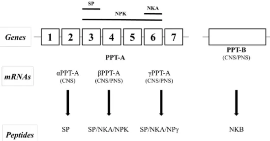
gene. The PPT-A gene also encodes for other tachykinins, including neurokinin A, neuropeptide K (NPK) and neuropeptide γ (NPγ) (Carter and Krause, 1990). Post-transcriptional modification of the PPT-A gene transcript results in three mRNAs, which are αPPT-A, βPPT-A, and γPPT-A (Carter and Krause, 1990). All three PPT-A mRNAs encode for the substance P precursor sequence, while the βPPT-A and γPPT-A are encoding for the NKA. NPK precursor sequence is only present on βPPT-A, and NPγ precursor sequencec is only present for γPPT-A (Nakanishi, 1987). PPT-B gene also originates from the same common ancestral gene as PPT-A. However, the PPT-B gene encodes for Neuropeptide B only. (Nakanishi, 1987) (Figure. 2).
Substance P is a neuropeptide in the central nervous system associated with stress regulation, anxiety-related behaviors, and pain (Ebner and Singewald, 2006). Plasma concentrations of Substance P have been used to evaluate muscle pain, soft tissue injury of human and castration pain of calves (Onuoha and Alpar, 1999; Coetzee et al., 2008; Park et al., 2018; Marti et al., 2017).
10
Figure 2 Schematic representation of the biosynthesis of substance P and related peptides (Harrison and Geppetti, 2001). PPT-A = pre-protachykinin-A. PPT-B = pre-protachykinin-B. αPPT-A = α pre-protachykinin-pre-protachykinin-A. βPPT-A = β pre-protachykinin-βPPT-A. γPPT-βPPT-A = γ pre-protachykinin-βPPT-A. CNS = central nervous system. PNS = peripheral nervous system. NKA = neurokinin A. NPK = neuropeptide K. NPr = neuropeptide γ. NKB = neuropeptide K. SP = substance P.
11 2.3. Acute phase protein
Acute phase reaction is to counteract infection and injuries in order to recover the homeostasis as soon as possible. All the up-regulated proteins have been called “positive APP” and All the down-regulated proteins have been called “negative APP”. APP also are classified by function (Table 2).
Haptoglobin is synthesized by hepatocytes in response to inflammatory processes, inducing anti-inflammatory processes (Murate et al., 2004). Haptoglobin participates in defense mechanisms against acute changes such as infection, stress, damage, or injury (Jain et al., 2011). It has been suggested that haptoglobin can be used as an accurate indicator of inflammatory conditions in cattle (Horadagoda et al., 1999).
Several studies have been observed the increase of haptoglobin in cattle after castration. Park et al. (2018) observed that haptoglobin increased one day and 3 days after knife castration. Ting et al. (2003) reported an increase in haptoglobin concentration on the d1 and d3 after burdizzo castration in 13-mo-old calves. Ting et al. (2004) saw an increase in haptoglobin concentration on the third day after burdizzo castration in 2.5- to 3.5-mo-old calves. Brown et al. (2015) observed an increase in haptoglobin concentration for 72h after knife castration.
12
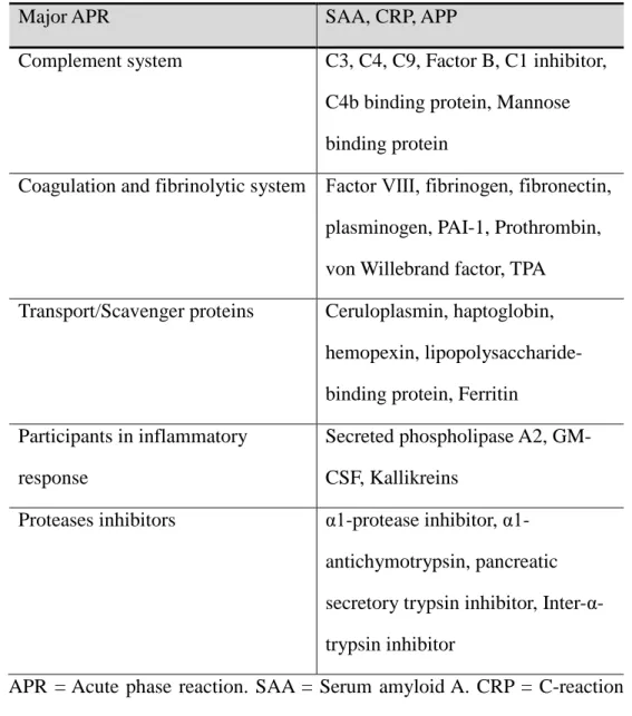
Table 2. Acute-phase Proteins (Ceciliani et al., 2002)
Major APR SAA, CRP, APP
Complement system C3, C4, C9, Factor B, C1 inhibitor, C4b binding protein, Mannose binding protein
Coagulation and fibrinolytic system Factor Ⅷ, fibrinogen, fibronectin, plasminogen, PAI-1, Prothrombin, von Willebrand factor, TPA Transport/Scavenger proteins Ceruloplasmin, haptoglobin,
hemopexin, lipopolysaccharide-binding protein, Ferritin
Participants in inflammatory response
Secreted phospholipase A2, GM-CSF, Kallikreins
Proteases inhibitors protease inhibitor, α1-antichymotrypsin, pancreatic secretory trypsin inhibitor, Inter- α-trypsin inhibitor
APR = Acute phase reaction. SAA = Serum amyloid A. CRP = C-reaction protein. APP = Acute phase protein. PAI-1 = Plasminogen activator inhibitor-1. TPA = Tissue plasminogen activator. GM-CSF = Granulocyte-macrophage colony-stimulating factor.
2.4. Cytokine
Cytokines are abroad category of small secreted protein released by cells and have a specific effect on the interactions and communications between
13
cells. Cytokines released by inflammatory leukocytes induce hepatocytes to synthesize and release acute phase proteins (Cheville, 1999). It is common for different cell types to secrete the same cytokine or for a single cytokine to act on several different cell types. Cytokines are redundant in their activity, meaning similar functions can be stimulated its target cells to make additional cytokines. They are often produced in a cascade, as one cytokine stimulates its target cells to make additional cytokines. Cytokines can also act synergistically or antagonistically.
Acute stress affects pro-inflammatory and anti-inflammatory cytokines. Previous studies have shown that glucocorticoids suppress the production of pro-inflammatory cytokines, such as TNF-α, IFNγ, and IL-2, in vitro and in vivo in mice and human (Beutler et al., 1986; Boumpas et al., 1993). Glucocorticoids, however, stimulate the production of anti-inflammatory cytokines, such as IL-10, and IL-4 in sclerosis patients and rat CD4++ T cells (Gayo et al., 1998; Ramierz et al., 1996).
There are few literatures about cytokine change under castration stress. Burdizzo castrated calves had higher IL-6 mRNA level than non-castrated calves at both 12 h and 24 h post-castration in the epididymis, whereas IL-1 mRNA level was not different (Pang et al., 2009). Leukocyte IL-6 mRNA level was not different between band castrated calves, burdizzo castrated calves, and non-castrated calves (Pang et al., 2011).
14
.
2.4.1. IL-1β
IL-1b is expressed by hematopoietic cells as a 31-kDa precursor, mostly in response to inflammatory stimuli. However, pro-IL-1b needs to be cleaved into its mature 17-kDa form for binding to the IL-1R (Mosley et al., 1987). IL‑1β binds to IL‑1 receptor type 1 (IL‑1R1) on target cells, which then interacts with the IL‑1 receptor accessory protein. This interaction recruits intracellular adapter molecules such as myeloid differentiation factor 88, IL-1 receptor-associated kinase 4 and TNF receptor-associated factor 6. Signaling through these pathways will result in activation of NFκB, as well as p38, c-Jun N-terminal kinase, extracellular signal-regulated kinase and mitogen-activated protein kinases (Dunne and O’Neill, 2003).
IL‑1β is a highly potent pro-inflammatory mediator at the tissue level that leads to vasodilatation, promotes the attraction of granulocytes to the inflamed tissue and induces the expression of prostaglandins (Zucali et al., 1986).
2.4.2. IL-1α
IL-1a is constitutively expressed as a 31-kDa precursor by epithelial cells, endothelial cells, and keratinocytes. IL-1a is released from damaged cells. It has been recently demonstrated that cleavage of IL-1a by a number of inflammatory proteases, including elastase, granzyme B, and mast cell
15
chymase, results in a dramatic increase in IL-1a bioactivity (Afonina et al., 2011). Thus, similar to IL-1b, the biological activity of IL-1a is also regulated via proteolytic processing.
2.4.3. IL-6
Bovine IL-6 is made up of 212 amino acids, including a 28-amino-acid signal peptide, and its gene has been mapped to chromosome 4. The core protein is 20kDa and the size of natural IL-6 is 21-26 kDa due to glycosylation. It was first described as B-stimulatory factor 2, which induces B lymphocytes to synthesis immunoglobulin (Hirano et al., 1986). IL-6 binds to the non-signaling membrane-bound IL-6 receptor (mIL-6R) for affecting on target cells (Tanaka and Kishimoto, 2014). After IL-6 is synthesized in a local lesion in the initial stage of inflammation, it moves to the liver through the bloodstream, followed by the rapid induction of an extensive range of acute phase proteins such as C-reactive protein (CRP), serum amyloid A (SAA), fibrinogen, haptoglobin, and a1-antichymotrypsin.
2.4.4. IL-1ra
IL-1ra is a 152-amino-acid protein that functions as an inhibitor of the IL-1α and IL-1β (Dinarello, 1998). The human gene for IL-1ra is on chromosome 2 in close proximity to the IL-1A and IL-1B. Genetic evidence indicates that IL-1ra deviated from an ancestral IL-1 gene as a partial duplication event early in vertebrate evolution (Dinarello, 1996). IL-1ra
16
inhibits the action of 1α and 1β by binding with the 1 receptor. IL-1ra binds with equal or greater affinity than does IL-1α and IL-1β to the type 1 membrane-bound IL-1 receptor (Simms et al., 1993). When IL-1ra binds to the IL-1 receptor, no signaling occurs.
IL-1ra is produced by monocytes and macrophages in > 100-fold excess than either IL-1α or IL-1β after lipopolysaccharide stimulation in human volunteers (Dinarello, 1998). Excess IL-1ra synthesis in relationship to IL-1α or IL-1β synthesis has been reported to increase susceptibility to human pathogens such as Lyme arthritis, tuberculosis, and a variety of other infectious diseases (Miller et al., 1993; Fang et al., 1999)
17
3. Literature cited
Amatayakul-Chantler, S. et al. Effects on performance and carcass and meat quality attributes following immunocastration with the gonadotropin releasing factor vaccine Bopriva or surgical castration of Bos indicus bulls raised on pasture in Brazil. Meat Sci. 95(1), 78-84 (2013).
Broom, D. M. Indicators of poor welfare. Br. Vet. J. 142(6), 524-526 (1986). Brown, A. C. et al. Effect of castration timing and oral meloxicam
administration on growth performance, inflammation, behavior, and carcass quality of beef calves. J. Anim. Sci. 93(5), 2460-2470 (2015). Carter, M. S. & Krause, J. E. Structure, expression, and some regulatory
mechanisms of the rat preprotachykinin gene encoding substance P, neurokinin A, neuropeptide K, and neuropeptide gamma. J. Neurosci. 10(7), 2203-2214 (1990).
Ceciliani, F., Giordano, A. & Spagnolo, V. The systemic reaction during inflammation: the acute-phase proteins. Protein Pept. Lett. 9(3), 211-223 (2002).
Council, F. A. W. Farm animal welfare in Great Britain: Past, present and future. (2009).
Chang, M. M., Leeman, S. E. & NIALL, H. D. Amino-acid sequence of substance P. Nat. New Biol. 232(29), 86 (1971).
Chase C. C. et al. Plasma cortisol and white blood cell responses in different breeds of bulls: a comparison of two methods of castration. J. Anim. Sci.
18 73, 975-98 (1995)..
Dinarello, C. A. Interleukin-1, interleukin-1 receptors and interleukin-1 receptor antagonist. Int. Rev. Immunol. 16(5-6), 457-499 (1998).
Dong, J. et al. Cortisol inhibits NF-κB and MAPK pathways in LPS activated bovine endometrial epithelial cells. Int. Immunopharmacol. 56, 71-77 (2018).
Dunne, A. & O'Neill, L. A. The interleukin-1 receptor/Toll-like receptor superfamily: signal transduction during inflammation and host defense. Sci. STKE. 2003(171), re3-re3 (2003).
Erspamer, V. Amphibian skin peptides in mammals—looking ahead. Trends Neurosci. 6, 200-201. (1983).
Fang, X. M., Schroder, S., Hoeft, A. & Stuber, F. Comparison of two polymorphisms of the interleukin-1 gene family: interleukin-1 receptor antagonist polymorphism contributes to susceptibility to severe sepsis. Crit. Care Med. 27(7), 1330-1334 (1999).
Faulkner P. M. et al. Performance and health of weanling bulls after butorphanol and xylazine administration at castration. J. Anim. Sci. 70:2970-2974 (1992).
Fell L. R., Wells R. & Shutt D. A. Stress in calves castrated surgically or by the application of rubber rings. Aust. Vet. J. 63, 16-18 (1986).
Fisher A. D. et al. Effect of castration method and the provision of local anesthesia on plasma cortisol, scrotal circumference, growth, and feed intake of bull calves. J. Anim. Sci. 74:2336-2343 (1996).
19
Fisher, A. D. et al. Effects of cortisol on in vitro interferon-γ production, acute-phase proteins, growth, and feed intake in a calf castration model. J. Anim. Sci. 75(4), 1041-1047 (1997).
Fordyce, G. et al. An evaluation of calf castration by intra‐testicular injection of a lactic acid solution. Aust. Vet. J. 66(9), 272-276 (1989).
Fraser, D. Understanding animal welfare. Acta Vet. Scand. 50(1) (2008). Gayo, A. et al. Glucocorticoids increase IL-10 expression in multiple
sclerosis patients with acute relapse. J. Neuroimmunol. 85(2), 122-130 (1998).
Harrison, S., & Geppetti, P. Substance p. Int. J Biochem. & cell Biol. 33(6), 555-576 (2001).
Gardner, D. G., Shoback, D. & Greenspan, F. S. Greenspan's basic & clinical endocrinology. McGraw-Hill Medical (2007).
Hirano, T. et al. Complementary DNA for a novel human interleukin (BSF-2) that induces B lymphocytes to produce
immunoglobulin. Nature, 324(6092), 73 (1986).
Kent, J. E. et al. Castration of calves: a study of methods used by farmers in the United Kingdom. Vet. Rec. 138(16), 384-387 (1996).
Miller, L. C. et al. Balance of synovial fluid IL-1β and IL-1 receptor antagonist and recovery from Lyme arthritis. Lancet, 341(8838), 146-148 (1993).
Molony V., Kent J. E. & Robertson I. S. Assessment of acute and chronic pain after different methods of castration of calves. App. An. Beh. Sci. 46,
33-20 48 (1995).
Mosley, B. et al. The interleukin-1 receptor binds the human interleukin-1 alpha precursor but not the interleukin-1 beta precursor. J. Biol. Chem. 262(7), 2941-2944 (1987).
Nakanishi, S. Substance P precursor and kininogen: their structures, gene organizations, and regulation. Physiol. Rev. 67(4), 1117-1142 (1987). Ramirez, F. et al. Glucocorticoids promote a TH2 cytokine response by CD4+
T cells in vitro. J. Immunol. 156(7), 2406-2412 (1996).
Robertson, I. S., Kent, J., & Molony, V. Effect of different methods of castration on behaviour and plasma cortisol in calves of three ages. Res. Vet. Sci. 56(1), 8-17 (1994).
Simms, J. E., Gayle, M. A. & Slack, J. L. Interleukin-1 signaling occurs exclusively via type 1 receptor. Proc. Natl. Acad. Sci. USA, 90, 6155-6159 (1993).
Stafford, K. Alleviating the pain caused by the castration of cattle. Vet. J. 173, 245-247 (2007).
Stafford K. et al. The cost of alleviating the pain caused by the castration of beef calves. Proc. N.Z. Soc. Anim. Prod. 65 (2005).
Tanaka, T. & Kishimoto, T. The biology and medical implications of interleukin-6. Cancer Immunol. Res. 2(4), 288-294 (2014).
Ting, S. T. L., Earley, B., Hughes, J. M. L. & Crowe, M. A. Effect of ketoprofen, lidocaine local anesthesia, and combined xylazine and lidocaine caudal epidural anesthesia during castration of beef cattle on
21
stress responses, immunity, growth, and behavior. J. Anim. Sci. 81(5), 1281-1293 (2003).
Ting, S. T. L., Earley, B. & Crowe, M. A. Effect of cortisol infusion patterns and castration on metabolic and immunological indices of stress response in cattle. Domest. Anim. Endocrin. 26(4), 329-349 (2004).
Ting S. et al. Effects of age of Holstein-Friesian calves on plasma cortisol, acute-phase proteins, immunological function, scrotal measurements and growth in response to Burdizzo castration. An. Sci. 80:377-386 (2005). Thüer, S. et al. Effect of local anaesthesia on short-and long-term pain
induced by two bloodless castration methods in calves. Vet. J. 173(2), 333-342 (2007).
v. Euler, U. S. & Gaddum, J. H. An unidentified depressor substance in certain tissue extracts. J. Physiol. 72(1), 74-87 (1931).
Zucali, J. R. et al. Interleukin 1 stimulates fibroblasts to produce granulocyte-macrophage colony-stimulating activity and prostaglandin E2. J. Clin. Invest. 77(6), 1857-1863 (1986).
22
Ⅲ. Materials and Methods
1. Ethical statement
All experimental procedures involving animals were approved by the Seoul National University Institutional Animal Care and Use Committees (SNUIACUC), Republic of Korea. The experiments were conducted in accordance with national guidelines provided by SNUIACUC.
2. Animals, treatments, and diets
Eight-month-old male Korean cattle calves (BW 255 ± 5.81kg; 8.18 ± 0.07 months of age or 245 ± 1.97 d) were used in 14 d experiment at the Seoul national university animal farm (Pyeongchang, Republic of Korea). An acclimation period began on d-14 lasted until d-1 of the experiment. During the acclimation period, calves were fed a concentrate (approximate 1.7% BW) and timothy (approximate 1.4% BW) and those feed were continuously fed to calves during the experiment. One calf died during the acclimation period. After the acclimation period, Calves were distributed into two groups by weight and age: sham group (control calves; n = 9) and castration group (n = 10). Calves in the same group were randomly divided into two pens (four or five heads/pen). Each pen (5 m x 10 m) had a water system and sawdust bedding, so all calves were able to drink water freely.
Calves were individually offered averagely 4.3kg/d of concentrate (1.7% of BW) and averagely 3.6kg/d of timothy hay (1.4% of BW) three times daily
23
at 8:00 am, 1:00 pm, and 6:00 pm. Calves were tied in a stanchion individually during feeding and calves consumed all the given concentrate. Hay intake was measured by subtracting orts from the hay provided. The composition of the timothy hay was 95.54% DM, 11.01% CP, and 1.42% crude fat; the composition of the concentrate was 92.0% DM, 15.57% CP, and 4.08% crude fat. Table 3 lists the ingredients of the concentrate and the chemical composition of the diets. Body weight was measured one day prior to the experiment and at the end of the experiment (d 14).
Calves were castrated for a period of 4 to 5 min while restrained in a squeeze chute. An incision in the scrotum with a Newberry castration knife (Syrvet Inc., Waukee, IA) in order to externalize the testicles was done and then twisting and cutting of the spermatic cords was done using a Henderson castrating tool. Sham-castrated calves were handled the same way as castrated calves for a similar amount of time but without making an incision.
24
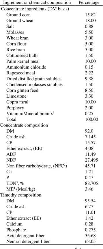
Table 3. Ingredients and chemical composition of diets for Korean cattle calves
Ingredient or chemical composition Percentage Concentrate ingredients (DM basis)
Ground corn 15.82 Ground wheat 18.00 Salt 0.88 Molasses 5.50 Wheat bran 3.00 Corn flour 5.00 Rice bran 3.00 Cottonseed hulls 1.50 Palm kernel meal 10.00 Ammonium chloride 0.15 Rapeseed meal 2.22 Dried distilled grain solubles 9.38 Condensed molasses solubles 1.50 Corn gluten feed 8.50 Limestone 3.30 Copra meal 10.00 Porphyry 2.00 Vitamin/Mineral premix1 0.25 Total 100.00 Concentrate composition DM 92.0 Crude ash 7.145 CP 15.57
Ether extract, (EE) 4.08
ADF 11.49
NDF 27.495
Non fiber carbohydrate, (NFC2) 45.71
Ca 1.21 P 0.47 TDN3, % 88.705 ME4 (Mcal/kg) 3.46 Timothy composition DM 95.54 Crude ash 6.77 CP 11.01
Ether extract (EE) 1.42
Calcium 0.28
Phosphate 0.275 Acid detergent fiber 35.68 Neutral detergent fiber 63.05
25
1Vitamin and mineral premix contained 2,650,000 IU vitamin A, 530,000 IU vitamin D3, 1,050 IU vitamin E, 10 g Niacin, 4.4 g Mn, 4.4 g Zn, 13.2 g Fe, 2.2 g Cu, 0.44 g I, and 0.44 g Co per kg of additive (Grobic-DC, Bayer Health Care, Leverkusen, Germany).
2 NFC (%) = 100 – (CP + EE + ash + NDF)
3 TDN (%) = NFC + CP + ((EE - 1) x 2.25) + NDF -7 (NRC, 2016) 4 ME (Mcal/kg) = (1.01 x (DE) – 0.45) + 0.0046 x (EE-3) (NRC, 2016)
3. Behavioral parameters
Immediately after 0.5 h sampling, calves were released to their pen. Behavior data were collected by live observation during the 3h immediately after 0.5h sampling. The observers sat behind pile of timothy hay out of sight of the calves, while recording starting and end time of behaviors. Behaviors measured included lying, eating, and drinking (Table 4).
4. Physiological parameters
Blood and saliva samples. Blood was collected by jugular venipuncture immediately before castration (h 0) and at h 0.5, h 6, d 1, d 3, d 7, and d 14 after castration. Two 10ml EDTA vacutainers (K2E, BD, NJ, USA) were used for collecting blood for plasma and leukocyte isolation. Two 10ml non-heparinized vacutainers (SST II Advance, BD, NJ, USA) were used for
26
collecting blood for serum. Both serum and plasma were separated by centrifugation at 1500 x g at 4°C for 15 min, and stored at -80°C until analysis. Saliva samples were collected from all the animals immediately before castration (0 h), 0.5 h, 6 h, and 1, 3, 7, 14 d after castration at the same moment of the blood sampling according to the method of Hernandex et al. (2014). Unfortunately, 6 h saliva samples were deficient for analysis.
ELISA analyses. Every ELISA analyses were conducted according to the manufacturer’s specifications. Plasma cortisol was analyzed using a salivary cortisol enzyme immunoassay kit (Salimetrics LLC, State College, PA, USA). Inter-assay and intra-assay coefficient of variability (CV) values were 9.85% and 6.64%, respectively. Plasma substance P was analyzed using a substance P ELISA kit (Enzo Life Sciences, NY, US). Inter-assay and intra-assay coefficient of variability (CV) values were 10.6% and 8.42%, respectively. Salivary cortisol was analyzed with the same enzyme immunoassay kit as for plasma samples. Inter-assay and intra-assay coefficient of variability (CV) values were 9.13% and 8.84%, respectively. Plasma haptoglobin was analyzed using a bovine haptoglobin enzyme immunoassay kit (GWB-A43096, GenWay Biotech, CA, USA). Inter-assay and intra-assay coefficient of variability (CV) values were 8.67% and 1.46%, respectively.
27
Table 4. Ethogram of behaviors recorded after castration modified from Marti et al. (2017)
Behavior Definition
Lying Either lateral (lying with hip and shoulder on the ground with at least 3 limbs extended) or ventral (lying in sternal recumbency with legs folded under the body or 1 hind or front leg extended) lying
Eating Ingesting hay or concentrate from the feeder Drinking Ingesting water from the water feeder
5. Leukocyte Isolation
Leukocyte was isolated according to the method of O’Loughlin et al. (2011). Briefly, red blood cells were lysed in a hypotonic solution and then restored in a hypertonic solution. The tubes were then centrifuged to collect the leukocyte pellet, suspended in 1mL of TRI Reagent (Sigma-Aldrich Ireland Itd., Dublin, Ireland) and stored in a sterile tube at -70°C.
6. Real-time polymerase chain reaction
Total RNA was isolated from leukocyte using TRIzol reagent according to the manufacturer’s instructions. The RNA concentration was measured using a Nanophotometer (Implen GmbH, Schatzbogen, München, Germany). The integrity of total RNA was double-checked through ethidium bromide staining of the 28S and 18S agarose gel electrophoresis bands and a Bioanalyzer (Agilent Technologies, Santa Clara, CA, USA): an RNA integrity number ≥ 9.0 was regarded as acceptable. RNA was stored at -70°C until analysis. RNA integrity number of almost h 0 samples was lower than 9. The
28
h 0 leukocyte sample, therefore, did not be used for analysis. The temperature of the centrifugation machine was higher than 4°C during h 0 leukocyte isolation. It may cause the RNA degradation of the h 0 leukocyte samples.
Total RNA was reverse-transcribed into cDNA using the iScript cDNA synthesis kit (Bio-Rad Laboratories Inc., Hercules, CA, USA) according to the manufacturer’s instructions. Reverse transcription was conducted in a 10-uL total reaction volume that contained 2ug RNA template, 2 10-uL of 5 x iScrip Reaction Mix, 0.5uL of iScript reverse transcriptase, and 2.5uL of nuclease-free water. The thermal parameters were: 25°C for 5min, 42°C for 30min, and 85°C for 5min.
The qPCR was performed as reported previously (Jeong et al., 2013) using QuantiTect SYBR Green RT-PCR Master Mix (Qiagen, Valencia, CA, USA). All primers were designed using integrated DNA technology, based on published sequences from the National Center for Biotechnology Information (Table 5). Primers were designed including exon-exon junction to prevent amplification of the DNA template. The melting temperatures of the primers were 57.0-61.5 °C. Thus, an annealing temperature of 55 °C was used for amplification of all of the genes. All qPCR analyses were conducted in a 25-μL total reaction volume that contained 20 ng cDNA, 12.5 25-μL SYBR Green RT-PCR Master Mix, and 1.25 μL of 10 μM primers. The thermal cycling parameters were: 95 °C for 15 min, followed by 40 cycles at 94 °C for 15 s, 55 °C for 30 s, and 72 °C for 30 s. In this study, we evaluated whether β-actin, GAPDH, and 18s rRNA are suitable reference genes. 18s RNA expression
29
was generally uniform. Therefore, all test gene expressions were normalized against 18s RNA expression. Because h 0 leukocyte samples were discarded, the means of all time control sample value were used as the calibrator according to the ∆∆CT method (Livak & Schmittgen, 2001) to enable graphing of fold changes. The 0.5 h and 14 d samples were also tested as the calibrator and the statistical results were not different.
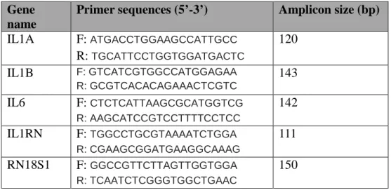
Table 5. Primer sequences and amplicon information forqRT-PCR
Gene name
Primer sequences (5’-3’) Amplicon size (bp)
IL1A F: ATGACCTGGAAGCCATTGCC R: TGCATTCCTGGTGGATGACTC 120 IL1B F: GTCATCGTGGCCATGGAGAA R: GCGTCACACAGAAACTCGTC 143 IL6 F: CTCTCATTAAGCGCATGGTCG R: AAGCATCCGTCCTTTTCCTCC 142 IL1RN F: TGGCCTGCGTAAAATCTGGA R: CGAAGCGGATGAAGGCAAAG 111 RN18S1 F: GGCCGTTCTTAGTTGGTGGA R: TCAATCTCGGGTGGCTGAAC 150
7. Statistical Analyses
All data were tested for normality using Proc Univariate (SAS, version 9.4, SAS Inst. Inc., Cary, NC). Blood, saliva, and mRNA data that were not normally distributed were log transformed. Behavior and growth performance data that were not normally distributed were root square +1 transformed. Blood, saliva, and mRNA data were analyzed using a repeated measures model PROC MIXED (SAS, version 9.4, SAS Inst. Inc., Cary, NC).
30
Fixed effect included treatment, time, and their interactions, whereas random effects included pen and calf within pen. Within the tree covariance structure (unstructured, compound symmetry, and autoregressive order one), the covariance structure with the lowest Schwarz’s Bayesian criterion was used for the analysis. When significant effects were found, Tukey-Kramer post-test was used to examine pair-wise differences for fixed effects. T-test was also conducted to compare castration group and non-castration group at each time points. Body weight, average daily gain (ADG), feed intake, feed efficiency, and behavioral data were analyzed using comparing group means model PROC TTEST (SAS, version 9.4, SAS Inst. Inc., Cary, NC). Correlation between concentration of blood and saliva parameters and relative cytokine mRNA levels were determined using the PROC CORR (SAS, version 9.4, SAS Inst. Inc., Cary, NC). Significant was indicated by P < 0.05.
31
Ⅳ. Results and Discussion
1. Growth performance
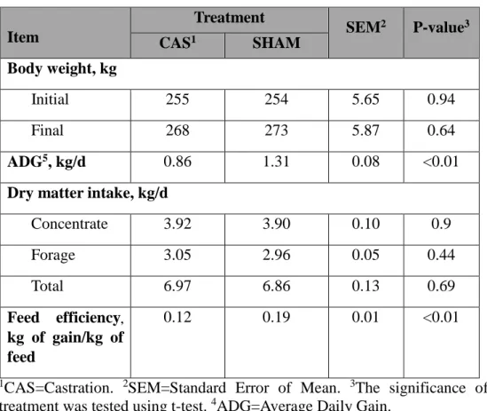
The effects of castration on growth performance parameters are presented in Table 6. Average dry matter intake was similar for both treatments (P = 0.69, t-test). Final body weight was also similar for both treatment (P = 0.64, t-test). Castrated calves had lower ADG (-34%, P < 0.05, t-test) and feed efficiency (0.12 vs 0.19, P < 0.05, t-test) than non-castrated calves.
Our results are agreement with results of the present study. Surgical castration reduced ADG of calves (8-9 months of age) during post-castration period until d 35 (Knight et al., 2000) and reduced ADG of calves (4 months of age) during post-castration period until d 49 (Marti et al., 2017). According to Bretschneider (2005), the reduction of ADG was independent of the method of castration. Several previous studies have reported that castration did not affect feed intake (Warnock et al., 2014; Stafford et al., 2002; Pang et al., 2006). The decreased feed efficiency of castrated calves was due to decreased ADG. Calves took a similar amount of feed and energy but showed different feed efficiency. Castrated calves used less energy for growth than non-castrated calves, indicating that castration caused utilization of energy sources for other functions not for growth. In the present study, indicators of stress, pain, and inflammation were different between castrated calves and non-castrated calves. These suggest that castrated calves may have greater
32
energy expenditure on stress responses or immune responses. Overall, our results suggest the decreased ADG of castrated calves may result from stress responses or immune responses.
Table 6. Effects of castration on growth performance in Korean cattle calves
Item Treatment SEM2 P-value3 CAS1 SHAM Body weight, kg Initial 255 254 5.65 0.94 Final 268 273 5.87 0.64 ADG5, kg/d 0.86 1.31 0.08 <0.01
Dry matter intake, kg/d
Concentrate 3.92 3.90 0.10 0.9 Forage 3.05 2.96 0.05 0.44 Total 6.97 6.86 0.13 0.69 Feed efficiency, kg of gain/kg of feed 0.12 0.19 0.01 <0.01
1CAS=Castration. 2SEM=Standard Error of Mean. 3The significance of treatment was tested using t-test. 4ADG=Average Daily Gain.
3.2. Behavior observation
The effects of castration on the behavior of calves from 0.5 to 3.5 h after castration are presented in Table 7. Castrated calves had differences in behavioral observations for lying and eating frequency, as well as eating
33
duration, from 0.5 to 3.5 h after castration. Castrated calves spent less time eating (-67%, P < 0.001, t-test) and had less eating frequency (1.5 vs. 3.4, P < 0.01, t-test) than non-castrated calves from 0.5 to 3.5h after castration. Lying frequency was high (8.2 vs. 1.2, P < 0.001, t-test) in castrated calves, but lying duration was not different (P = 0.96, t-test). Castrated calves did not maintain the lying posture compared to non-castrated calves (-78%, P < 0.05, t-test). No differences were seen in behavioral observations for drinking.
We could not precisely compare our findings to previous studies, as there is a lack of literature on the behavior of 8-mo-old calves after knife castration especially right after castration. Previous studies have reported that castration affects calves’ behaviors. Knife castration decreased lying and eating times of two-month-old calves and numerically decreased lying and eating times of 4-month-old calves compared with control calves during 2 to 4 h post-castration (Marti et al., 2017). Burdizzo castration increased standing behavior and decreased lying behavior of 13-month-old calves during the first 6 h after castration (Ting et al., 2003). According to Moya et al. (2014), the feeding behavior of 7-month-calves did not affect by either band or surgical castration during the first week after castration. When combined with the previous results, castration had short-term negative effects on lying and eating behaviors which is agreement with our results.
Lying behavior is important for both adult cattle and calves. Housed cattle are motivated to lie down for approximately 12 hours a day (Jensen et
34
al., 2004; Jensen et al., 2005) and housed calves spent approximately 18 hours a day lying down (Chun at el., 2002; Panivivat et al., 2004). In this study, castrated calves had a difference in lying frequency, but not lying duration. When we consider lying duration as the motivation desire for lying down, castrated calves and non-castrated calves were similarly motivated to lie down. It is possible that castrated calves experienced discomfort when the incision touched surface. The discomfort experienced may have caused that calves did not maintain lying position. Therefore, our results suggest knife castrated calves experienced more discomfort than non-castrated calves. Greater lying frequency and smaller eating time may be evidence of discomfort.
35
Table 7. Effect of castration on behaviors from 0.5 h to 3.5 h after castration
1CAS=Castration. 2SEM=Standard Error of Mean. 3The significance of treatment was tested using t-test.
Item Treatment SEM2 P-value3 CAS1 SHAM Eating Frequency(no./3h)
1.5
3.4
0.34
<0.01
Duration(min/3h)12.2
37.1
3.74
<0.001
Duration/visit(min/no.)6.9
13.3
1.79
0.09
Drinking Frequency(no./3h)1.7
1.1
0.27
0.27
Duration(min/3h)3.5
1.3
0.73
0.14
Duration/visit(min/no.)1.78
1.1
0.38
0.38
Lying Frequency(no./3h)8.2
1.2
1
<0.001
Duration(min/3h)71.6
70.4
10.26
0.96
Duration/visit(min/no.)10.1
46.2
7.36
0.02
36
3.3. Saliva and plasma cortisol
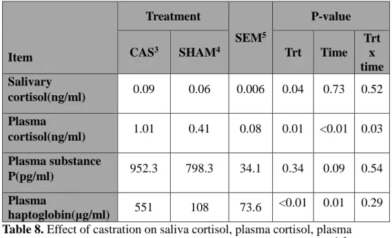
The effects of castration on the saliva cortisol (SC) and the plasma cortisol (PC) concentration are presented in Table 8.SC concentrations had a treatment effect (P < 0.001, repeated measures two-way ANOVA). PC concentrations had a time effect (P < 0.01, repeated measures two-way ANOVA) and treatment effect (P < 0.05, repeated measures two-way ANOVA). A treatment x time interaction (P < 0.05, repeated measures two-way ANOVA) was also observed for PC concentration. Immediately following castration (h 0.5), castrated calves had both elevated SC (P < 0.05, t-test) and PC concentrations (P < 0.001, t-test) compared with non-castrated calves (Figure 3). Elevated concentration of both cortisol returned to h 0 concentration at h 6 after castration.
Cortisol concentration has been used as an indicator of the degree of pain and physiological stress suffered by castrated cattle (AVMA, 2007; Stafford, 2007). In agreement with our findings, previous studies have reported increases in PC and SC concentration in castrated calves within a short time period: at one hour after surgical castration in 2-mo-old calves (Melendez et al., 2018), at h 0.5 after surgically castrated 6.3-mo-old calves (Park et al., 2018a), within 2 hours after burdizzo-castrated 13-mo-old calves (Ting et al., 2003), at h 0.5 after surgically castrated 3-mo-old calves (Sutherland et al., 2013), at 1 hour after band castrated 219-day-old calves (González et al., 2010). The short-term elevation of cortisol concentration immediately after
37
castration could be due to a rapid clearance of the cortisol from the bloodstream (Pumb, 1994).
Plasma cortisol concentrations were also elevated in the non-castrated calves at h 0.5 compared to h 0 (Figure 3). Similar to our results, Park et al. (2018a) had reported an increase in cortisol concentration in non-castrated groups. Park et al. (2018a) suggested that elevation of non-castrated groups could be due to handling procedures such as collecting blood. In our study, shame treatment and handling procedures might have induced stress, indicating cortisol response was not specifically associated with nociception. Cortisol concentration may be a general stress indicator.
The two profiles were similar and there was a significant and positive correlation between SC and PC concentration (r = 0.69, P < 0.001, pearson correlation; Table 10; Figure 3). To our knowledge, there are no studies investigating the relationship between SC and PC after castration of bulls. Albeit the inducer of increasing cortisol concentration was different, previous studies observed that SC concentration reflected blood cortisol concentration when ACTH was injected to cattle (Negrao et al., 2004; Hernandez et al., 2014; Schwinn et al., 2016). Vining (1987) confirmed that blood free cortisol is diffused passively to saliva across the acinar cells of the lumen of the salivary gland. When combined our results with the previous results, saliva cortisol may reflect plasma cortisol under castration stress. In this study, however, we analyzed cortisol concentration at certain time. Further study
38
with sampling at short intervals is needed to investigate the accurate relationship of saliva cortisol and plasma cortisol.
Thus, cortisol concentration may be a general stress indicator and the positive correlation between saliva and plasma cortisol concentration after castration suggests a SC as a potential stress indicator instead of PS since collection of salivary sample is easier and may give less stress compared with collection of blood sample.
Table 8. Effect of castration on saliva cortisol, plasma cortisol, plasma substance P, and plasma haptoglobinduring the 14-d postcastration1, 2. 1Means of all samples (immediately before castration (h 0), h 0.5, h 6, d1, d3, d7, and d 14 after castration). 2The significance of fixed effect (castration, time, and their interaction) was tested using repeated measure two-way ANOVA.
3CAS=Castration. 4SHAM=Sham treatment. 5SEM=Standard Error of Mean. Item
Treatment
SEM5
P-value
CAS3 SHAM4 Trt Time
Trt x time Salivary cortisol(ng/ml) 0.09 0.06 0.006 0.04 0.73 0.52 Plasma cortisol(ng/ml) 1.01 0.41 0.08 0.01 <0.01 0.03 Plasma substance P(pg/ml) 952.3 798.3 34.1 0.34 0.09 0.54 Plasma haptoglobin(μg/ml) 551 108 73.6 <0.01 0.01 0.29
39
Figure 3. Effect of castration on plasma concentrations of cortisol, substance P, and haptoglobin and saliva cortisol concentrations in Korean cattle calves.Male calves were allocated into two groups: CAS = castration (n=10); SHAM = sham (n=9). Values represent the mean + SEM. The differences between values was tested using the Tukey-Kramer post-test. An asterisk (*) indicates that the
*
*
*
0 1 2 3 4 h 0 h 0.5 h 6 d 1 d 3 d 7 d 14 P las m a cor ti sol , n g/ m lTime after castration
CAS SHAM 0.0 0.1 0.2 0.3 h 0 h 0.5 d 1 d 3 d 7 d 14 S al iva c or ti sol , n g/ m l
Tiem after castration
CAS SHAM
*
‡ 0 400 800 1200 1600 h 0 h 0.5 h 6 d 1 d 3 d 7 d 14 Subs ta nc e P , pg /m lTime after castration
CAS SHAM
*
0 500 1000 1500 2000 2500 h 0 h 0.5 h 6 d 1 d 3 d 7 d 14 H ap togl ob in , μ g/ m lTime after castration
CAS SHAM †
*
‡*
‡*
‡ †40
values differ at each time point (P < 0.05). A double cross (‡) indicates that the value differs from the value at h 0 within the castration group (P < 0.05). A single cross (†) indicates that the value differs from the value at h 0 within the sham group (P < 0.05).
41
3.4. Plasma substance P
The effects of castration on the plasma substance P (SP) concentration are presented in Table 8. No treatment or time effects were observed (P > 0.05). SP concentration was higher (P < 0.05, t-test) in castrated calves than in non-castrated calves d 1 after castration (Figure 3).
Substance P consists of 11 amino acids and is modified from preprotachykinin. Substance P is a neuropeptide in the central nervous system associated with stress regulation, anxiety-related behaviors, and pain (Ebner and Singewald, 2006). Plasma concentrations of SP have been used to evaluate muscle pain, soft tissue injury of human and castration pain of calves (Coetzee et al., 2008; Park et al., 2018a; Marti et al., 2017; Onuoha and Alpar, 1999).
Several previous studies have reported the relationship between castration and substance P concentration. However, there are inconsistent results among studies. Coetzee et al. (2008) reported that mean SP concentration was higher in castrated calves than non-castrated calves during 4 hours after castration. Mintline et al. (2014) observed SP concentrations of castrated calves were highest immediately before castration and dropped by d 3 after castration. Meléndez et al. (2017) reported SP concentrations of castrated calves reached a peak at d 5 after castration. Repenning et al. (2013) reported no differences in SP concentration at 24h post-castration compared to baseline samples taken 24h prior to castration, while SP concentrations of
42
castrated calves and sham castrated calves were not different at 24h post-castration. Park et al. (2018a) observed SP concentrations of castrated calves were higher than SP concentrations of uncastrated calves 6h after castration. These differences could be due to the age of calves used in experiments. Calves castrated at eight wk had lower levels than those castrated at six mo (Dockweiler et al., 2013). Marti et al. (2017) observed that four-mo-old calves presented a greater number of indicators of pain compared with 2-mo-old calves and 1-wk-old calves. According to Zempoalteca el a. (2018), 21-d-old rat had more developed peripheral nervous system than 7-d-old rat. Poor nerve development of younger calves may result in those differences. Substance P may not only reflect acute pain but also stress. According to Lieberman et al. (2012), plasma SP concentration was associated with anxiety, depression, and fatigue in human. Meléndez et al. (2017) also suggested extra handling could have increased SP level. These and our result imply the potential weaknesses of substance P as a pain indicator of castration.
3.5. Plasma haptoglobin and leukocyte cytokine gene expression
The effects of castration on the plasma haptoglobin are presented in Table 8. Haptoglobin concentrations had a time effect (P < 0.05, repeated measures two-way ANOVA) and treatment effect (P < 0.001, repeated measures two-way ANOVA). Castrated calves had greater concentrations than non-castrated calves on d1 (P < 0.05, t-test) and d 3 (P<0.01, t-test) after
43 castration (Figure 3).
Acute-phase proteins like haptoglobin increase in response to trauma, infection or inflammation (Hughes et al., 2014). Haptoglobin is synthesized by hepatocytes in response to inflammatory processes, inducing anti-inflammatory processes (Murate et al., 2004). Haptoglobin participates in defense mechanisms against acute changes such as infection, stress, damage, or injury (Jain et al., 2011). It has been suggested that haptoglobin can be used as an accurate indicator of inflammatory conditions in cattle (Horadagoda et al., 1999). Therefore, independent of the castration method, haptoglobin would be expected to increase as local tissue trauma was produced.
We observed an increase in haptoglobin concentration on the d 1 and d 3 after castration, and it is consistent with many previous studies. Ting et al. (2003) reported an increase in haptoglobin concentration on the d1 and d3 after burdizzo castration in 13-mo-old calves. Ting et al. (2004) saw an increase in haptoglobin concentration on the third day after burdizzo castration in 2.5- to 3.5-mo-old calves. Brown et al. (2015) observed an increase in haptoglobin concentration for 72h after knife castration in 214-day-old calves . Warnock et al. (2012) reported that surgically castrated calves had increased haptoglobin concentration 2-d post-castration in 200-day-old calves. Roberts et al., (2018) observed an increase in haptoglobin concentration on the d1 and d4 after surgical castration in 190-day-old calves. Our previous study also reported an increase in haptoglobin concentration on
44
the d1 and d3 after surgical castration of Korean cattle calves (Park et al., 2018a). Castration increased of haptoglobin concentration and haptoglobin is one of the inflammation indicators. Therefore, castration induces inflammatory responses in calves.
The effects of castration on relative mRNA levels of cytokines are presented in Table 9. A treatment x time interactions (P < 0.05) were observed for IL6, IL1A, and IL1RN mRNA levels. The relative mRNA levels of IL-6, IL-1A, IL-1B had a time effect (P < 0.001) and treatment effect (P < 0.01). Expression of IL-6, IL-1α, IL1RN was higher (P < 0.05) in castrated calves than in non-castrated calves at h 6 and d 1 after castration but not at other times. Expression of IL-1β was higher (P < 0.05) in castrated calves than in non-castrated calves only h 6 after castration (Figure 4).
Cytokines are a broad category of small secreted protein released by cells and affect the interactions and communications between cells. Cytokines are predominantly made by leukocytes such as helper T cells and macrophages. IL-1α, IL-1β, and IL-6 promote inflammation and are called proinflammatory cytokines. IL-6 secretion is induced by a variety of stimulating signals, including lipopolysaccharide, IL-1, and tumor necrosis factor, and platelet-derived growth factor (Di Paolo et al., 2016; Cahill and Rogers, 2018; Tosato and Jones, 1990). After IL-6 is synthesized in a local lesion in the initial stage of inflammation, it moves to the liver through the bloodstream, followed by the rapid induction of an extensive range of acute phase proteins (Heinrich et
45
al. 1990). IL1RN, a member of IL-1 family, suppresses the activity of IL1 and is one of the anti-inflammatory cytokines.
Previous study reported that the relative quantity of cytokine mRNA was not different between banding or burdizzo castrated calves and non-castrated-calves (Pang et al., 2006). These differences could be due to castration methods. Pang et al. (2006) used banding and burdizzo castration methods which do not make an incision in the scrotum. In our study, we used knife castration which makes wounds on stratum and ductus deferens. From these wounds, inflammatory responses may be induced.
The relative expression of IL-1A, IL-1B, and IL-6 of non-castrated calves was elevated d 1 after castration (Figrue 4). The haptoglobin concentration of non-castrated calves was also elevated d 1 after castration. Sporer et al. (2008) observed expression changes in genes involved in immune responses after transportation. Stress generated during sham treatment may affect cytokine expression of non-castrated calves.
Leukocyte mRNA levels of IL-1α and IL-1β showed positive correlations with leukocyte mRNA levels of IL-6 (r=0.96, P<0.001; r=0.55, P<0.001; Table 10). 1 is known to induce 6 production and the correlation of IL-1 and IL-6 was observed (Schindler et al., IL-1990). According to Tsakiri et al. (2008), IL-1 induces IL-6 production through the sphingomyelinase/Scr kinase pathway. mRNA expression of IL-6 and IL-1α decreased together in luteal phase endometrium of women with recurrent miscarriage (Jasper et al.,
46
2007). Considering these results and the relationship between IL-1 and IL-6, the positive correlation may result from IL-6 induction by IL-1.
Leukocyte mRNA levels of IL-1RN was highly related to the leukocyte mRNA levels of IL-6 and IL-1β (r=0.67, P<0.001; r=0.83, P<0.001; Table
10). IL-1ra is a specific inhibitor of the IL-1α and IL-1β. IL-1ra blocks the
action of IL-1α and IL-1β by inhibiting the binding of IL-1α and IL-1β to interleukin 1 receptor (Arend and Gabay, 2000). Due to IL-1ra production by the liver, circulating 1ra concentration is almost 100-fold greater than IL-1 concentration (Gabay et al., IL-1997; Gabay et al., 200IL-1). IL-IL-1ra levels are raised in the circulation of patients with a variety of inflammatory, infectious, and post-surgical conditions (reviewed by Ared et al., 1998). There are several results that both IL-1ra and IL-6 or IL-1β increased. According to Biasucci et al. (1999), patients with acute coronary syndrome showed elevating levels of both IL-1ra and IL-6. Children with cerebral malaria showed higher serum IL-1ra level than community children (John et al., 2008). John et al. (2006) also reported that children with cerebral malaria had increased levels of IL-6 and IL-1β. Increased IL-1ra level following IL-6 infusion was reported (Steensberg et al., 2003). Several studies suggested that maintenance of a balance between IL-1 and IL-1ra may be important in preventing the development of inflammatory diseases (Horai et al., 2000; Nicklin et al., 2000, Hirsch et al., 1996). IL-1ra expression may have increased for balancing with increased IL-1 level caused by castration. Overall, this is the first report that