Korean J Gastroenterol Vol. 61 No. 3, 170-173 http://dx.doi.org/10.4166/kjg.2013.61.3.170 pISSN 1598-9992 eISSN 2233-6869
CASE REPORT
Korean J Gastroenterol, Vol. 61 No. 3, March 2013 www.kjg.or.kr
내과적 치료로 회복된 급성 췌장염에 병발된 간문맥 내 가스 1예
동시헌, 조현근, 백정훈, 강버들, 김미성
1, 조재희, 이정훈
2, 천송욱
관동대학교 의과대학 명지병원 내과학교실, 방사선학교실1, 외과학교실2
A Case of Successful Treatment of Portal Venous Gas Caused by Acute Pancreatitis
Shi Heon Dong, Hyeon Geun Cho, Jeong Hoon Baek, Beo Deul Kang, Mi Sung Kim1, Jae Hee Cho, Jeong Hoon Lee2 and Song Wook Chun
Departments of Internal Medicine, Radiology1, Surgery2, Myongji Hospital, Kwandong University College of Medicine, Goyang, Korea
Hepatic portal venous gas (HPVG) has been considered a rare entity associated with a poor prognosis. Portal vein gas is most commonly caused by mesenteric ischemia but may have a variety other causes. HPVG can be associated with ischemic bowel disease, inflammatory bowel disease, intra-abdominal abscess, small bowel obstruction, acute pancreatitis, and gastric ulcer. Because of high mortality rate, most HPVG requires emergent surgical interventions and intensive medical management.
We experienced a case of hepatic portal venous gas caused by acute pancreatitis and successfully treated with medical management. (Korean J Gastroenterol 2013;61:170-173)
Key Words: Hepatic portal vein; Superior mesenteric vein; Gas; Acute pancreatitis
Received April 13, 2012. Revised July 16, 2012. Accepted July 20, 2012.
CC This is an open access article distributed under the terms of the Creative Commons Attribution Non-Commercial License (http://creativecommons.org/licenses/
by-nc/3.0) which permits unrestricted non-commercial use, distribution, and reproduction in any medium, provided the original work is properly cited.
교신저자: 조현근, 412-270, 고양시 덕양구 화정동 697-24, 명지병원 내과
Correspondence to: Hyeon Geun Cho, Department of Internal Medicine, Myongji Hospital, Kwandong University College of Medicine, 697-24 Hwajeong-dong, Deogyang- gu, Goyang 412-270, Korea. Tel: +82-31-810-5412, Fax: +82-31-810-5109, E-mail: [email protected]
Financial support: None. Conflict of interest: None.
서 론
간문맥 내 가스는 주로 허혈성 장질환에 의한 장괴사에서 관찰되는 매우 드문 소견으로, 높은 사망률과 관련된다. 대부 분 허혈성 장질환에 의한 장괴사에서 주로 관찰되지만, 염증 성 장질환, 복강 내 농양, 소장 폐쇄, 위궤양의 천공 및 복부 외상, 내시경 처치 후에도 발생한다고 보고되었다.1 그외에도 다양한 복강 내 질환에 의해서도 야기될 수 있는데 국내에서 는 내과적 집중 치료에도 불구하고 다발성 장기부전으로 사망 한 중증의 괴사 췌장염에 동반된 간문맥 내 가스 증례가 두 예 보고되었다.1
이번 증례는 급성 복통으로 내원한 58세 남자로, 복부 전산 화단층촬영에서 급성 췌장염과 그에 병발한 간문맥 및 상장간 막정맥 내 공기음영을 발견하였으나 당시 활력징후가 불안정 하여 외과적 수술을 하지 않고 내과적 집중 치료만 시행하여
생존한 국내 증례 1예를 경험하였기에 문헌 고찰과 함께 보고 하는 바이다.
증 례
58세 남자 환자가 내원 12시간 전부터 시작된 복부 통증을 주소로 내원하였다. 환자는 과거력에서 특이 병력은 없었고 60 갑년의 흡연력과, 2병 정도의 소주를 매일 복용한 음주력이 있 었다. 특이 가족력은 없었다. 활력징후는 혈압 70/40 mmHg, 맥박수 110회/분, 체온 36oC, 호흡수 20회/분이었고, 급성 병 색을 띠고 있었으며 입술과 혀는 건조하였다. 복부는 부드럽 고 팽대되어 있었고 장음은 감소되어 있었다. 복부의 전반적 인 압통이 있으나 복부강직은 없었고 장기비대나 종괴는 촉지 되지 않았다.
검사실 소견에서, 일반혈액검사는 백혈구 3,200/mm3 (중성
Dong SH, et al. Successful Treatment of Portal Venous Gas Caused by Acute Pancreatitis
171
Vol. 61 No. 3, March 2013
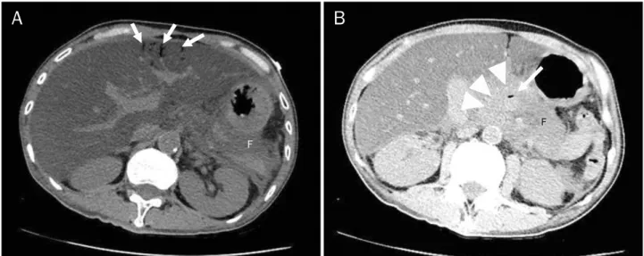
Fig. 1. Acute pancreatitis with portal and superior mesenteric venous gas. (A) CT showed portal vein air (arrows) in the peripheral portion of left hepatic lobe. Loculated fluid collection (F) in the lesser sac and retroperitoneal space. Note hepatomegaly with severe fatty liver indicating alcoholic liver disease. (B) CT revealed air in the superior mesenteric vein (arrow) and swelling of the pancreas (arrowheads) with peripancreatic fluid (F).
구 43%, 림프구 51%, 단핵구 6%), 혈색소 16.0 g/dL, 혈소판 33,000/mm3였으며, 일반화학검사는 Na+ 117 mEq/L, K+ 3.8 mEq/L, Cl− 79 mEg/L, 혈중요소질소/크레아티닌 20.7/2.2 mg/dL, AST/ALT 334/82 U/L, 총빌리루빈 3.5 mg/dL, 총단백 8.8 g/dL, 알부민 4.0 g/dL, 아밀라아제 720 U/L, 리파아제 2,990 U/L였고, 프로트롬빈 시간, 활성화부분트롬보플라스틴 시간은 각각 12.5초(11.9-13.9초), 37.3초(29.6-41.0초)였다.
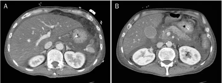
동맥혈 가스검사에서 pH 7.35, pCO2 19 mmHg, pO2 80 mmHg, HCO3 10.5 mmol/L, 산소포화도 95%였다. 아밀라 아제와 리파아제 상승으로 급성 췌장염을 의심하여 복부 전산 화단층촬영을 계획하였으나, 당시 환자의 혈청 크레아티닌이 상승되어 있어 조영제를 투여하여 조영 증강을 할 경우 조영 제 유발 신증이 발생할 것을 우려하여 조영 증강 없이 복부 전산화단층촬영을 시행하였다. 검사 결과 급성 췌장염의 소견 에 합당한 미만성 췌장부종 및 액체 저류 소견이 보였으며, 병발한 간문맥 및 상장간막정맥 내의 공기음영도 관찰되었다 (Fig. 1). 조영 증강을 하지 않아 정확한 평가는 어려우나 돌창 자의 장벽비후 소견이 있고 간문맥 및 상장간막정맥 내의 공 기음영은 장경색의 가능성을 시사하므로 외과적 처치 및 치료 가 필요하다고 판단하였으나, 당시 환자의 혈압이 70/40 mmHg로 활력징후가 불안정하여 외과적 치료가 어려울 것으 로 보고 중환자실에서 내과적 치료를 먼저 계획하였다. 환자 는 금식을 하며 수액 및 항생제를 투여받았다. 내원 3일째 활 력징후가 안정화되며 혈중요소질소/크레아티닌이 6.2/0.8 mg/dL로 감소하여 추적 조영 증강 복부 전산화단층촬영을 시행한 결과, 돌창자의 장벽비후와 간문맥 및 상장간막정맥 내 공기음영은 소실되었으며 췌장부종 및 액체 저류는 남아있
었다(Fig. 2). 이후 환자는 복부통증에 호전을 보이고 일반화 학검사에서도 아밀라아제 및 리파아제 수치에 점차 감소를 보 여 입원 6일째 식이를 시작하였고 입원 18일째 퇴원하여 현재 외래에서 추적 관찰 중이다.
고 찰
간문맥 내 가스는 Wolfe와 Evans2에 의해 1955년에 괴사 성 장염을 동반한 영아에서 최초로 보고되었다.
간문맥 내 가스의 발생 기전은 정확하게 알려져 있지는 않 지만 장관 또는 가스를 생성하는 박테리아를 통해서 공기가 간문맥 계로 들어가 발생할 것으로 추정된다.3 간문맥 내 가스 의 존재는 복부 내 병변에 의해 이차적으로 생기는 현상이므 로 반드시 원인 질환을 찾아 치료해야 한다. 간문맥 내 가스는 흔히 허혈성 장염, 수술 후 합병증, 크론병, 복강 내 농양, 장 폐쇄에 의하여 발생하며 드물게는 대장암, 위궤양, 급성 췌장 염, 간문맥염, 기타 감염성 질환, 구불창자 게실염에 의해서 발생한다.4
간문맥 내 가스를 동반한 환자는 예전에는 사망률이 75-90%
로 매우 높았지만 전산화단층촬영에 의해 초기 진단률이 빨라 지면서 사망률은 현재 감소되는 추세를 보이고 있다.5
복부 전산화단층촬영은 간문맥 내 가스의 진단에 가장 널 리 쓰이는 검사 방법으로, 간문맥 내에 말초에서부터 가지를 치는 양상으로 보이는 가스음영이 존재할 경우 진단이 가능하 다.4,5
간문맥 내 가스의 경우는 혈액의 흐름으로 인해 간외막부 터 2 cm 내에 방사선 투과성(radiolucency)을 보이나 담도
172
동시헌 등. 내과적 치료로 회복된 급성 췌장염에 병발된 간문맥 내 가스The Korean Journal of Gastroenterology
Fig. 2. Follow-up CT after 2 days. CT scans showed absence of air in the portal vein (A) and superior mesenteric vein (B). Diffuse pancreatic swelling with fluid collection in the perihepatic and retroperitoneal space suggested acute pancreatitis.
공기 음영은 간외막부터 2 cm 이상 떨어져 방사선 투과성을 보이므로 구별이 가능하다.6
예후는 간문맥 내 가스가 진단된 182명의 환자들의 임상경 과를 분석한 Kinoshita 등7의 보고에 따르면, 자료와 임상경 과가 명확하지 않은 20명을 제외한 162명 중에서 39%의 총 사망률을 보였으며, 그 중 장괴사로 인한 경우는 75%의 높은 사망률을 보였다. 장관 확장, 복강 내 농양, 위궤양으로 인한 경우는 약 20-30%의 사망률을 보였으며, 궤양성 대장염, 크론 병, 내시경 합병증, 복강 내 종양, 췌장염, 담도염으로 인한 사망은 없어 췌장염의 경우는 비교적 예후가 좋은 것으로 보 이나,7 국내에서 보고된 급성 췌장염에 병발한 간문맥 내 가스 가 발견된 두 예의 환자는 모두 사망하였다.1
급성 췌장염에 병발한 간문맥 내 가스의 보고는 1961년에 Wiot와 Felson8에 의해 최초로 이루어졌으며, 급성 췌장염에 서의 간문맥 내 가스 발생기전은 췌장 효소에 의해 직접적인 장점막 손상이 발생할 뿐만 아니라 장간막 허혈에 의해 이차적 인 장점막 손상으로 가스가 장관을 통해 간문맥 내로 유입되는 것으로 생각된다. 이번 증례 역시 저혈압에 의한 이차적인 장 점막 손상 및 췌장 효소에 의한 직접적인 장관 손상으로 인해 장관 내의 가스가 간문맥 내로 유입됐을 것으로 추정된다.
국내에서는 간문맥 내 가스에 대한 총 10명의 증례가 보고 되었으며 수술적 치료 후에 회복된 경우는 3건, 수술 후 사망 한 경우는 1건, 보존치료 후 회복된 경우는 1건, 보존치료 후 사망한 경우는 5건이었다. 이중 급성 췌장염에 병발한 2예가 있었으나 보존 치료에도 불구하고 사망하였다.1
환자는 당시 간문맥 및 상장간막정맥 내 가스 소견으로 장 허혈 혹은 장경색을 의심하여 응급개복 수술 등이 필요할 것 으로 생각되었으나 저혈압 소견 등 활력징후가 불안정한 이차 적인 패혈성 쇼크 상태로 외과적 치료가 불가능한 상태로 판
단하여 내과적 치료만 시행하였으며, 급성 중증 췌장염이 호 전되면서 간문맥 및 상장간막정맥 내 가스소견도 소실된 것으 로 보인다.
간문맥 내 가스가 발견된 환자 중 크론병 환자에서 장천공 에 병발하여 발생하고 수술적 치료 없이 내과적 치료만으로 회복된 증례가 있었으나9 급성 췌장염에 병발한 간문맥 내 가 스 중 내과적 치료로 회복된 증례는 국내에서는 보고된 바가 없다. 따라서 이 증례는 중증의 급성 췌장염 환자에서 간문맥 및 상장간막정맥 내의 가스라는 드문 합병증이 발생했을 때 내과적 치료만으로도 호전될 수 있음을 보여주는 증례라는 점 에서 의미가 있으므로 문헌 고찰과 함께 보고하는 바이다.
REFERENCES
1. Park HC, Lee WS, Joo SY, et al. Hepatic portal venous gas asso- ciated with acute pancreatitis: reports of two cases and review of literature. Korean J Gastroenterol 2007;50:131-135.
2. Wolfe JN, Evans WA. Gas in the portal veins of the liver in infants;
a roentgenographic demonstration with postmortem anatomi- cal correlation. Am J Roentgenol Radium Ther Nucl Med 1955;
74:486-488.
3. Sebastià C, Quiroga S, Espin E, Boyé R, Alvarez-Castells A, Armengol M. Portomesenteric vein gas: pathologic mecha- nisms, CT findings, and prognosis. Radiographics 2000;20:
1213-1224.
4. Iannitti DA, Gregg SC, Mayo-Smith WW, et al. Portal venous gas detected by computed tomography: is surgery imperative? Dig Surg 2003;20:306-315.
5. Liebman PR, Patten MT, Manny J, Benfield JR, Hechtman HB.
Hepatic-portal venous gas in adults: etiology, pathophysiology and clinical significance. Ann Surg 1978;187:281-287.
6. Faberman RS, Mayo-Smith WW. Outcome of 17 patients with portal venous gas detected by CT. AJR Am J Roentgenol 1997;
Dong SH, et al. Successful Treatment of Portal Venous Gas Caused by Acute Pancreatitis
173
Vol. 61 No. 3, March 2013 169:1535-1538.
7. Kinoshita H, Shinozaki M, Tanimura H, et al. Clinical features and management of hepatic portal venous gas: four case reports and cumulative review of the literature. Arch Surg 2001;136:
1410-1414.
8. Wiot JF, Felson B. Gas in the portal venous system. Am J Roentgenol Radium Ther Nucl Med 1961;86:920-929.
9. Ha NR, Lee HL, Lee OY, et al. A case of Crohn's disease presenting with free perforation and portal venous gas. Korean J Gastroen- terol 2007;50:319-323.