한국인 성인 쇄골의 해부학적 계측 및 쇄골과 Pre-Contoured Anatomical Plate의 적합성에 대한 분석
김형준 • 남경모 • 강대명 • 오성학 • 고덕환
건국대학교 의학전문대학원 충주병원 정형외과학교실
Anatomical Analysis of Clavicles in Korean Adults and Compatibility of Pre-Contoured Anatomical Plates
Hyeungjune Kim, M.D., Kyoungmo Nam, M.D., Daemyung Kang, M.D., Sunghak Oh, M.D., and Dukhwan Kho, M.D.
Department of Orthopedic Surgery, Konkuk University Chungju Hospital, Konkuk University School of Medicine, Chungju, Korea
Purpose: Due to the complex anatomy of clavicles, clavicular plates are not always compatible with clavicular fractures. The purpose of
this study was to analyze basic data on the anatomy of the clavicle in order to determine compatibility between clavicles of Korean adults and pre-contoured anatomical plates.Materials and Methods: We analyzed the anatomy of 600 clavicles of 300 patients who underwent three-dimensional (3D) computed
tomography of clavicles in the emergency room of Konkuk University Chungju Hospital, between July 2010 and July 2011, using Andermahr's method; in addition, the compatibility between 3D axial images of clavicles and sectional images of pre-contoured anatomical plates was also examined using Adobe Photoshop.Results: The mean length of the clavicle was 146.21±4.98 mm, the mean width was 9.63±1.67 mm, and the mean thickness was 9.54±1.67
mm. The location of the maximum superior bow was 36.17±0.60 mm from the lateral end of the clavicle and the mean magnitude was 5.88±0.62 mm. The mean depth of medial curvature was 15.89±1.33 mm, and the mean depth of the lateral curvature of the clavicle was 11.73±1.66 mm. The compatibility between clavicles and plates was 79% as above a fair compatibility in the 50% range of clavicles and 48% as above a fair compatibility in the 60% to 70% range of clavicles. On the contrary, in application of medial and lateral plates in the 60% to 70% range of clavicles, above a fair compatibility had increased to 67%.Conclusion: A more adequate pre-contoured anatomical plate is required for satisfactory improvement of the compatibility of clavicles of
Korean adults.Key words: clavicle, pre-contoured plate, Korea, adult
서 론
쇄골 골절은 성인 전체 골절의 2%-5%를 차지하며 shoulder girdle
골절 중 35%-44%에 해당하는 비교적 흔한 골절이다.1,2) 전통적 으로 쇄골 골절의 치료는 8자형 석고 또는 붕대 고정 등의 보존 적 치료를 우선으로 하였고, 수술적 치료는 개방형 골절, 신경 혈 관의 손상, 연부조직의 중첩 등 제한된 적응증에 대하여 시행하 였다.3-7) 그러나 최근 여러 문헌에서 보존적 치료는 단축, 부정유 합, 불유합, 운동제한 등의 합병증과 불안정한 정복, 장기간의 고 정 등의 환자 만족도의 문제가 발생하며, 견고한 내고정을 통한 조기 관절운동 회복을 위해 수술적 치료의 필요성을 보고하고 있
Copyright © 2013 by The Korean Orthopaedic Association
“This is an Open Access article distributed under the terms of the Creative Commons Attribution Non-Commercial License (http://creativecommons.org/licenses/by-nc/3.0/) which permits unrestricted non-commercial use, distribution, and reproduction in any medium, provided the original work is properly cited.”
The Journal of the Korean Orthopaedic Association Volume 48 Number 5 2013 Received January 23, 2013 Revised June 5, 2013
Accepted September 2, 2013
Correspondence to: Dukhwan Kho, M.D.
Department of Orthopedic Surgery, Konkuk University Chungju Hospital, 82 Gugwon- daero, Chungju 380-704, Korea
TEL: +82-43-840-8250 FAX: +82-43-844-7300 E-mail: [email protected]
다.8,9) 수술적 치료는 점진적인 수술기법의 발달로 골편간 나사 고정, 원형강선 고정술, 골수강 내 핀 고정술, 금속판 고정술 등 의 방법이 적용되고 있으며, 이 중 금속판 고정술은 피질골 간 압 박력을 통한 견고한 고정, 회전력에 대한 저항 등의 장점이 있어 높은 골유합률을 보고하고 있다. 금속판 고정술 시 쇄골의 복잡 한 S자 모양의 해부학적 특성으로 쇄골과 금속판과의 적합성이 맞지 않는 경우가 있어 비교적 윤곽을 맞추기 쉬운 재건 금속판 (reconstruction plate; AO Synthes, Paoli, PA, USA)이 사용되었으 나, 재건 금속판의 경우 염전 및 축성 저항력이 낮아 골절 부위에 서 변형되어 불유합, 부정유합의 원인이 된다고 보고되고 있다.10) 최근에는 low profile, beveled edges 형태로 낮은 연부조직 자극과 해부학적 적합성을 가진 pre-contoured anatomical plate (Acumed, Hillsboro, OR, USA)가 개발되어 사용 중이나, 실제 수술 시 쇄골 의 윤곽과 일치하지 않는 경우가 많다. 본 연구에서는 한국인 쇄 골의 해부학적 기초자료를 분석하여 쇄골 골절에 사용되는 pre- contoured anatomical plate와의 형태학적 적합성을 조사하였다.
대상 및 방법
1. 연구 대상
2010년 7월부터 2011년 7월까지 건국대학교 충주병원 응급실로 내원하여 3차원 흉부 전산화 단층 촬영(multi-detector computed tomography, MDCT. Light Speed 16; Toshiba Medical Systems Co., Tokyo, Japan)을 시행받은 환자 중 쇄골에 골절 및 기타 골질환
이 없는 300명, 600예를 대상으로, 건국대학교 연구윤리위원회로 부터 연구에 대한 승인을 받은 이후에 후향적 연구를 시행하였 다. 연령은 평균 42.3세(20-65세)였고 신장 및 체중은 남자는 평균 163.7 cm, 75.2 kg, 여자는 154.7 cm, 65.5 kg이었다. 환자에 따른 편 차(bias)를 줄이기 위하여 성별, 연령대별 분포를 균등 분산하였으 며, 골질환이 있는 경우와 쇄골의 외상력이 있는 경우는 대상에 서 제외하였다.
2. 쇄골 계측
쇄골 계측은 MDCT 영상에서 쇄골 영상만을 재형성한 후, Toshi- ba사(Toshiba Medical Systems Co.)의 Aquilion 64 system 프로그램 을 이용하여 실제 쇄골 크기로 보정작업을 거친 후, axial enface view는 쇄골 전장의 너비가 최대치가 되는 시점의 영상을, coro- nal enface view는 두께가 최대치가 되는 시점의 영상을 선택하여 Andermahr 등11)의 계측 방법을 이용하여 분석하였다. 쇄골의 3차 원 axial enface view에서 해부학적 지침으로 쇄골의 내측단(medial edge, A)과 외측단(lateral edge, B), 전방 측단(anterior edge, C, D)과 후방 측단(posterior edge, E, F)을 표시하였고(Fig. 1), 쇄골의 길이 (length, L1), 너비(width), 내측 만곡의 깊이(depth of medial curve, L2), 외측 만곡의 깊이(depth of lateral curve, L3)를 측정하였다. 쇄 골의 3차원 coronal enface view에서 쇄골의 두께(thickness)와 상 방극점(the point of the maximum superior bow)을 측정하였다.
길이(L1)는 내측 단에서 외측 단까지 최단 직선거리로 측정하 였고(Fig. 1), 너비는 쇄골 길이를 10등분한 후, 각 분점(dividing point)에서 내측단과 외측단을 연결한 선의 수직으로 거리를 측정 하였으며(Fig. 2), 두께는 쇄골 길이를 10등분하여 각 분점에서 쇄 골과의 수직 거리를 측정하였다(Fig. 3). 내측 만곡의 깊이는 쇄골 의 2개의 후방 측단을 잇는 직선거리에서 쇄골 피질까지 최대 거
Figure 1. Measurement of dimensions and curvature of the right clavicle.
A, medial edge; B, lateral edge; C, D, anterior edge; E, F, posterior edge;
L1, length of the clavicle; L2, depth of the medial curve of the clavicle;
L3, depth of the lateral curve of the clavicle; Ant., anterior; Post., posterior; Med., medial; Lat., lateral.
Figure 2. Clavicular width (W; mm) measured at 10% intervals of total length using Aquilion 64 3-dimensional software.
Figure 3. Clavicular thickness (T; mm) measured at 10% interval of total length with using Aquilion 64 3-dimensional software.
Figure 4. Measurement method used to determine the location (D1) and magnitude (D2) of the maximum superior clavicular bow. Measurements were performed using Aquilion 64 3-dimensional software. A, medial edge; B, lateral edge; D1, location of the maximum superior clavicular bow; D2, magnitude of the maximum superior clavicular bow.
리를 측정하였고, 외측 만곡의 깊이는 쇄골의 2개의 전방 측단을 잇는 직선거리에서 쇄골 피질까지 거리의 최대 거리를 측정하였 다(Fig. 1). 상방극점은 쇄골 내측단과 쇄골 외측단을 이은 직선에 서 쇄골 피질까지 최대 거리를 측정하였다(Fig. 4). 측정자 간 오 차를 줄이기 위해 정형외과 전문의 2인에 의해 3회 반복 측정 후 표준 편차 내의 측정치만 결과에 포함시켰다.
3. 쇄골과 pre-contoured anatomical plate의 적합성 Pre-contoured anatomical plate는 현재 국내에서 많이 사용되는 해부학적 금속판 중 하나이다. 쇄골 간부 골절에 많이 사용되는 8 hole 금속판의 경우 straight, standard, curved의 3가지 형태가 있는 데 이 중 standard형이 가장 많이 사용되며 적합성이 높은 것으로 보고되고 있다.12) 쇄골과 pre-contoured anatomical plate의 적합성 은 가늠자를 이용하여 8 hole standard 금속판을 단순 방사선 촬영 을 한 후, 실제 크기로 보정하고 Adobe Photoshop (Adobe System, San Jose, CA, USA)을 이용하여 쇄골의 3차원 영상을 기준으로 금 속판 영상을 자유롭게 회전 및 이동하여 쇄골과의 해부학적 일치 도, 금속판의 돌출(overhanging), 금속판 나사 구멍의 돌출 유무를 분석하였다. 쇄골에 대해 금속판 적용 위치는 쇄골의 3차원 axial enface view에서 쇄골 전장을 10등분한 후, 쇄골 골절이 가장 많이 발생하는 중간부 50% 위치와 외측 60%-70% 위치에 적용하였으 며, 적합성은 적절함(good), 보통(fair), 부적절함(poor)의 세 단계 로 분류하였다. 적절함(good)은 금속판이 쇄골의 해부학적 구조 와 일치하며 전후방 돌출이 없는 경우, 보통(fair)은 금속판 전방 혹은 후방 일측으로 돌출이 발생하나 나사 구멍의 돌출이 발생하 지 않은 경우, 부적절함(poor)은 금속판 및 금속판 나사구멍 모두 돌출이 발생하는 경우로 정의하였다.
4. 통계 처리
측정된 결과를 남자와 여자, 우측과 좌측으로 구분하여 평균값 과 표준편차로 표기하였고 SPSS 통계 프로그램(SPSS software version 12.0; SPSS GMbH, Muenchen, Germany)을 이용한 paired t-test 분석을 시행하였으며 유의 수준은 0.05 이하로 하였다.
결 과
1. 쇄골의 해부학적 구조(Table 1-8) 1) 길이(length) (Table 1)
쇄골의 평균 길이는 146.21±4.98 mm로 측정되었으며 남성은 150.66±2.25 mm, 여성은 141.78±2.24 mm로 측정되었다. 남성의 경우 우측 쇄골의 평균 길이는 148.12±3.14 mm, 좌측 쇄골의 평 균 길이는 153.20±3.11 mm였고, 여성의 경우 우측 쇄골의 평균 길이는 140.46±3.22 mm, 좌측 쇄골의 평균 길이는 143.10±3.24 mm였다. 남성이 여성보다 평균 9 mm 더 길었고 좌, 우 비교에서 는 남성이 평균 5 mm, 여성이 평균 3 mm 정도 좌측이 우측보다 더 길었다.
2) 너비(width) (Table 2)
쇄골의 중앙부의 평균 너비는 9.63±1.67 mm로 측정되었으며, 남 성은 평균 11.19±0.65 mm, 여성은 8.08±0.58 mm로 측정되었다.
쇄골 전장을 10% 간격으로 측정한 평균 쇄골의 너비는 50%-70%
영역에서 변화가 컸으며(Fig. 5), 남성이 여성보다 길었으며 전장 에서 측정된 너비의 길이도 남성이 여성보다 길었다. 또 좌측 쇄 골의 너비가 쇄골 내, 외측단을 제외한 전장에서 우측 쇄골의 너 비보다 길었으며 이는 남, 여 모두에서 동일하게 측정되었다.
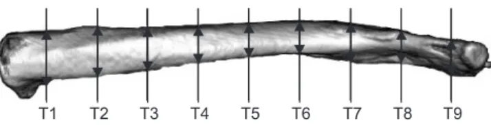
Table 1. Mean Geometric Parameters (mm)
Mean Length Width 50% Thickness 50% Medial depth Lateral depth MSB distance Magnitude of MSB
MaleRight 148.12±3.14 10.55±0.86 11.12±0.91 16.36±1.75 15.27±1.39 35.67±0.87 6.20±0.86 Left 153.20±3.11 11.83±0.85 10.37±0.88 14.94±1.82 10.83±1.46 36.33±0.80 5.63±0.87 Female
Right 140.46±3.22 8.05±0.89 8.46±0.86 17.43±1.78 10.93±1.50 36.71±0.82 6.40±0.91 Left 143.10±3.24 8.13±0.83 8.23±0.87 14.84±1.76 9.92±1.47 35.99±0.84 35.99±0.84 Total 146.21±4.98 9.63±1.67 9.55±1.36 15.89±1.33 11.74±1.66 36.17±0.61 5.88±0.62 Male 150.66±2.25 11.19±0.65 10.75±0.67 15.65±1.32 13.05±0.94 36.00±0.57 5.91±0.63 Female 141.78±2.24 8.09±0.58 8.35±0.58 16.13±1.31 10.42±1.09 36.35±0.60 5.85±0.62 Right 144.29±4.98 9.30±1.53 9.79±1.60 16.90±1.84 13.10±2.61 36.19±0.99 6.30±0.89 Left 148.15±5.97 9.98±2.04 9.30±1.39 14.89±1.79 10.38±1.53 36.16±0.84 5.47±0.91 Values are presented as mean±standard deviation. MSB, maximum superior clavicular bow.
Table 2. Clavicular Width (W; mm) Measured at 10% Intervals of Total Length from the Clavicular Medial End
Mean W1 W2 W3 W4 W5 W6 W7 W8 W9
Male
Right 15.80±0.85 11.61±0.80 10.85±0.82 10.74±0.80 10.55±0.86 12.06±0.91 16.16±0.83 16.33±0.90 25.24±0.90 Left 17.10±0.88 14.33±0.85 13.03±0.84 12.40±0.91 11.83±0.85 13.24±0.89 17.61±0.94 18.52±0.86 21.32±0.83 Female
Right 15.25±0.88 8.82±0.88 8.22±0.84 8.02±0.88 8.05±0.89 9.68±0.88 12.92±0.89 13.21±0.90 17.97±0.88 Left 13.40±0.92 8.35±0.89 7.95±0.86 8.17±0.85 8.13±0.83 9.51±0.86 13.45±0.83 13.87±0.89 18.83±0.89 Total 15.39±1.24 10.78±2.28 10.01±2.02 9.83±1.85 9.64±1.67 11.12±1.65 15.03±1.96 15.48±2.05 20.87±2.49 Male 16.45±0.59 12.97±0.58 11.94±0.60 11.57±0.62 11.19±0.65 12.65±0.65 16.88±0.67 17.42±0.64 23.28±0.59 Female 14.32±0.68 8.59±0.63 8.09±0.65 8.10±0.65 8.09±0.59 9.60±0.61 13.18±0.61 13.54±0.65 18.45±0.68 Right 15.52±0.90 10.22±1.63 9.53±1.56 9.38±1.61 9.30±1.53 10.87±1.49 14.54±1.84 14.77±1.80 21.60±3.75 Left 15.25±2.06 11.34±3.12 10.49±2.68 10.28±2.29 9.98±2.04 11.37±2.06 15.52±2.26 16.19±2.49 20.13±1.47 Values are presented as mean±standard deviation.
Figure 5. Clavicular width (W; mm) measured at 10% interval of total length.
Figure 6. Clavicular thickness (T; mm) measured at 10% interval of total length.
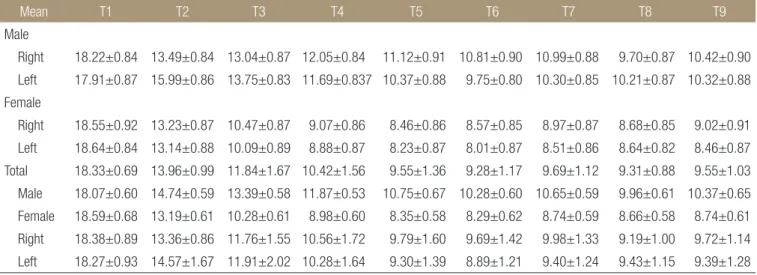
Table 3. Clavicular Thickness (T; mm) Measured at 10% Intervals of Total Length from the Clavicular Medial End
Mean T1 T2 T3 T4 T5 T6 T7 T8 T9
Male
Right 18.22±0.84 13.49±0.84 13.04±0.87 12.05±0.84 11.12±0.91 10.81±0.90 10.99±0.88 9.70±0.87 10.42±0.90 Left 17.91±0.87 15.99±0.86 13.75±0.83 11.69±0.837 10.37±0.88 9.75±0.80 10.30±0.85 10.21±0.87 10.32±0.88 Female
Right 18.55±0.92 13.23±0.87 10.47±0.87 9.07±0.86 8.46±0.86 8.57±0.85 8.97±0.87 8.68±0.85 9.02±0.91 Left 18.64±0.84 13.14±0.88 10.09±0.89 8.88±0.87 8.23±0.87 8.01±0.87 8.51±0.86 8.64±0.82 8.46±0.87 Total 18.33±0.69 13.96±0.99 11.84±1.67 10.42±1.56 9.55±1.36 9.28±1.17 9.69±1.12 9.31±0.88 9.55±1.03 Male 18.07±0.60 14.74±0.59 13.39±0.58 11.87±0.53 10.75±0.67 10.28±0.60 10.65±0.59 9.96±0.61 10.37±0.65 Female 18.59±0.68 13.19±0.61 10.28±0.61 8.98±0.60 8.35±0.58 8.29±0.62 8.74±0.59 8.66±0.58 8.74±0.61 Right 18.38±0.89 13.36±0.86 11.76±1.55 10.56±1.72 9.79±1.60 9.69±1.42 9.98±1.33 9.19±1.00 9.72±1.14 Left 18.27±0.93 14.57±1.67 11.91±2.02 10.28±1.64 9.30±1.39 8.89±1.21 9.40±1.24 9.43±1.15 9.39±1.28 Values are presented as mean±standard deviation.
3) 두께(thickness)
쇄골의 중앙부의 평균 두께는 9.55±1.36 mm로 측정되었으며 남 성은 10.75±0.67 mm, 여성은 8.35±0.58 mm로 측정되었다. 쇄골 을 10% 간격으로 나누어 측정한 쇄골의 두께는 내측단이 가장 두 꺼웠으며 쇄골 전장의 60% 지점까지 점차 감소하다 외측단까지 균일한 두께를 유지하였다(Fig. 6). 내, 외측단을 제외한 전장에서 남성의 쇄골 두께가 여성보다 두꺼우며 내, 외측단의 경우 평균 쇄골의 두께에서 여성이 남성보다 두꺼웠다(Table 3).
4) 내측 만곡의 깊이(depth of medial clavicular curve)
남성의 경우, 쇄골 내측 만곡의 깊이는 평균 15.65±1.32 mm, 우측 내측 만곡의 깊이는 평균 16.36±1.75 mm, 좌측 내측 만곡의 깊이
는 평균 14.94±1.81 mm로 측정되었다. 여성의 경우, 쇄골 내측 만 곡의 깊이는 평균 16.13±1.30 mm, 우측 내측 만곡의 깊이는 평균 17.42±1.77 mm, 좌측 만곡의 깊이는 평균 14.83±1.76 mm로 측정 되었다.
5) 외측 만곡의 깊이(depth of lateral clavicular curve)
남성의 경우, 쇄골 외측 만곡의 깊이는 평균 13.04±0.93 mm, 우측 외측 만곡의 깊이는 평균 15.26±1.39 mm, 좌측 외측 만곡의 깊이 는 평균 10.82±1.46 mm로 측정되었다. 여성의 경우, 쇄골 외측 만 곡의 깊이는 평균 10.42±1.09 mm, 우측 외측 만곡의 깊이는 10.92
±1.49 mm, 좌측 외측 만곡의 깊이는 평균 9.92±1.47 mm로 측정 되었다.
6) 쇄골 상방극점(the point of the maximum superior bow) 쇄골의 상방극점은 외측단에서 내측으로 평균 36.17±0.60 mm 위치에서 평균 5.88±0.62 mm로 측정되었으며, 남성의 경우 평균 35.99±0.57 mm 위치에서 5.91±0.63 mm로 측정되었고, 여성의 경우 평균 36.35±0.59 mm 위치에서 평균 5.85±0.61 mm로 측정 되었다.
Table 4. Quality of Fitness: 50% Portion of Clavicle
Good Fair Poor Total
Male 126 114 60 300
Female 60 174 66 300
Clavicle
Right 96 144 60 300
Left 90 144 66 300
Total 186 288 126 600
Values are presented as number.
Table 5. Quality of Fitness: 60 to 70% Portion of Clavicle
Good Fair Poor Total
Male 84 88 128 300
Female 41 79 180 300
Clavicle
Right 67 87 146 300
Left 58 80 162 300
Total 125 167 308 600
Values are presented as number.
Table 6. Quality of Fitness: 60 to 70% Portion of Clavicle; Plate Applieds to the Medial and Lateral Sides in Opposition
Good Fair Poor Total
Male 94 109 97 300
Female 71 128 101 300
Clavicle
Right 89 121 90 300
Left 76 116 108 300
Total 165 237 198 600
Values are presented as number.
Table 7. Statistical Analysis: Male vs. Female (mm)
Male Female p-value
Length 150.66±2.25 141.78±2.24 0.01 Width 50% 11.19±0.65 8.09±0.58 0.01 Thickness 50% 10.75±0.67 8.35±0.58 0.01 Medial depth 15.65±1.32 16.13±1.31 0.12 Lateral depth 13.05±0.94 10.42±1.09 0.02 MSB distance 36.00±0.57 36.35±0.60 0.28 Magnitude of MSB 5.91±0.63 5.85±0.62 0.37 Values are presented as mean±standard deviation. MSB, maximum superior clavicular bow.
Table 8. Statistical Analysis: Left vs. Right (mm)
Left Right p-value
Length 148.15±5.97 144.29±4.98 0.01 Width 50% 9.98±2.04 9.30±1.53 0.35 Thickness 50% 9.30±1.39 9.79±1.60 0.29 Medial depth 14.89±1.79 16.90±1.84 0.22 Lateral depth 10.38±1.53 13.10±2.61 0.03 MSB distance 36.16±0.84 36.19±0.99 0.71 Magnitude of MSB 5.47±0.91 6.30±0.89 0.56 Values are presented as mean±standard deviation. MSB, maximum superior clavicular bow.
2. 쇄골과 pre-contoured anatomical plate의 적합성 쇄골의 50% 영역에 금속판을 적용시켰을 때 쇄골과 pre-con- toured anatomical plate의 적합성은 적절함(good)이 186예(31%), 보통(fair)이 288예(48%), 부적절함(poor)이 126예(21%)로 나타났 고, 실제 수술 시 금속판을 고정할 수 있는 보통 이상의 일치도 는 79%로 나타났다(Table 4). 쇄골의 60%-70% 영역에 금속판을 적용시켰을 때, 적합성은 적절함(good)이 125예(21%), 보통(fair) 이 167예(28%), 부적절함(poor)이 308예(51%)로 나타났고, 실제 수 술 시 금속판을 고정할 수 있는 보통 이상의 일치도는 48%로 나 타났다(Table 5). 저자들의 임상적 경험상, 쇄골의 60%-70% 영역 의 골절에서 금속판의 적합성이 맞지 않는 경우 금속판의 내, 외 측을 반대로 적용시켰을 때 적합성이 맞는 경우가 많아 금속판의 내측과 외측을 반대로 적용하여 분석한 결과, 적절함(good)이 165 예(27%), 보통(fair)이 237예(40%), 부적절함(poor)이 198예(33%)로 나타났고, 실제 수술 시 금속판을 고정할 수 있는 보통 이상의 일 치도는 67%로 증가하였다(Table 6).
고 찰
쇄골 간부 골절의 수술적 치료는 점진적인 수술기법의 발달로 골 편간 나사고정, 원형강선 고정술, 골수강 내 핀 고정술, 금속판 고 정술 등의 방법이 사용되고 있으며, 이 중 관혈적 정복 후 금속판 고정술 또는 경피적 골수강 내 핀 고정술이 가장 흔히 사용되고 있다. 골수강 내 핀 고정술은 절개가 작고 연부조직 박리가 적다 는 장점이 있으나 회전력에 대한 고정력이 약하고 감염, 불유합 의 가능성이 높은 단점이 있다. 금속판 고정술은 견고한 고정을 통한 조기 관절운동으로 재활이 빠르고 단축을 극복할 수 있다는 장점이 있으나 금속판 돌출, 연부조직 손상의 문제가 있다. 이와 같은 장, 단점을 바탕으로 단순한 쇄골 간부 골절에서는 금속판 고정술과 골수강 내 핀 고정술의 우위에 대해서는 다양한 임상적 결과가 보고되고 있으나13-15) 전위성 쇄골 분쇄 골절에 대해서는 골절편 사이에 연부조직이 감입되어 있는 경우가 많아 관혈적 정 복 및 금속판 고정술이 더 선호되고 있다.16)
금속판 고정술 시, 쇄골의 복잡한 S자 모양의 해부학적 특성으 로 쇄골과 금속판과의 적합성이 맞지 않는 경우가 있어 비교적 윤곽을 맞추기 쉬운 재건 금속판(reconstruction plate)이 사용되었 으나 재건 금속판의 경우, 염전 및 축성 저항력의 강도가 낮아 골 절 부위에서 변형되어 불유합, 부정유합의 원인이 되며 피부로 돌출되어 환자의 불편함과 통증의 빈도가 높다는 문제가 있어, 최근에는 pre-contoured anatomical plate가 개발되어 사용되고 있 다. Chandrasenan 등17)은 pre-contoured anatomical plate는 쇄골과 적합성이 맞도록 설계되어 수술 시 contouring을 할 필요가 없어 수술 시간을 줄일 수 있고 강도가 높아 금속판 파단을 줄일 수 있 다고 하였고, VanBeek 등18)은 금속판이 low profile, beveled edges
형태이므로 술 후 피부로 돌출되지 않아 금속판 제거를 위한 재 수술의 빈도를 줄일 수 있다고 하였다.
최근 외국에서 개발된 pre-contoured anatomical plate를 국내에 수입하여 사용하고 있으나, 서양인의 해부학적 기준에 따라 만들 어졌고 쇄골의 복잡한 해부학적 특성과 개인별 다양성으로 실제 수술 시 쇄골과 금속판과의 적합성이 맞지 않는 경우가 많다. 적 합성이 맞지 않는 원인은 성별, 쇄골의 길이, 외측 만곡의 깊이 등 이 보고되고 있으며 쇄골이 길이가 짧고 폭이 좁으며 외측 만곡 의 깊이가 깊지 않은 경우 적합성이 맞지 않다고 보고되고 있다.
Daruwalla 등19)과 Duprey 등20)은 남성이 여성보다 쇄골의 길이, 폭, 외측 만곡의 깊이가 더 크다고 하였고, Andermahr 등11)은 200예의 쇄골의 해부학적 형태 연구에서 쇄골은 개인별 편차가 있으며 좌 측 쇄골이 우측 쇄골보다 더 길다고 하였다. Huang 등12)은 금속판 을 해부학적 위치에서 외측으로 조금만 위치시켜도 쇄골과 적합 성을 얻지 못한다고 하였고, 특히 백인 여성의 쇄골에는 해부학 적 모양이 맞지 않는다고 하였다. 본 연구에서도 쇄골의 길이는 남성이 여성보다 평균 9 mm 더 길었고, 좌측과 우측의 비교에서 는 남성이 평균 5 mm, 여성이 평균 3 mm 정도 좌측이 더 길었으 며 외측 만곡의 깊이는 평균 3 mm 정도 남성이 여성보다 깊었다.
금속판과의 적합성은 쇄골의 50% 지점에서 적절함(good)을 나타 낸 경우가 남성 126예, 여성 60예였고, 60%-70% 영역에서는 남성 84예, 여성 41예로 남성에서 50% 정도 적합성이 높은 것으로 나타 났다.
국내에 수입된 pre-contoured anatomical plate는 서양인을 기 준으로 제작되었기 때문에 서양인과의 해부학적 차이에 의해 적 합성이 맞지 않는 경우가 있다. Andermahr 등11)은 서양인의 쇄골 의 해부학적 연구에서, 쇄골의 길이는 평균 151 mm, 평균 내측 만 곡의 깊이는 우측 17 mm, 좌측 16 mm, 평균 외측 만곡의 깊이는 우측 13 mm, 좌측 12 mm라고 하였고, 본 연구의 결과와 비교해 볼 때, 한국인 여성은 서양인에 비해 길이는 평균 5 mm, 외측 만 곡의 깊이는 최대 2 mm까지 차이를 나타내며, 남성에서도 외측 만곡의 깊이의 좌, 우 편차가 평균 5 mm로 나타나 pre-contoured anatomical plate와의 적합성이 맞지 않을 가능성이 높음을 알 수 있었다.
쇄골 골절은 약 68%-82%까지 중간부위에서 많이 발생한다고 보고되고 있다.19) 쇄골 중간 부위에서 골절이 많이 발생되는 원 인에 대해, Andermahr 등11)은 쇄골의 너비가 50% 지점에서 가장 좁으며 쇄골의 두께가 60% 지점에서 가장 얇아지기 때문에, 쇄 골 중간부에서 골절이 가장 잘 일어난다고 하였다. 본 연구에서 도 쇄골의 너비는 50% 지점에서 가장 적으며 변화폭도 커지기 시 작하였고, 두께는 내측에서부터 감소하여 50% 지점부터 변화폭 이 적어져 60% 지점에서 최소값을 나타내었으며, 쇄골의 너비와 두께는 50%-70% 영역에서 가장 큰 변화를 나타내었다. 따라서, 본 연구에서는 쇄골의 해부학적 변화가 가장 큰 50%-70% 영역
을, 50% 지점과 60%-70% 영역으로 나누어 쇄골과 금속판과의 적 합성에 대해 조사하였으며 실제 수술 시, 금속판을 적용할 수 있 는 보통(fair) 이상의 적합성은 50% 지점에서 79%, 60%-70% 영역 에서는 48%로 나타났다. Pre-contoured anatomical plate의 적합 성이 맞지 않을 때, 많은 경우 내측과 외측을 반대로 적용시켜 적 합성을 얻는 경우가 있으며 실제로 수술 시 사용되고 있다. 쇄골 의 60%-70% 영역에서, 금속판의 내측과 외측을 반대로 적용하여 분석한 결과, 보통 이상의 일치도는 48%에서 67%로 증가하였다 (Fig. 7). 따라서, 금속판을 60%-70% 영역에 적용시킬 경우, pre- contoured anatomical plate의 내측과 외측 구분은 의미가 없으며 금속판을 선택할 때, 쇄골의 길이와 만곡 정도가 중요하다고 생 각하며, 쇄골의 해부학적 특징과 위치에 적합한 금속판의 개발이 필요하다고 생각한다.
본 연구의 제한점은 응급실에 내원한 환자에 대해 후향적으로 연구를 시행하였기 때문에 직업, 우세 수부 등 기초 자료가 부족
하였고 한국인을 대표하기에는 증례수가 적고, 연구 대상이 지역 적으로 국한되어 있다는 것이며, 계측 시 쇄골의 상하 방향에 대 해 계측은 하였으나 이에 대해 분석을 하지 못했다는 점이다. 이 와 같은 제한점은 추후 연구와 meta-analysis를 통해 보완될 수 있 으리라 생각한다.
결 론
한국인 쇄골에 대한 해부학적 연구 결과, 길이와 외측 만곡에서 성별, 좌측과 우측, 서양인과 차이가 있었고, 국내에 수입된 pre- contoured anatomical plate는 쇄골의 60%-70% 위치에 적용하였을 때 적합성이 떨어지는 경우가 많았다. Pre-contoured anatomical plate가 가지고 있는 제 기능을 위해 길이와 외측 만곡을 고려한 다양한 제원이 준비되어야 할 것으로 생각한다.
REFERENCES
1. Nordqvist A, Petersson C. The incidence of fractures of the clavicle. Clin Orthop Relat Res. 1994;300:127-32.
2. Moseley HF. The clavicle: its anatomy and function. Clin Or- thop Relat Res. 1968;58:17-27.
3. Eskola A, Vainionpää S, Myllynen P, Pätiälä H, Rokkanen P.
Surgery for ununited clavicular fracture. Acta Orthop Scand.
1986;57:366-7.
4. Kwon KW, Ahn DJ. A clinical study on surgical treatment of clavicular non-unions. J Korean Orthop Assoc. 1987;22:1127- 31.
5. Neer CS 2nd. Fractures of the distal third of the clavicle. Clin Orthop Relat Res. 1968;58:43-50.
6. Rowe CR. An atlas of anatomy and treatment of midclavicular fractures. Clin Orthop Relat Res. 1968;58:29-42.
7. Wilkins RS, Jjhnston RM. Ununited fractures of the clavicle. J Bone Joint Surg Am. 1983;65:773-8
8. Hill JM, McGuire MH, Crosby LA. Closed treatment of dis- placed middle-third fractures of the clavicle gives poor results.
J Bone Joint Surg Br. 1997;79:537-9.
9. Zlowodzki M, Zelle BA, Cole PA, Jeray K, McKee MD;
Evidence-Based Orthopaedic Trauma Working Group. Treat- ment of acute midshaft clavicle fractures: systematic review of 2144 fractures: on behalf of the Evidence-Based Orthopaedic Trauma Working Group. J Orthop Trauma. 2005;19:504-7.
10. Iannotti MR, Crosby LA, Stafford P, Grayson G, Goulet R. Ef- fects of plate location and selection on the stability of midshaft clavicle osteotomies: a biomechanical study. J Shoulder Elbow Figure 7. (A) Intra-operative photograph of a clavicle with a plate applied
to the medial (Med.) and lateral (Lat.) sides in opposition. (B) Digital images of a clavicle with a plate applied as original. (C) Digital image of a clavicle with a plate applied in a Lat. mid-shaft clavicle fracture. (D) Digital image of a clavicle with a plate applied to the Med. and Lat. sides in opposition. Ant., anterior; Post., posterior.
Surg. 2002;11:457-62.
11. Andermahr J, Jubel A, Elsner A, et al. Anatomy of the clavicle and the intramedullary nailing of midclavicular fractures.
Clin Anat. 2007;20:48-56.
12. Huang JI, Toogood P, Chen MR, Wilber JH, Cooperman DR.
Clavicular anatomy and the applicability of precontoured plates. J Bone Joint Surg Am. 2007;89:2260-5.
13. Kloen P, Sorkin AT, Rubel IF, Helfet DL. Anteroinferior plat- ing of midshaft clavicular nonunions. J Orthop Trauma.
2002;16:425-30.
14. Mullaji AB, Jupiter JB. Low-contact dynamic compression plating of the clavicle. Injury. 1994;25:41-5.
15. Schwarz N, Höcker K. Osteosynthesis of irreducible frac- tures of the clavicle with 2.7-MM ASIF plates. J Trauma.
1992;33:179-83.
16. Ferran NA, Hodgson P, Vannet N, Williams R, Evans RO.
Locked intramedullary fixation vs plating for displaced and shortened mid-shaft clavicle fractures: a randomized clinical trial. J Shoulder Elbow Surg. 2010;19:783-9.
17. Chandrasenan J, Espag M, Dias R, Clark DI. The use of anatomic precontoured plates in the treatment of midshaft clavicle fractures. J Bone Joint Surg Br 2009;91:179-80.
18. VanBeek C, Boselli KJ, Cadet ER, Ahmad CS, Levine WN.
Precontoured plating of clavicle fractures: decreased hard- ware-related complications? Clin Orthop Relat Res. 2011;469:
3337-43.
19. Daruwalla ZJ, Courtis P, Fitzpatrick C, Fitzpatrick D, Mullett H. Anatomic variation of the clavicle: A novel three-dimen- sional study. Clin Anat. 2010;23:199-209.
20. Duprey S, Bruyere K, Verriest JP. Influence of geometrical personalization on the simulation of clavicle fractures. J Bio- mech. 2008;41:200-7.
한국인 성인 쇄골의 해부학적 계측 및 쇄골과 Pre-Contoured Anatomical Plate의 적합성에 대한 분석
김형준 • 남경모 • 강대명 • 오성학 • 고덕환
건국대학교 의학전문대학원 충주병원 정형외과학교실
목적: 쇄골은 복잡한 해부학적 특성으로 인해 수술 시 금속판과 적합성이 맞지 않는 경우가 있다. 본 연구에서는 한국인 쇄골의 해부 학적 자료를 분석하여 pre-contoured anatomical plate와의 형태학적 적합성을 조사하였다.
대상 및 방법: 2010년 7월부터 2011년 7월까지 건국대학교 충주병원 응급실로 내원하여 3차원 흉부 컴퓨터 단층촬영을 시행한 300명을 대상으로 쇄골의 길이, 너비, 두께, 내, 외측 만곡의 깊이, 상방극점을 Andermahr의 방법을 이용하여 계측하였다. Pre- contoured anatomical plate의 적합성은 Adobe Photoshop를 이용하여 쇄골의 50%, 60%-70% 구간을 기준으로 적절함, 보통, 부적 함으로 나누어 조사하였다.
결과: 쇄골의 평균 길이는 146.21±4.98 mm, 평균 폭은 9.63±1.67 mm, 평균 두께는 9.54±1.67 mm로 나타났다. 쇄골 상방극 점은 쇄골 외측단에서 내측으로 평균 36.17±0.60 mm, 평균 위치에서 5.88±0.62 mm로 측정되었으며, 쇄골의 내측 만곡의 깊이 는 평균 15.89±1.33 mm, 외측 만곡의 깊이는 평균 11.73±1.66 mm로 측정되었다. Pre-contoured anatomical plate의 적합성은 50% 지점에서 보통(fair) 이상의 적합성이 79%로 나타났고 60%-70% 영역에서는 48%로 나타났다. 60%-70% 영역에서 금속판의 내, 외측을 반대로 적용시켰을 때, 보통 이상의 적합성은 67%로 증가하였다.
결론: 서양인의 쇄골에 맞게 제작된 pre-contoured anatomical plate는 한국인의 쇄골과 해부학적으로 맞지 않아 금속판 적용을 하 기 힘든 경우가 많으므로 향후 한국인의 쇄골에 알맞은 pre-contoured anatomical plate의 개발이 필요하다고 생각한다.
색인단어: 쇄골, 선굽힘 판, 한국, 성인
Copyright © 2013 by The Korean Orthopaedic Association
“This is an Open Access article distributed under the terms of the Creative Commons Attribution Non-Commercial License (http://creativecommons.org/licenses/by-nc/3.0/) which permits unrestricted non-commercial use, distribution, and reproduction in any medium, provided the original work is properly cited.”