introduction
One of the important goals of total knee arthroplasty (TKA) is to increase the functional flexion angle without creating insta- bility. The range of flexion after TKA is probably influenced by many factors, including patient factors, prosthesis design, surgi- cal technique, and rehabilitation. In terms of patient factors, the preoperative flexion angle is known to greatly influence the post- operative flexion angle1,2). In terms of surgical factors, changes in the femoral posterior condylar offset (PCO), tibial posterior slope
angle (PSA), joint line height (JLH), femoral roll back, posterior cruciate ligament (PCL) tension, gap balancing, and patellar tracking have been analyzed3-8). However, the effects of the PCO, PSA, and JLH on the postoperative flexion angle after TKA are still debated4-6,9-11), and the identification of independent factors that affect the postoperative flexion angle is mostly difficult be- cause many variables are involved5).
Restoration of the PCO and PSA has been shown to delay tibio- femoral impingement and increase the flexion angle. Bellemans et al.6) reported that the magnitude of PCO was found to correlate with the flexion angle after cruciate-retaining TKA (CR-TKA).
Other previous studies4,12) argued that it did not correlate with the flexion angle because there were lots of variables affecting the flexion angle and a systematic analysis might be difficult to detect the complex effects of confounding variables with conventional radiographic measurements. It is difficult to perform restoration of the PCO and PSA in every knee because of mismatching be- tween the component and bone geometry, flexion gap tightness, and wide variation in the preoperative PSA1,13). We thought that it would be necessary to evaluate the change of PSA, PCO, and JLH
Changes in Femoral Posterior Condylar Offset, Tibial Posterior Slope Angle, and Joint Line Height after
Cruciate-Retaining Total Knee Arthroplasty
Sang Jun Song, MD, Dae Kyung Bae, MD, Kang Il Kim, MD, and Ho Yeon Jeong, MD
Department of Orthopaedic Surgery, Kyung Hee University School of Medicine, Seoul, Korea
purpose: Changes in the femoral posterior condylar offset (PCO), tibial posterior slope angle (PSA), and joint line height (JLH) after cruciate- retaining total knee arthroplasty (CR-TKA) were evaluated to determine their influence on the flexion angle.
materials and methods: A total of 125 CR-TKAs performed on 110 patients were retrospectively reviewed. Pre- and postoperative PCO, PSA, and JLH were compared using correlation analysis. Independent factors affecting the postoperative flexion angle of the knee were analyzed.
results: The PCO was 28.2±2.0 mm (range, 24.5 to 33.1 mm) preoperatively and 26.7±1.8 mm (range, 22.2 to 31.2 mm) postoperatively (r=0.807, p<0.001). The PSA was 10.4°±4.9° (range, 1.6° to 21.2°) preoperatively and decreased to 4.9°±2.0° (2.2° to 10.7°) postoperatively (r=–0.023, p=0.800).
The JLH was 16.2±3.0 mm (range, 10.2 to 27.5 mm) preoperatively and 16.1±2.6 mm (range, 11.1 to 24.8 mm) postoperatively (r=0.505, p<0.001).
None of the independent factors affected the flexion angle (p>0.291).
conclusions: Although the PCO and JLH did not change significantly after CR-TKA, the PSA decreased by 5.5° with a small range of variation.
Restoration of the PCO and JLH could promote optimization of knee flexion in spite of the decreased PSA after CR-TKA.
Keywords: Knee, arthroplasty, femoral posterior condylar offset, tibial posterior slope angle, Joint line height pISSN 2234-0726 · eISSN 2234-2451
Knee Surgery & Related Research
Received July 27, 2015; Revised (1st) September 1, 2015;
(2nd) October 25, 2015; Accepted January 18, 2016 Correspondence to: Dae Kyung Bae, MD
Department of Orthopaedic Surgery, Kyung Hee University School of Medicine, 26 Kyungheedae-ro, Dongdaemun-gu, Seoul 02447, Korea Tel: +82-2-958-8366, Fax: +82-2-964-3865
E-mail: [email protected]
27
This is an Open Access article distributed under the terms of the Creative Commons Attribution Non-Commercial License (http://creativecommons.org/licenses/by-nc/4.0/) which permits unrestricted non-commercial use, distribution, and reproduction in any medium, provided the original work is properly cited.
Copyright © 2016 KOREAN KNEE SOCIETY www.jksrr.org
28
Song et al. Changes in PCO, PSA, and JLH after CR-TKAand to analyze their influences on the postoperative flexion angle concomitantly.
The purposes of this study were to evaluate the changes in the PCO, PSA, and JLH after CR-TKA and to analyze the influence of independent factors on the postoperative flexion angle of the knee. We hypothesized that the PCO, PSA, and JLH would not change significantly after CR-TKA and that several independent factors would affect the postoperative flexion angle6).
materials and methods
1. Subjects
All patients who underwent CR-TKA with a single type of pros- thesis between August 2007 and July 2009 were retrospectively reviewed. During this period, 125 CR-TKAs were performed on 110 patients. Three patients were lost to follow-up before post- operative 2 years, leaving 107 patients (122 TKAs) available for review. Informed consent was obtained from all patients before the review, and no patient refused to participate in this study. The inclusion criterion was primary TKA using the PFC Sigma CR prosthesis (Johnson & Johnson, Raynham, MA, USA) because of degenerative osteoarthritis. The exclusion criteria were TKA using other prostheses, PCL-substituting TKA, or TKA due to secondary osteoarthritis, hemophilic arthropathy, or partial an- kylosis with a range of motion (ROM) of <90°. The average age of the patients (90 females and 17 males) was 67.2 years (range, 50 to 81 years). The average body mass index was 25.4 kg/m2 (range, 18.7 to 36.0 kg/m2). Fifty-seven right knees and 65 left knees were treated. The follow-up period averaged 5.8 years (range, 2.0 to 7.9 years).
2. methods
Clinical and radiographic results obtained from these patients were thoroughly evaluated. Patients were contacted on a regu- lar basis and postoperative examinations were performed at 3 months, 6 months, and 1 year after surgery, and annually thereaf- ter. Data were collected from the chart review and radiographs.
The Knee Society knee score and function score were used to evaluate pain and function, including ROM, in the preoperative and latest follow-up sessions. The flexion angle was measured us- ing a long-armed goniometer. Changes in the Knee Society knee and function scores and the flexion angle at the latest follow-up versus baseline were evaluated with paired t-tests.
Preoperative and postoperative mechanical axes (MAs) were measured on full-length hip-knee-ankle, weight-bearing antero- posterior (AP) radiographs of the leg (orthoroentgenogram).
The MA for coronal plane alignment was defined as the angle between the femoral and tibial MAs. Serial preoperative and postoperative AP and lateral views of the knee were obtained and reviewed to assess limb alignment, component positioning, PCO, PSA, and JLH1,6). A detailed analysis of the radiographs was performed to determine the femoral angle (α), tibial angle (β), femoral flexion angle (γ), and tibial PSA (δ) using the Knee Society roentgenographic evaluation method14). Pre- and post- operative true lateral views of the knee, in which the distal and posterior femoral condyles overlapped exactly, were obtained.
Radiographic measurements of the PCO and PSA were taken on these lateral radiographs using a picture archiving and com- munication system. The PCO was defined as the thickness of the posterior condyle, projected posteriorly to the tangent of the posterior cortex of the femoral shaft (Fig. 1). Pre- and postopera- tive PCOs were compared after correction for magnification us- ing a reference measurement of the diameter of the femoral shaft 10 cm proximal to the femoral articular surface6). The reference line of the PSA was defined as the line connecting the center of the medullary canal 10 and 20 cm distal to the tibial plateau (Fig.
2)1). The preoperative PSA was defined as the angle between the perpendicular line of reference and a line connecting the anterior and posterior borders of the medial tibial plateau. The postop- erative PSA was defined as the angle between the perpendicular line of reference and the undersurface of the tibial component.
The JLH was defined as the shortest distance between the fibular
Fig. 1. The measuring method of the femoral posterior condylar offset (a).
head and lateral femoral condyle. It was measured as the distance between a line perpendicular to the mechanical axis of the tibia at the apex of the fibular head and a parallel line to the first line at the level of the distal aspect of the lateral femoral condyle (Fig. 3).
In addition, the postoperative flexion angle and the amount of increase therein with regard to changes in the PCO, PSA, and JLH were analyzed (Pearson’s correlation analysis)2). Knees showing postoperative PCO reduction within 3 mm of the pre- operative value were assigned to the inlier group (PCO-inlier, n=111). The other 11 knees were assigned to the outlier group (PCO-outlier). There was no knee with an increase in PCO post- operatively. The cut-off value of 3mm was selected with the con- sideration about standard deviation of PCO change in previous studies and the radiographic measurement accuracy4,12). Changes in the flexion angle were compared between the PCO-inlier and PCO-outlier groups. Knees within 3° of the average postoperative PSA of 4.9° were assigned to the inlier group13,15-17) (PSA-inlier, n=108), in which the postoperative PSA ranged from 1.9° to 7.9°.
Fourteen knees were assigned to the outlier group, in which post- operative PSA ranged from 0.2° to 1.9°.
To reduce observational bias, two independent investigators repetitively performed all radiographic measurements. The intra- and interobserver reliabilities of all measurements were assessed using the intraclass correlation coefficient (ICC). These reliabilities were confirmed by ICC values exceeding 0.85 for all measurements (Pearson’s correlation analysis).
3. Surgical technique
All TKAs were performed by the single surgeon (Bae) using a midline skin incision and medial parapatellar arthrotomy. A tourniquet was used in all knees. Bone cuts were made using a modified measured resection technique. The size of the femoral component was selected using the anterior-referencing method, considering the frequent flexion tightness during CR-TKA18) and relatively high AP/ ML ratio of the used femoral component (PFC Sigma). For cases in which the AP dimension of the femoral condyles fell between size grades, the smaller femoral component was used to avoid the tendency of flexion gap tightness in CR- TKA18). However, efforts were made to reduce the alteration of PCO by shifting the AP cutting guide 2 mm backward using Sigma instrument. In cases of mediolateral overhang, the size of the femoral component was reduced by one grade. Thirty two knees were downsized due to between size grades of the femoral component, and three knees were downsized due to mediolateral overhang. The extramedullary tibial cutting guide was used. The tibial slope was usually set to 3° of the posterior slope in the sagit- tal plane. All osteophytes were removed. Any contracted medial or lateral soft tissue was evaluated carefully with palpation and then released selectively. The flexion and extension gaps were also assessed with a spacer block or trial component. In cases of flexion tightness or lift-off and/or disturbance of rollback during Fig. 2. The measuring method of the tibial posterior slope angle by
proximal tibial medullary canal-reference. It was calculated as 90° (b). Fig. 3. The measuring method of the joint line height. The distance (c) is measured between a line perpendicular to the mechanical axis of the tibia at the apex of the fibular head and a parallel line to the first line at the level of the distal aspect of the lateral femoral condyle.
30
Song et al. Changes in PCO, PSA, and JLH after CR-TKAflexion, the PCL was released or the posterior slope of the tibia was increased as necessary. The tibial cut surface was trimmed and adjusted using a sharp electronic saw with confirmation of sagittal alignment. All patellae were resurfaced. All components were cemented onto cleansed and dried surfaces. There was no knee in which it was necessary to manipulate under anesthesia because of postoperative ROM limitation.
4. Statistical analysis
Statistical analyses were performed using SPSS ver. 18.0 (SPSS Inc., Chicago, IL, USA). The pre- and postoperative PCO, PSA, and JLH were compared using correlation analysis19). The changes in the flexion angle were compared between the PCO- inlier and PCO-outlier groups using Student’s t-test. They were also compared between the PSA-inlier and PSA-outlier groups (Student’s t-test). The correlation between the preoperative flex- ion angle and the preoperative PCO or PSA was analyzed. The correlation between the preoperative PCO and PSA was also analyzed. A p-value of <0.05 was considered to indicate statistical significance.
results
1. clinical results
The average Knee Society knee score was 55.1±10.9 before sur- gery and 93.7±3.4 at the final follow-up examination (p<0.001).
The average function score was 55.4±9.3 before surgery and 92.0±4.8 at the final follow-up examination (p<0.001). The aver- age flexion contracture was 5.1°±6.3° before surgery and 0.6°±2.1°
at the final follow-up examination (p<0.001). The average further flexion was 133.2°±8.6° before surgery and 132.9°±6.9° at the fi- nal follow-up examination (p=0.200).
2. radiographic results
The average MA was 10.0°±4.9° varus preoperatively and 0.3°±1.8° varus postoperatively. According to the roentgeno-
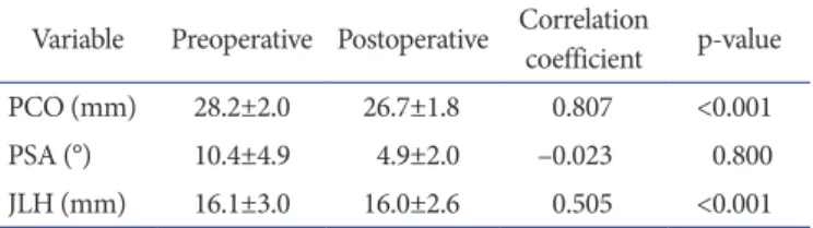
graphic evaluation method of the American Knee Society, the mean α, β, γ, and δ angles were 95.8°±0.9°, 90.2°±0.9°, 2.1°±1.1°, and 85.2°±2.2°, respectively. The average PCO was 28.2±2.0 mm preoperatively and 26.7±1.8 mm postoperatively (r=0.807, p<0.001). The average PSA was 10.4°±4.9° preoperatively and 4.9°±2.0° postoperatively (r=–0.023, p=0.800). The average JLH was 16.2±3.0 mm preoperatively and 16.1±2.6 mm postopera- tively (r=0.505, p<0.001) (Table 1).
3. correlation between changes in pco, pSa, and Jlh and the change in flexion angle
Changes in the PCO, PSA, and JLH after TKA did not influ- ence the postoperative flexion angle (p=0.878, 0.715, and 0.486, respectively) or the change therein (p=0.486, 0.146, and 0.540, respectively). The average postoperative flexion angle and change therein did not differ significantly between the PCO-inlier and PCO-outlier groups (p=0.323 and 0.571, respectively). The average postoperative flexion angle did not differ significantly between the PSA-inlier and PSA-outlier groups (p=0.986). The average flexion angle increased by 0.9°±11.9° in the PSA-inlier group, but decreased by 0.6°±6.8° in the PSA-outlier group; how- ever, this difference was not significant (p=0.718). The only inde- pendent factor that affected the postoperative flexion angle was the preoperative flexion angle (r=0.541, p=0.039).
The preoperative PCO was significantly correlated with the pre- operative flexion angle (r=0.226, p=0.012), but the preoperative PSA was not (p=0.067). The preoperative PCO was significantly correlated with the preoperative PSA (r=–0.362, p<0.001).
discussion
The most important finding of the present study was that the PCO and JLH did not change significantly after CR-TKA.
However, the postoperative PSA decreased by as much as 5.5°
on average, with a small range of variation. The average amount of decrease in the PCO after TKA was 1.5 mm, despite the use of an anterior reference guide system in the present study. We used the PFC Sigma instrument, in which PCO alteration can be reduced by adjusting the drill guide position and size of cut- ting block. The sizes of the femoral components used were 1.5, 2, 2.5, and 3, which are separated by 2–3-mm increments in the AP dimension. Although femoral components cannot exactly match femoral size in every knee, the authors’ efforts to reduce PCO alteration by shifting the AP cutting guide slightly back- ward reduced the decrease in the PCO. The restoration of normal JLH is important to regain normal knee kinematics and increase Table 1. Correlations between Preoperative and Postoperative PCO,
PSA, and JLH after Cruciate-Retaining Total Knee Arthroplasty Variable Preoperative Postoperative Correlation coefficient p-value
PCO (mm) 28.2±2.0 26.7±1.8 0.807 <0.001
PSA (°) 10.4±4.9 4.9±2.0 –0.023 0.800
JLH (mm) 16.1±3.0 16.0±2.6 0.505 <0.001
PCO: posterior condylar offset, PSA: posterior slope angle, JLH: joint line height.
mid-flexion stability20,21). The change in the JLH was 0.1 mm in the present study, which employed careful determination of the femoral and tibial bone resection levels and confirmation with caliper-obtained measurements of resected condylar thickness22). The change in the PSA was larger in the present study than in other studies5,13). Our preoperative PCO and PSA data were simi- lar to those of an anatomical study of normal knees in which the PSA exhibited greater variability than the PCO, and the PSA and PCO were correlated23). Patients with high preoperative PCOs tended to have small PSAs. We could imagine the concomitant effect of PCO and PSA on the individual flexion angle.
Although the preoperative PCO was significantly correlated with preoperative flexion angle in the present study, there are lots of factors affecting the flexion angle. Degenerative changes around the knee joint including osteophytes and loose bodies could also influence the flexion angle as mechanical obstacles.
However, they were removed clearly during TKA procedure and those qualified variables could not be evaluated in the present study.
A possible reason for the decrease in the PSA after TKA might be the wide variation in preoperative PSA values. While the variation in preoperative PCO and JLH values was small (Table 1), the standard deviation of the preoperative PSA was 4.9° and the values ranged from 1.6° to 21.2°. Several authors1,23) have also reported variable ranges of anatomical PSA in normal knees. The restoration of the PSA after TKA has been shown to delay tibio- femoral impingement, decrease the PCL load, enhance femoral roll back, and improve the flexion angle of the knee24,25). However, an excessive PSA in TKA has potentially deleterious effects15,26). It may induce AP instability, cause anterior subluxation of the tibial component, move the joint contact force location to eccentric positions, increase the wear on the posterior portion of the poly- ethylene surface, and cause aseptic loosening27-29). Because the posterior horn of the meniscus is thicker than the anterior horn, the biomechanical PSA determined with consideration of this structural feature differed from bony PSA measured on radio- graphs30). A previous study16) demonstrated the importance of the lateral PSA as a guide in order not to cut the tibia at more than 8°. We thought that the restoration of the original PSA in every case is impossible because of the high degree and wide range of variation in the preoperative PSA, and the excessively steep slope doesn’t have to be restored with the consideration of the possible various complications27-29). Postoperative PSAs are recommended variously according to prosthesis type due to diversity in the posterior offset of the femoral component and the slope of the polyethylene insert1). The proper cutting angle for the PSA is still
controversial, and a wide range of optimal angles has been pro- posed1). Generally, the PSA can be more reasonably set to 3° to 5°
of the posterior slope in the sagittal plane during the resection of the proximal tibia, and the tibial cut surface should be adjusted using a sharp electronic saw after confirmation of flexion and ex- tension gap balancing, PCL tension, and femoral roll back1).
Another important finding of the present study is that changes in the PCO, PSA, and JLH did not affect the postoperative flexion angle after CR-TKA. The only independent factor that affected the postoperative flexion angle was the preoperative flexion angle. There have been lots of previous studies about the impact of PCO and PSA on the postoperative flexion angle1,2,4,6). It was reported that the postoperative PCO and PSA influenced the postoperative flexion angle in some studies2,6) or they did not in other studies1,4,17). The restoration of the PCO delays tibiofemoral impingement and increases knee flexion, especially in cases with paradoxical femoral roll-forward6,25). However, the postoperative flexion angle is influenced by various factors, such as the severity of preoperative deformity, flexion angle of the femoral compo- nent, alteration of the PSA and flexion gap, PCL tension, soft- tissue balance, type of prosthesis, and rehabilitation2,3). Thus, the comparative analysis of clinical outcomes according to postopera- tive PCO is complicated by inherent statistical bias. Furthermore, the alteration in the postoperative PCO was small (average, –1.5 mm) in the present study, and only 11 outliers showed a decrease of more than 3 mm. Thus, the comparison of the flexion angle in relation to the postoperative PCO should be carefully interpreted.
The comparison of the flexion angle in relation to the postopera- tive JLH and PSA should also be carefully interpreted for similar reasons.
This study was also limited by its retrospective design and lack of a control group. Most patients were women, and all had osteo- arthritic knees with varus deformities. This female predominance in the distributions of osteoarthritis and varus deformity is com- mon in our populations because of the differences in disease de- mographics among ethnic groups and gender differences in the incidence of bowed legs. Another limitation was that the femoral component size and final PSA were determined by the predic- tion and experience of the corresponding author. However, the surgeon already had the sufficient experience of TKA (more than 200 TKAs annually), and his decision for sizing and balancing could improve the quality of our data. The strengths of the pres- ent study include the prospective collection of a single surgeon’s data and minimal loss to follow-up despite the retrospective na- ture of the study.
32
Song et al. Changes in PCO, PSA, and JLH after CR-TKAconclusions
The PCO and JLH did not change significantly after CR-TKA in the present study. The PSA decreased by 5.5° with a small range of variation. Those amounts of their changes did not affect the postoperative flexion angle after CR-TKA in the present study.
The Knee Society knee and function scores were satisfactory and the mean further flexion exceeded 130°, with admitted PSA alteration after CR-TKA. Restoration of the PCO and JLH could make the optimization of knee flexion in spite of the decreased PSA after CR-TKA. It is questionable whether surgeons should always restore the original PSA despite the wide range in this angle.
conflict of interest
No potential conflict of interest relevant to this article was re- ported.
references
1. Bae DK, Song SJ, Yoon KH, Noh JH, Moon SC. Compara- tive study of tibial posterior slope angle following cruciate- retaining total knee arthroplasty using one of three implants.
Int Orthop. 2012;36:755-60.
2. Fujimoto E, Sasashige Y, Masuda Y, Hisatome T, Eguchi A, Masuda T, Sawa M, Nagata Y. Significant effect of the poste- rior tibial slope and medial/lateral ligament balance on knee flexion in total knee arthroplasty. Knee Surg Sports Trauma- tol Arthrosc. 2013;21:2704-12.
3. Jia YT, Wang L, Zhang Y, Zhao C, Sun ZH, Liu J. Does mis- match of the femoral component aspect ratio influence the range of knee flexion after posterior-stabilized total knee arthroplasty? Chin J Traumatol. 2012;15:152-7.
4. Seo SS, Ha DJ, Kim CW, Choi JS. Effect of posterior condy- lar offset on cruciate-retaining mobile TKA. Orthopedics.
2009;32(10 Suppl):44-8.
5. Bellemans J, Robijns F, Duerinckx J, Banks S, Vanden- neucker H. The influence of tibial slope on maximal flexion after total knee arthroplasty. Knee Surg Sports Traumatol Arthrosc. 2005;13:193-6.
6. Bellemans J, Banks S, Victor J, Vandenneucker H, Moemans A. Fluoroscopic analysis of the kinematics of deep flexion in total knee arthroplasty: influence of posterior condylar off- set. J Bone Joint Surg Br. 2002;84:50-3.
7. Dennis DA, Kim RH, Johnson DR, Springer BD, Fehring
TK, Sharma A. The John Insall Award: control-matched evaluation of painful patellar Crepitus after total knee ar- throplasty. Clin Orthop Relat Res. 2011;469:10-7.
8. Del Gaizo DJ, Della Valle CJ. Instability in primary total knee arthroplasty. Orthopedics. 2011;34:e519-21.
9. Kansara D, Markel DC. The effect of posterior tibial slope on range of motion after total knee arthroplasty. J Arthroplasty.
2006;21:809-13.
10. Kim JH. Effect of posterior femoral condylar offset and posterior tibial slope on maximal flexion angle of the knee in posterior cruciate ligament sacrificing total knee arthro- plasty. Knee Surg Relat Res. 2013;25:54-9.
11. Youm YS, Cho SD, Eo J, Park KB, Lee SH. Can we use the posterior condylar off set as a predictive factor for overhang of the implant in total knee arthroplasty? Knee Surg Relat Res. 2011;23:149-52.
12. Ishii Y, Noguchi H, Takeda M, Sato J, Toyabe S. Posterior condylar offset does not correlate with knee flexion after TKA. Clin Orthop Relat Res. 2013;471:2995-3001.
13. de Boer JJ, Blankevoort L, Kingma I, Vorster W. In vitro study of inter-individual variation in posterior slope in the knee joint. Clin Biomech (Bristol, Avon). 2009;24:488-92.
14. Ewald FC. The Knee Society total knee arthroplasty roent- genographic evaluation and scoring system. Clin Orthop Relat Res. 1989;(248):9-12.
15. Wasielewski RC, Galante JO, Leighty RM, Natarajan RN, Rosenberg AG. Wear patterns on retrieved polyethylene tibial inserts and their relationship to technical consider- ations during total knee arthroplasty. Clin Orthop Relat Res.
1994;(299):31-43.
16. Kuwano T, Urabe K, Miura H, Nagamine R, Matsuda S, Satomura M, Sasaki T, Sakai S, Honda H, Iwamoto Y. Im- portance of the lateral anatomic tibial slope as a guide to the tibial cut in total knee arthroplasty in Japanese patients. J Orthop Sci. 2005;10:42-7.
17. Shi X, Shen B, Kang P, Yang J, Zhou Z, Pei F. The effect of posterior tibial slope on knee flexion in posterior-stabilized total knee arthroplasty. Knee Surg Sports Traumatol Ar- throsc. 2013;21:2696-703.
18. In Y, Kim JM, Woo YK, Choi NY, Sohn JM, Koh HS. Fac- tors affecting flexion gap tightness in cruciate-retaining total knee arthroplasty. J Arthroplasty. 2009;24:317-21.
19. Hsu WH, Hsu RW, Weng YJ. Effect of preoperative defor- mity on postoperative leg axis in total knee arthroplasty: a prospective randomized study. Knee Surg Sports Traumatol Arthrosc. 2010;18:1323-7.
20. Porteous AJ, Hassaballa MA, Newman JH. Does the joint line matter in revision total knee replacement? J Bone Joint Surg Br. 2008;90:879-84.
21. Cope MR, O’Brien BS, Nanu AM. The influence of the pos- terior cruciate ligament in the maintenance of joint line in primary total knee arthroplasty: a radiologic study. J Arthro- plasty. 2002;17:206-8.
22. Bae DK, Song SJ, Yoon KH, Noh JH, Lee CH. Intraop- erative assessment of resected condyle thickness in total knee arthroplasty. Knee Surg Sports Traumatol Arthrosc.
2012;20:2039-46.
23. Cinotti G, Sessa P, Ripani FR, Postacchini R, Masciangelo R, Giannicola G. Correlation between posterior offset of femo- ral condyles and sagittal slope of the tibial plateau. J Anat.
2012;221:452-8.
24. Banks S, Bellemans J, Nozaki H, Whiteside LA, Harman M, Hodge WA. Knee motions during maximum flexion in fixed and mobile-bearing arthroplasties. Clin Orthop Relat Res.
2003;(410):131-8.
25. Massin P, Gournay A. Optimization of the posterior condy- lar offset, tibial slope, and condylar roll-back in total knee
arthroplasty. J Arthroplasty. 2006;21:889-96.
26. Migaud H, De Ladoucette A, Dohin B, Cloutier JM, Gou- geon F, Duquennoy A. Influence of the tibial slope on tibial translation and mobility of non-constrained total knee pros- thesis. Rev Chir Orthop Reparatrice Appar Mot. 1996;82:7- 13.
27. Jojima H, Whiteside LA, Ogata K. Effect of tibial slope or posterior cruciate ligament release on knee kinematics. Clin Orthop Relat Res. 2004;(426):194-8.
28. Catani F, Leardini A, Ensini A, Cucca G, Bragonzoni L, Toksvig-Larsen S, Giannini S. The stability of the cemented tibial component of total knee arthroplasty: posterior cru- ciate-retaining versus posterior-stabilized design. J Arthro- plasty. 2004;19:775-82.
29. Bai B, Baez J, Testa N, Kummer FJ. Effect of posterior cut an- gle on tibial component loading. J Arthroplasty. 2000;15:916- 20.
30. Jenny JY, Rapp E, Kehr P. Proximal tibial meniscal slope: a comparison with the bone slope. Rev Chir Orthop Repara- trice Appar Mot. 1997;83:435-8.