Coracoclavicular Ligament Suture Augmentation with Anatomical Locking Plate Fixation for Distal Clavicle Fracture
Tae Kang Lim*, Min Soo Shon1*, Hyung Gon Ryu2, Jae Sung Seo2, Jae Hyun Park, Young Ko3, Kyoung-Hwan Koh4
Department of Orthopaedic Surgery, Eulji Hospital, Eulji University School of Medicine, 1Department of Orthopaedic Surgery, National Medical Center, 2Department of Orthopaedic Surgery, Seoul Medical Center, Seoul, 3College of Nursing, Gachon University, Incheon, 4Department of Orthopaedic Surgery, Inje University Ilsan Paik Hospital, Goyang, Korea
Background: For Neer type IIB fracture of distal clavicle with coracoclavicular ligament injury, various surgical treatments have been used in literatures. However, there was no consensus on the optimal treatment. The aim of this study is to report the clinical and radio- logical results of open reduction and internal fixation of unstable distal clavicle fracture and suture augmentation of disrupted coracocla- vicular ligament.
Methods: A prospective study was performed in 23 patients with Neer type IIB distal clavicle fracture in Seoul Medical Center, Eulji Hospital, and National Medical Center. Firstly, suture anchors are inserted in the base of coracoid process and preliminary reduction was achieved by tie-off of three suture limbs around the clavicle. Then, the final fixation was completed with anatomical locking plate. Bony union and the distance between coracoclavicular ligaments were evaluated. Clinical results and complications including stiffness and sec- ondary procedures were evaluated.
Results: Bony union was achieved in all cases except one (22 of 23). At mean 14.9 months, no significant difference in the mean cora- coclavicular distance was observed compared to uninjured shoulder (8.2 ± 7.9 mm versus 7.3 ± 3.4 mm, p=0.14). Pain visual ana- logue scale, American Shoulder and Elbow Surgeons score, Constant score, and Disabilities of the Arm, Shoulder and Hand score were 0.5, 83.4, 78.5, and 6.2, respectively. Revision surgery was performed in one case of nonunion. Four patients who complained of skin irritation underwent implant removal.
Conclusions: In cases of an unstable distal clavicle fracture with coracoclavicular ligament disruption, satisfactory clinical results were obtained by locking plate fixation and coracoclavicular ligament suture augmentation concurrently.
(Clin Shoulder Elbow 2014;17(4):175-180)
Key Words: Shoulder fractures; Clavicle; Plate; Coracoclavicular joint
Introduction
Clavicle fractures account for 2.6% of all fractures and this means an annual incidence rate of 64 per 100,000 popula- tions.1,2) In particular, distal clavicle fracture represents approxi- mately 10 to 15% of all clavicle fractures.3) Of these, especially, Neer type IIB clavicle fracture is inherently unstable because coracoclavicular (CC) ligament injury is accompanied, and high nonunion or malunion rate of more than 20% after nonoperative
treatment has been reported.1,4) Therefore, surgical treatment is generally considered for Neer type IIB clavicle fracture.3,5)
Various surgical procedures for the treatment of this have been reported and in recent years. Treatment options include transacromial K-wires,6) temporary CC screw fixation,7) tension band suturing,8) and arthroscopic stabilization.9) The procedure using a hook-plate10-12) or an anatomical locking plate has been increasingly used.13) However, each of these techniques come with specific benefits and disadvantages or complication such as Clinics in Shoulder and Elbow
Copyright © 2014 Korean Shoulder and Elbow Society. All Rights Reserved. pISSN 2383-8337
Clinics in Shoulder and Elbow Vol. 17, No. 4, December, 2014 http://dx.doi.org/10.5397/cise.2014.17.4.175
Received August 9, 2014. Revised October 14, 2014. Accepted October 17, 2014.
Correspondence to: Kyoung-Hwan Koh
Department of Orthopaedic Surgery, Inje University Ilsan Paik Hospital, Juhwa-ro, 170 Ilsanseo-gu, Goyang 411-706, Korea Tel: +82-31-910-7995, Fax: +82-31-910-7967, E-mail: [email protected]
*Tae Kang Lim and Min Soo Shon contributed equally to this article as co-first authors.
Financial support: None. Conflict of interests: None.
non-union, implant failure,14) pin migration,6) bony defects and subacromial impingement.10) A consensus has not been achieved yet on the issue of optimal surgical technique for Neer type IIB clavicle fracture.
Small distal fragment often precludes adequate screws pur- chase, which is the case in old-aged patients with osteoporosis.15) Furthermore, superior displacement of medial fragment by mus- cle action is aggravated by disrupted CC ligament.16) We pos- tulated that a small distal fragment can be fixed with a locking plate and superiorly displacing force by disrupted CC ligament can be successfully neutralized with use of suture augmentation.
Accordingly, the purpose of this study is to report the clinical and radiological results of open reduction and internal fixation of unstable distal clavicle fracture and suture augmentation of disrupted CC ligament.
Methods
Patient Collection
From March 2011 to December 2012, a prospective study was performed in the patients, who need surgical treatment, with an unstable distal clavicle fracture by using in three different hospitals. For the radiographic evaluation, an antero-posterior radiograph and a lordotic view of the clavicle were taken. The inclusion criteria was acute type IIB distal clavicle fractures ac- cording to the Neer classification and we excluded the patients
who had damage on the ipsilateral upper extremity, a history of fracture or surgery in the ipsilateral shoulder joint, other medical problem or poor general condition that are inevitable to delay surgery for clavicle injuries. During the study period, 23 patients who meet the criteria were analyzed (19 men, 4 women; mean age 48 ± 25).
This Study was approved by the Institutional Review Board of Seoul Medical Center (Seoul MC IRB study number assigned:
2012-028).
Surgical Technique
All patients received CC ligament suture augmentation and internal fixation of fracture with a locking plate. The surgery was performed by three different surgeons. However, anesthetic and surgical techniques, and post-surgical rehabilitation were equally provided to all patients. The patient was placed in beach chair position for the surgery under general anesthesia. The standard sabre-cut or transverse incision was made on the site of a distal clavicle fracture and the fracture site was exposed. After the frac- ture site was confirmed, the dorsal surface of coracoid process base was exposed in order to perform CC ligament suture aug- mentation. We used 2 methods for CC ligament fixation. Firstly, the temporary reduction on the fracture site was achieved by manual reduction and the sling procedure was performed using
#2 Ethibond (Ethicon, Somerville, NJ, USA) looped around the coracoid process and tied over a clavicle. Another method was
Fig. 1. Photograph taken after insertion of triple loaded suture anchor into
the basal part of coracoid process. Fig. 2. Sutures was looped around a clavicle and tied off in maintaining the reduction of a medial fragment of the clavicle.
Fig. 3. (A) Preoperative radiograph of un- stable distal clavicle fracture. (B) Radiograph showing final application of anatomical lock- ing plate and suture augmentation.
A B
that of using suture anchor. The 4.5-mm bio-absorbable screws (CrossFT suture anchor; ConMed Linvatec, Largo, FL, USA) load- ed with three strands of suture was inserted into the basal part of coracoid process (Fig. 1). Sutures was looped around a clavicle and tied off in maintaining the reduction of a bone fragment (Fig. 2). The small distal fragment was fixed using 3.5-mm lock- ing compression Clavicle Shaft Plate (Synthes, Paoli, PA, USA) and locking screw which were designed to fit for the anatomical shape of the distal clavicle (Fig. 3) after the final confirmation of anatomical reduction of fracture.
Rehabilitation
After the surgery, the surgery site was protected by wearing a sling for approximately 3 weeks. Range of motion (ROM) ex- ercise of finger and wrist was permitted right after the surgery.
Scapular stabilizing exercise and elbow flexion/extension exer- cise were allowed to perform intermittently. Three weeks after the surgery, sling was taken off and the patients started perform- ing passive ROM exercise. As recovering passive ROM, active ROM exercise and strengthening exercise were performed gradually.
Evaluation
At final follow-up, the bony union was evaluated and the CC distance in both injured and uninjured shoulder was measured by radiographic examinations. For the radiographic evalua- tion, an antero-posterior radiograph and a lordotic view of the clavicle were taken. For analyzing the period of bony union, we defined the bony union as the period when there was no pain at the fracture site and callus formation was observed on three sides among 4 sides of cortical bone.4) The CC distance was defined as measuring the shortest distance between the
peak of coracoid and the lower boundary of clavicles.13) Clinical results were evaluated by visual analogue scale score for pain (PVAS), American Shoulder and Elbow Surgeons (ASES) score,17) Constant score,18) Disabilities of the Arm, Shoulder and Hand (DASH) score,19) and ROM of the shoulder joint. Moreover, we assessed the presence of complications during the surgery or the postoperative period. We additionally investigated whether any secondary surgery, including implant removal, was performed or not due to any reason. In order to confirm the clinical results of patients who did not visit the hospital after the union was achieved, we called them at least one year after the surgery and questioned about PVAS, ASES score and any other discomfort.
In this process, we recommended the patients, who complained of discomfort, to see a doctor for re-examination. It was impos- sible to evaluate ROM objectively because the patients mostly did not visit the hospital 3 months after the surgery. However, we intended to figure out it even indirectly using the notion of patient defined stiffness and performed the telephone survey.
The patient defined stiffness is a clinically more reliable method and its criteria are not a threshold system on physical examina- tions by the inspector but the discomfort that the patient feels during ROM exercise.20) Patients were questioned whether they had the discomfort during ROM exercise after their surgery and we regarded that the patients have postoperative stiffness if they complained of it.
Statistical analysis was performed using PASW Statistics ver.
18.0 (IBM Co., Armonk, NY, USA) and p-values below 0.05 were considered significant.
Results
As a result of radiographic evaluation, the bony union was
C
A B
Fig. 4. (A) Initial postoperative radiograph in a patient showed nonunion. (B) Radiograph at 3 months after surgery showing nonunion in same patient. (C) Another case with screw loosening.
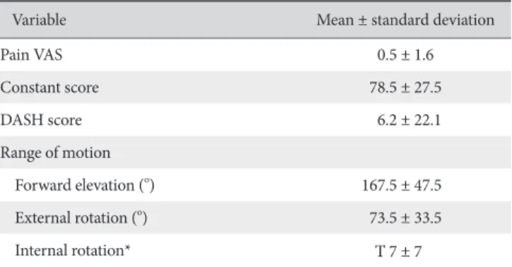
achieved in all cases except for 1 case (95.7% of union rate). At final follow-up (mean 14.9 months; range 12 to 23 months after the surgery), there was no significant difference in the mean CC distance (p=0.14). It was 8.2 ± 7.9 mm on the affected side and 7.3 ± 3.4 mm on the unaffected side. The preoperative mean distance between the fractured clavicle and the coracoid process was 15.1 ± 7.7 mm on the affected side. Except for 1 patient with nonunion, all patients achieved the bony union at the mean 2.8 months after the surgery. At 14.9 months follow- up, we obtained good clinical results as follows: the results of ROM were 168 degree of forward elevation, 74 degree of exter- nal rotation and T7 of internal rotation. In addition, PVAS, ASES, constant score and DASH score were 0.5, 83.4, 78.5, and 6.2, respectively. Complications after the surgery were nonunion in 1 patient (Fig. 4B), screw loosening in 1 patient (Fig. 4C). In one patient, nonunion developed at 3 months postoperatively, and underwent re-surgery using bone graft and a locking plate and could succeed to acquire the bony union finally. Implant remov- al was carried out in 4 patients who complained of skin irritation due to the plates. There were no other major complications such as infection. Also, all patients did not complain of stiffness during follow-up period. Results are summarized in Table 1 and 2.
Discussion
This study proved that the successful results can be achieved by performing conoid ligament suture augmentation with lock- ing plate fixation for unstable distal clavicle fracture (Neer type IIB) with conoid ligament disruption. Surgical treatment has been recommended for an unstable distal clavicle fracture and many studies have introduced several surgical methods.15) Recently, open reduction and internal fixation using a hook plate has been more widely used for Neer type IIB fracture of distal clavicle because it is easier and faster than other surgical methods with excellent results.10) On the other hand, this method may induce the complications such as subacromial impingement, rotator cuff damage, acromion fracture, and hook cut-out.3) There are some patients who suffer from severe shoulder pain after hook plate fixation. Because a forward elevation is limited over a period of time, the patients sometimes complain of postopera- tive stiffness.3) In order to prevent these complications, removal should be performed as soon as possible after the bony union
is achieved.15) Even though the results of this surgery are favor- able, this is the insurmountable disadvantage to have the surgery twice (implant removal) comparing with other treatment that usually solve the problems by undergoing the surgery only once.
In this study, we achieved the successful results from open re- duction and internal fixation using anatomical locking plate and conoid ligament suture augmentation for Neer type IIB fracture of distal clavicle. In this surgical procedure, primary stability of fracture reduction is obtained by performing conoid ligament suture augmentation first and it helps the plate insertion easier.
Conoid augmentation sutures may neutralize superior displac- ing force and facilitate healing in the early postoperative period.
In addition, secondary operations such as implant removal are not necessary unless there is complication, such as skin irrita- tion by plate. There is also no risk of the complications such as erosion or cut-through of acromion as like those of the surgery using hook plate. The surgical procedure using a hook plate is the method to lead the bony union by achieving the indirect reduction of fracture site and neutralizing the superior displacing force. However, the procedure of this study is the way to induce fracture healing by anatomic reduction and fixation of fracture.
Similar methods have been reported recently. Modern locking T-plate and suture anchors or looped sutures for CC augmentation provided over 94% union rates with few com- plications.13,16,21) Furthermore, a recent biomechanical study demonstrated that CC fixation adjunct to locking plate improved stability of type IIB fractures in cadaveric specimens.22) Neverthe- less, there was a case of nonunion in our case series (Fig. 4B).
The initial reduction was not acquired successfully in this case (Fig. 4A). While the gap was remained significantly in the lower margin of fracture, the upper margin was contacted well. The plate fixation and CC augmentation suture were done in this situation and the nonunion was occurred. This represents how important the achievement of initial reduction is. However, in this case, we consider that the reduction of lower margin was Table 1. Comparisons of Coracoclavicular Distance between Injured and
Uninjuried
Variable Mean ± standard deviation
Injured (mm) 8.2 ± 7.9
Uninjured (mm) 7.3 ± 3.4
p-value: 0.14.
Table 2. Clinical Results
Variable Mean ± standard deviation
Pain VAS 0.5 ± 1.6
Constant score 78.5 ± 27.5
DASH score 6.2 ± 22.1
Range of motion
Forward elevation (o) 167.5 ± 47.5
External rotation (o) 73.5 ± 33.5
Internal rotation* T 7 ± 7
VAS: visual analogue scale, DASH: Disabilities of the Arm, Shoulder and Hand.
*Internal rotation was measured according to the place on the back that could be reached by the thumb.
also difficult to achieve even though the indirect reduction was attempted with a hook plate. A locking screw was pulled out in another case. In our opinion, motion of acromioclavicu- lar joint existed during ROM exercise and this could affect to screw pull-out. Furthermore, in this case, screw position that inserted in the acromioclavicular joint could affect to pull-out, too. However, the fracture healing was completed successfully.
On the other hand, the patients included in this study did not complain of postoperative stiffness because they could perform ROM exercise relatively early after the surgery. This early ROM is also considered as another advantage of our surgical treatment comparing to the surgery with a hook plate that can causes stiff- ness significantly due to the motion limitation before implant removal.
This study has limitation of the small sample size and the short follow-up period. Even though we collected the data pro- spectively, the long term follow-up for ROM and clinical score was difficult because the patient rarely visited the hospital after the bony union was achieved and the discomfort was disap- peared. Also, there is another limitation that this is not a compar- ative study with the surgery using a hook plate or other surgeries.
Those patients involved in this study might be heterogeneous because three surgeons were involved and they had some minor differences in their surgical procedures and rehabilitation. How- ever, the results from three surgeons in three different hospitals could provide an external validity of our technique in this study.
Conclusion
In cases of an unstable distal clavicle fracture (Neer type IIB) with CC ligament disruption, the satisfactory bony union and clinical results were obtained by the surgical approach to con- duct locking plate fixation and CC ligament suture augmentation concurrently.
References
1. Flinkkilä T, Heikkilä A, Sirniö K, Pakarinen H. TightRope ver- sus clavicular hook plate fixation for unstable distal clavicular fractures. Eur J Orthop Surg Traumatol. 2014 [Epub ahead of print].
2. Lee SK, Park JS, Choy WS. Locking compression plate distal ulna hook plate as alternative fixation for fifth metatarsal base fracture. J Foot Ankle Surg. 2014;53(5):522-8.
3. Oh JH, Kim SH, Lee JH, Shin SH, Gong HS. Treatment of distal clavicle fracture: a systematic review of treatment modalities in 425 fractures. Arch Orthop Trauma Surg. 2011;131(4):525-33.
4. Nunez FA Jr, Li Z, Campbell D, Nunez FA Sr. Distal ulna hook plate: angular stable implant for fixation of distal ulna. J Wrist Surg. 2013;2(1):87-92.
5. Neer CS 2nd. Fracture of the distal clavicle with detachment of
the coracoclavicular ligaments in adults. J Trauma. 1963;3:99- 110.
6. Flinkkilä T, Ristiniemi J, Lakovaara M, Hyvönen P, Leppilahti J.
Hook-plate fixation of unstable lateral clavicle fractures: a re- port on 63 patients. Acta Orthop. 2006;77(4):644-9.
7. Fazal MA, Saksena J, Haddad FS. Temporary coracoclavicular screw fixation for displaced distal clavicle fractures. J Orthop Surg (Hong Kong). 2007;15(1):9-11.
8. Badhe SP, Lawrence TM, Clark DI. Tension band suturing for the treatment of displaced type 2 lateral end clavicle fractures.
Arch Orthop Trauma Surg. 2007;127(1):25-8.
9. Nourissat G, Kakuda C, Dumontier C, Sautet A, Doursounian L.
Arthroscopic stabilization of Neer type 2 fracture of the distal part of the clavicle. Arthroscopy. 2007;23(6):674.e1-4.
10. Kashii M, Inui H, Yamamoto K. Surgical treatment of distal clavicle fractures using the clavicular hook plate. Clin Orthop Relat Res. 2006;447:158-64.
11. Lee SK, Park JS, Choy WS. LCP distal ulna hook plate as alter- native fixation for fifth metatarsal base fracture. Eur J Orthop Surg Traumatol. 2013;23(6):705-13.
12. An WJ, Sun JB, Ye P, Guo WW. Comparative study on the treatment of acromioclavicular joint dislocation: coracocla- vicular ligament reconstruction combined with hook plate fixation or suture-anchor fixation. Zhonghua Wai Ke Za Zhi.
2013;51(4):349-53.
13. Herrmann S, Schmidmaier G, Greiner S. Stabilisation of verti- cal unstable distal clavicular fractures (Neer 2b) using locking T-plates and suture anchors. Injury. 2009;40(3):236-9.
14. Haidar SG, Krishnan KM, Deshmukh SC. Hook plate fixation for type II fractures of the lateral end of the clavicle. J Shoulder Elbow Surg. 2006;15(4):419-23.
15. Yoo JH, Chang JD, Seo YJ, Shin JH. Stable fixation of distal clav- icle fracture with comminuted superior cortex using oblique T-plate and cerclage wiring. Injury. 2009;40(4):455-7.
16. Kalamaras M, Cutbush K, Robinson M. A method for internal fixation of unstable distal clavicle fractures: early observations us- ing a new technique. J Shoulder Elbow Surg. 2008;17(1):60-2.
17. Ye JK, Yu BJ, Ye FS, Hong JY, Wang W, Tong PJ. Case-control study on therapeutic effects between modified Weaver-Dunn surgery and clavicular hook plate fixation in the treatment of acromioclavicular joint dislocation. Zhongguo Gu Shang.
2014;27(1):4-8.
18. Kaku N, Hara K, Tabata T, Tsumura H. Influence of the volume of bone defect, bone grafting methods, and hook fixation on stress on the Kerboull-type plate and screw in total hip arthro- plasty: three-dimensional finite element analysis. Eur J Orthop Surg Traumatol. 2014 [Epub ahead of print].
19. Gu X, Cheng B, Sun J, Tao K. Arthroscopic evaluation for omal- gia patients undergoing the clavicular hook plate fixation of distal clavicle fractures. J Orthop Surg Res. 2014;9:46.
20. Chen CY, Yang SW, Lin KY, et al. Comparison of single cora-
coclavicular suture fixation and hook plate for the treatment of acute unstable distal clavicle fractures. J Orthop Surg Res.
2014;9:42.
21. Webber MC, Haines JF. The treatment of lateral clavicle frac- tures. Injury. 2000;31(3):175-9.
22. Madsen W, Yaseen Z, LaFrance R, et al. Addition of a suture anchor for coracoclavicular fixation to a superior locking plate improves stability of type IIB distal clavicle fractures. Arthros- copy. 2013;29(6):998-1004.