DOI : 10.3341/jkos.2008.49.6.871
구상돌기절제술을 함께 시행한 코경유 눈물주머니코안연결술의 임상적 고찰
김정림․양재욱
인제대학교 의과대학 안과학교실, 인제대학교 의과대학 부산백병원 안과학연구재단
목적 : 구상돌기절제술이 코경유 눈물주머니코안연결술 시 눈물주머니를 찾는 지표로써의 유용성과 수술의 성공률에
미치는 영향에 대해 알아보고자 한다.
대상과 방법 : 2003년부터 2006년까지 코눈물관 막힘을 진단받고 코경유 눈물주머니코안연결술과 함께 구상돌기절
제술을 시행 받은 후 6개월간 경과관찰이 가능하였던 97명, 102안을 대상으로 수술 전 코 안 상태를 관찰하고 코 안 이상소견이 있었던 경우와 없었던 경우로 나누어 수술 후 증상 호전 정도와 염색약소실검사를 이용하여 수술성공률을 분석하였다.
결과 : 수술 전 코 안에 이상소견이 있었던 경우가 102안 중 64안으로 62.7%였고 수술성공률은 90.6%로 코 안에 이상소견이 없었던 경우와 비교하였을 때 통계적으로 유의한 차이를 보이지 않았다(p>0.99). 술 후 안와 지방탈출, 뇌 척수액누출, 지연성 출혈 등의 합병증은 보이지 않았다.
결론 : 코경유 눈물주머니코안연결술을 시행할 때 구상돌기의 앞쪽에 절제술을 시행하는 것은 눈물주머니를 노출시키
는 것을 용이하게 하고 정확한 위치에 골공을 형성하게 함으로써 수술성공률을 높이는데 기여할 것으로 사료된다.
<한안지 49(6):871-877, 2008>
<접수일 : 2007년 11월 20일, 심사통과일 : 2008년 2월 26일>
통신저자 : 양 재 욱
부산시 진구 개금동 633-165 인제대학교 부산백병원 안과 Tel: 051-890-6016, Fax: 051-890-6329 E-mail: [email protected]
* 본 논문의 요지는 2007년 대한안과학회 제98회 추계학술대회 에서 구연으로 발표되었음.
눈물흘림을 호소하는 코눈물관 막힘 환자의 수술적 치 료로 눈물주머니(lacrimal sac)와 코오목(nasal fossa) 사이에 눈물주머니오목(lacrimal fossa)을 절제하여 코눈물관 통로의 영구적인 지름길을 만들어 주는 눈물 주머니코안연결술(dacryocystorhinostomy)이 널리 이용되고 있다. 내측눈구석 부위의 피부절개를 이용한 눈 물주머니코안연결술(external dacryocystorhinostomy) 은 접근의 용이성과 높은 성공률로 인해 많이 시행되 어 왔으며 코경유 눈물주머니코안연결술(endonasal dacryocystorhinostomy)은 내시경이 발달하면서 피부반흔이 없고 혈종생성이 적으며 회복이 빠른 장점 과 함께 피부절개와 비슷한 성공률을 보이므로 최근 많 이 시술되고 있다.1-3 하지만 코경유 눈물주머니코안연
결술을 시행하는데 있어서 코 안에 이상이 있는 경우 골공(osteotomy) 위치와 수술적 접근법에 관한 확립 된 지침이 없다.
눈물주머니오목의 구조는 앞쪽으로 두꺼운 위턱뼈 (maxillary bone)의 이마돌기(frontal process)와 뒤쪽으로 얇은 눈물뼈(lacrimal bone)로 구성되며 안 쪽으로 벌집굴 공기집(ethmoidal air cell)과 중간코 선반(middle turbinate)과 경계를 이루고 있다. 코 안에서 보았을 때 눈물주머니는 안와의 눈물주머니오목 에 위치하며 중간코선반 앞부분 아래에 위치하는데 이 곳은 눈물주머니코안연결술 시 골공이 만들어 지는 부 위로 벌집뼈(ethmoid bone)와 중간코선반등의 구조 에 해부학적 변이가 많이 나타날 수 있으므로 이 부위 를 골공을 만드는 지표로 사용하기는 어렵다. 그러나 구상돌기(uncinate process)는 비강측벽의 전상방 에서 후하방으로 향하는 약 19~32 mm 길이의 낫 모양 으로 생긴 얇은 뼈로 앞쪽으로는 공통눈물소관(common canaliculi) 위치에서 눈물뼈의 안쪽에 부착하므로 눈 물주머니코안연결술 시 골공을 형성하는 지표로 사용할 수 있다.4-6
저자는 코눈물관폐쇄를 보이는 환자에서 코경유 눈 물주머니코안연결술과 구상돌기절제술을 함께 시행받
은 환자 97명, 102안을 후향적으로 분석하여 코경유 눈물주머니코안연결술을 시행할 때 눈물주머니를 찾는 지표로써 구상돌기의 유용성과 구상돌기절제술을 시행 하는 것이 수술의 성공률에 미치는 영향에 대해 알아보 고자 하였다.
대상과 방법
2003년 1월부터 2006년 12월까지 본원 안과에서 코눈물관 막힘을 진단받고 코경유 눈물주머니코안연결 술과 함께 구상돌기절제술을 시행 받았으며 6개월 이상 경과관찰이 가능하였던 97명, 102안을 대상으로 술 전 코 안 상태, 수술의 성공률과 합병증에 관한 후향적인 분석을 시행하였다.
코눈물관 폐쇄의 진단은 식염수관류검사(lacrimal irrigation)를 시행하여 눈물기관폐쇄 여부를 검사하고 더 듬자검사(probing)를 시행하여 눈물소관의 막혀있는 부위 를 알아보았으며 눈물길조영술(dacryocystography) 을 통해 확진하였다. 술 전 코내시경을 이용하여 코 안 검사를 통해 이상소견과 눈물배출을 방해하는 질환이나 폐쇄성 원인이 있는지를 관찰하여 코 안에 이상이 있었 던 경우를 A군, 코 안에 이상이 없는 경우를 B군으로 분류하였다.
수술은 전신마취 하에서 4% lidocaine과 1:1,000 으로 희석된 epinephrine 용액을 적신 거즈를 중비도 를 포함한 코 안에 팩킹하여 코점막을 충분히 수축시킨 다음 아래눈물점(lower puntum)을 확장시킨 후 초 자체 수술용 20 gauge 굴곡광원의 끝을 아래눈물점을 통해 눈물주머니 속에 밀어 넣고 코 안에 4 mm, 0°내 시경을 진입 시킨 후 광원이 가장 잘 투영되는 중비도 의 코점막, 중간코선반의 일부 및 코중격에 국소마취제 를 주입하고 광원이 투영되는 눈물주머니부위의 코점막 앞쪽, 구상돌기의 상부부착부위 앞쪽에서 하부로 Freer 골막거상기를 이용하여 절개를 가한 후 중비도 점막을 벗기고 사골겸자로 점막편을 일부 제거한 다음 코 안의 해부학적인 상태에 맞추어 구상돌기 앞부분을 절제하였다.
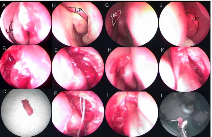
Paradoxical medial rotation 된 중간코선반와 구상돌기가 돌출되어 중비도가 좁아져 있는 경우는 돌 출된 구상돌기의 앞부분을 절제하여 쉽게 중비도를 넓 히고 골공을 형성하여 눈물주머니를 노출시켜 내벽을 제거하고 실리콘관을 삽입할 수 있었다(Fig. 1A, B, C).
위턱뼈의 이마돌기와 중간코선반 사이에 구상돌기가 숨겨져 있고 투과조명으로 눈물주머니의 위치를 확인하 기 어려운 경우에 중간코선반을 freer 골막거상기로
medialization 시켜 구상돌기를 노출시키고 앞부분을 절제한 후 골편을 제거하여 눈물주머니의 노출시키고 정확한 위치에 골공을 형성 할 수 있었다(Fig 1D, E, F, G, H, J, K).
구상돌기 절제 부위에 Kerrison punch로 골천공 을 만든 후 골편은 완전히 제거하였다(Fig. 1I, L).
눈물주머니 표면이 노출된 후 광원을 상하로 움직여 눈 물주머니의 위치와 크기를 파악하고 광원의 앞쪽으로 눈 물주머니 표면에 수직으로 아래에서 위로 keratome 을 사용하여 최소 5 mm의 수직절개를 가하고 절개선 의 위와 아래 끝에서 앞쪽으로 절개를 연장시켜 “ㄷ”자 절편을 형성한 후 사골겸자를 이용하여 절편을 제거하 였다. 굴곡광원이 코 안으로 비쳐 보이면 내시경과 굴 곡 광원의 끝을 이용하여 눈물주머니절개부위를 다시 확인하고 주사기법으로 식염수의 통과를 확인한 후 실 리콘 관의 양쪽 끝을 상, 아래눈물점을 통해 코안으로 통과시키고 양끝을 6회 정도 매듭으로 결찰하여 누공의 3~5 mm에 위치시켰다. 실리콘관의 끝이 코 안에 있 음을 확인 후 수술용 장갑의 손가락부분을 잘라 내부에 Merocel (Xomed, USA)로 패킹한 뒤 술 후 1일째 에 제거하였다.
술 후 첫 한달은 매주 1회, 2개월째는 2주에 1회 그 이후는 매달 1회 관찰하였으며 실리콘 관은 보통 술 후 3개월에 제거하였다. 술 후 항생제와 스테로이드 안약 을 점안하고 코 안에 가피형성을 줄이기 위해 환자 스 스로 생리식염수로 비강세척을 한 달간 하루에 1~2회 실시하도록 하였다.
주기적 외래경과관찰 시마다 환자의 증상 호전 여부 를 문진하고, 주사기법으로 누기의 개통여부를 확인하 였으며 코 안을 코내시경으로 검사하여 가피와 분비물 을 제거하였고, 골공 주위의 막성조직, 육아조직이 형 성된 경우 적절한 치료를 하였으며, 중간코선반 및 코 중격 유착 유무확인 및 치료를 시행하였다.
수술 후 눈물흘림증상 호전 정도와 염색약 소실검사 (dye disappearance test)를 이용하여 결과를 후향 적으로 분석하였다. 염색약 소실검사는 염색약 점안 후 5분 뒤의 결과를 0에서 +4까지 등급으로 나누는데 염 색약이 결막낭 내에 전혀 남아있지 않으면 0, 염색약이 거의 그대로 남아있으면 +4로 하였다. 본 연구에서는 수술의 성공을 최종관찰까지 눈물흘림증상이 없어지고 염색약소실검사상 +1 이하인 경우로 하였다. 술 후 경 과관찰을 통하여 A군과 B군의 성공률과 수술 실패의 원인으로 생각되는 후기 합병증 유무를 확인하여 비교 하였다. 통계적 분석은 SPSS통계프로그램을 이용한 Fisher's exact test를 사용하였다.
Table 1. Characteristics of patients Characteristics
Age (years) 48.5±12.8
Male : Female (eyes) 12:90
Follow up (months) 10.1±2.0
Intubation period (months) 4.8±3.4
Figure 1. Endoscopic sequence of a typical endonasal dacryocystorhinostomy procedure, including primary resection of the anterior uncinate process. (A) Preoperative appearance of a left nasal fossa showing the uncinate process (UP) protruding under the mucosa, between the nasal ridge of the maxillary bone (MB) and the middle turbinate (MT), below the transillumination spot. (B) Resection of the anterior part of the UP, and osteotomy of the MB expose the lacrimal sac. (C) Well positioned silicone tube in the nasal cavity. (D) Right nasal fossa: the UP is located between the nasal ridge of the MB and the MT, lateral to the septum. MT is medialization by Freer's elevator. (E) Anterior part of UP is resected by Blakesley forceps. (F) Lacrimal sac is exposed. J. Preoperative appearance of a left nasal fossa : the UP is located between the nasal ridge of the MB and the MT, lateral to the septum. (G) Preoperative appearance of a right nasal fossa: the UP is located between the nasal ridge of the MB and the MT, lateral to the septum. (H) Resection of the anterior part of the UP, and osteotomy of the MB expose the lacrimal sac. (I) Resected UP bony segment. (K) Resected bone segment of UP is medialization by suction tip and lacrimal fossa is exposed. (L) Resected UP bony segment.
결 과
97명, 102안 중 단안 92안, 양안 10안이었고 남자 12안, 여자 90안이었다. 연령은 15세에서 74세로 평 균 48.5±12.8 세였으며 평균 관찰기간은 10.1±2.0개 월이었다(Table 1).
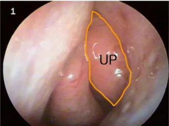
술 전 코 안에 이상소견을 보였던 경우는 102안 중 64안, 62.7%로 중간코선반의 비후가 21안으로 20.6%, 코중격편위가 있었던 경우가 19안, 18.6%, 벌집뼈 공 기세포의 전방돌출이 있었던 경우가 14안으로 13.7%, 중간코선반의 전방돌출이 5안으로 4.9%, 구상돌기가 코 안으로 돌출되어 있는 경우가 5안 4.9%였다(Fig.
2, Table 2).
술 후 눈물흘림 증상이 소실되었던 경우는 102안 중 95안으로 93.1%였고 염색약소실검사상 +1 이하인 경우가 92안으로 90.2%의 성공률을 보였다. A군의 성 공률은 89.5%, B군의 성공률은 90.6%로 두 군간의 통계학적으로 유의한 차이를 보이지 않았다(p>0.99, Table 3).
Table 2. Nasal cavity abnormalities
Number (eyes) %
Nasal septal deviation 19 18.6
Agger nasi 14 13.7
Middle turbinate hypertrophy 21 20.6
Obtruding middle turbinate 5 4.9
Prominent uncinate process 5 4.9
Total 64 62.7
Table 3. The success rate of group A and B
Success Failure Total
Group A 34 (89.5%) 4 (10.5%) 38
Group B 58 (90.6%) 6 (9.4%) 64
Total 92 (90.2%) 10 (9.8%) 102
* p-value>0.99; Group A=there was no nasal cavity abnormality before endonasal dacryocystorhinostomy; Group B=there was nasal cavity abnormality which was not treated;
* p-value=statistical significance was tested by Fisher's exact test.
Table 4. Late complication of endonasal dacryocystorhinos- tomy
Gruop A Group B Total Membranous obstruction 2 (50%) 3 (50%) 5 (50%) Granuloma 1 (25%) 2 (33.2%) 3 (30%)
Synechiae 0 (0%) 1 (16.6%) 1 (10%)
Canalicular stenosis 1 (25%) 0 (0%) 1 (10%)
Total 4 (100%) 6 (100%) 10 (100%)
Group A=there was no nasal cavity abnormality before endonasal dacryocystorhinostomy; Group B=there was nasal cavity abnormality which was not treated.
Figure 2. Prominent uncinate process. Left narrowed nasal space with septal deviation. the swelling just posterior to the lacrimal ridge could be the middle turbinate or a medially bent uncinate process.
코 안의 이상 유무와 수술 실패원인과의 관계는 A군 에서는 막성폐쇄로 인한 골공폐쇄가 2안, 5.3%, 육아 종으로 인한 골공폐쇄가 1안으로 2.6%, 눈물소관폐쇄 및 기타원인이 1안으로 2.6%이었으며 B군에서는 막성 폐쇄로 인한 골공폐쇄가 3안, 4.7%, 육아종으로 인한 골공폐쇄가 2안으로 3.1%, 중간코선반 및 코중격 유착 이 1안이었다(Table 4).
안와 지방탈출, 뇌척수액누출, 지연성 출혈 등의 합 병증은 보이지 않았다.
고 찰
눈물흘림을 주소로 눈물관막힘을 진단받은 환자의 치료법으로 시행되어온 눈물주머니코안연결술에는 피 부를 통한 접근방법과 코내시경을 이용하는 접근방법이 있다. 전통적인 피부를 통한 접근법은 Toti1에 의해 처 음 소개된 이후 수술 시야가 좋고 수술 중 발견되는 눈 물소관 및 공통눈물소관의 문제를 해결하기 용이하며 코점막과 눈물주머니를 서로 연결봉합함으로써 육아종 형성이 적어 90~96%의 높은 성공률이 보고되어 왔 다. 하지만 내측눈구석 피부절개를 하게 되므로 수술부
위 피부에 반흔을 남길 수 있고 수술 중 과도한 조작으 로 인한 주위조직의 손상과 출혈증가, 수술 중 코안을 직접 볼 수가 없다는 단점이 있다. 반면에 코경유를 통 한 접근법은 피부반흔이 없고 혈종생성이 적으며 회복 이 빠른 장점과 최근에는 내시경의 발달로 인해 코 안 의 시야 확보가 가능하게 되어 코 안 이상소견을 보다 잘 관찰할 수 있게 되었으며 피부절개법과 비슷한 82~
95%의 성공률이 보고되고 있다.3,4,8-14
코경유 눈물주머니코안연결술의 성공률에 영향을 미 치는 인자는 코중격편위, 중간코선반의 비후나 전방돌 출, 벌집뼈 공기세포의 전방돌출 등 코 안의 해부학적 이상소견과 수술 후 유착, 그리고 골공의 위치 등이 있
다.8,9,15-17 특히 코 안 구조 이상이나 질환 등으로 수술
결과가 좋지 않을 것으로 판단되는 경우에는 코경유 눈 물주머니코안연결술 시행 시 수술기구 접근이 어렵고 충분한 크기의 골공형성과 눈물주머니벽 제거가 힘들어 지게 된다.18 이런 경우에는 microdrill을 이용하여 골 공을 형성하는 것이 도움이 되지만 코 안에 화상이 발 생할 수 있고 시행하는 도중 시야가 가려져서 주위 조 직에 손상을 줄 수 있는 등의 문제점이 있어 피부절개 를 통한 방법을 고려하거나 코 안에 이상소견을 수술 전이나 수술 중에 치료해야 한다는 보고가 있었
다.9,19-23 하지만 최근에 코 안의 이상이 있는 경우 술
전 이비인후과 수술 없이 코경유 눈물주머니코안연결술
을 시행하였고 술 후 안과 및 이비인후과적 치료를 통 해 골공 부위의 막성폐쇄나 육아종을 제거함으로써 유 착을 방지하였을 때 코 안의 이상유무가 수술의 성공률 에 미치는 영향은 유의한 상관관계가 없었으며 코 안의 이상의 종류에 따른 수술 성공률에도 차이가 없었다는 보고와 코 안의 해부학적 이상소견이 있는 경우 구상돌 기절제술을 시행하여 중비도를 확장시키면 수술기구의 조작이 용이해지므로 주위조직에 불필요한 조작을 적게 하고 출혈과 수술 후 염증 등의 합병증을 줄여서 수술 의 성공률을 높일 수 있다는 보고가 있다.24,25 본 연구 에서는 총 102안 중 64안에서 코 안의 이상소견이 있 었고 이런 경우는 코 안을 충분히 수축시켜 술 전 코 안 의 이상을 교정하지 않고 코경유 눈물주머니코안연결술 과 함께 구상돌기 앞부분 절제술을 시행하여 중비도를 확장시킴으로써 수술시야를 넓히고 수술기구의 조작을 용이하게 하여 술 중 출혈, 술 후 염증과 유착 등의 합 병증이 줄일 수 있어서 코 안 이상이 없는 경우와 통계 적으로 차이가 없는 수술성공률을 보일 수 있었을 것이 다(p>0.99).
Mcdonogh and Meiring26는 코내시경으로 벌집 절제술 시 구상돌기가 눈물뼈의 안쪽에 부착하는 것을 관찰하였고, Yung and Hardman-Lea27는 위턱굴 과 구상돌기사이에 봉합의 틈으로 눈물뼈와 눈물주머니 를 노출시킬 수 있다고 보고하였다. 코경유 눈물주머니 코안연결술 시행 시 수술 결과에 영향을 미치는 결정적 인 단계는 3~5 mm 직경의 크기의 골공을 만드는 것 으로 공통눈물소관 위치에서 눈물주머니와 코안을 연결 시켜 주어야 장기간 효능을 유지할 수 있다.24,28,29 구상 돌기는 공통눈물소관 위치에서 눈물뼈 안쪽에 부착되므 로 구상돌기의 앞쪽을 절제하는 것은 코경유 눈물주머 니코안연결술시 정확한 위치에 골공을 형성할 수 있게 해 준다.30 본 연구에서 구상돌기의 앞부분을 절제하여 눈물주머니를 쉽게 노출시킬 수 있었으며 코 안의 해부 학적 이상으로 눈물주머니의 정확한 위치를 찾기 어려 웠던 경우에서도 정확한 위치에 골공을 형성함으로써 수술시간을 단축시키고 코 안의 해부학적 이상이 없었 던 경우와 통계적으로 유의한 차이가 없는 수술의 성공 률을 보였다(p>0.99). 이처럼 눈물뼈, 눈물주머니오목 과 구상돌기의 해부학적 위치 관계를 고려해 보면 코경 유 눈물주머니 코안연결술을 시행할 때 구상돌기는 눈 물주머니를 찾는 지표로 사용될 수 있을 것이다.
코경유 눈물주머니코안연결술 시행 시 일반적으로 구상돌기의 앞쪽에 있는 상악동의 이마돌기의 코능선 (nasal ridge)에서 눈물주머니의 정확한 위치를 찾기 전 골공을 형성하기 시작하여 코오목까지 넓혀간
다.11,31-39 이런 방법은 방향을 잘못 잡아 안와 지방탈
출, 혈종, 기종, 점막 화상, 하사근 부착부의 손상, 앞 사골동맥의 손상, 시신경의 손상을 포함한 부작용의 위 험이 있다.36,39 하지만 구상돌기는 위턱뼈나 눈물뼈가 부착된 위치와 상관없이 질감이나 크기에 의해 구분할 수 있고 쉽게 박리되므로 구상돌기의 부착지점 앞쪽을 절제하여 골공을 형성하는 것은 눈물주머니를 노출시키 는 것이 용이하게 하고 안와와 두 개내 합병증을 줄일 수 있을 것이다.24,40-42 또한 구상돌기 앞부분 절제술을 시행할 때는 골편을 완전히 제거하여 술 후 남은 골편 에 의해 골공이 막히거나 술 후 염증을 일으키는 방지 해야 한다.43 본 연구에서 코경유 눈물주머니코안연결 술 시 구상돌기 앞쪽 절제술을 함께 시행하여 골공을 형성함으로써 102안 중 안와 지방탈출, 앞사골동맥의 손상, 하사근 부착부의 손상, 시신경손상 등의 합병증 이 발생한 경우는 없었고 골공 형성시 골편을 완전히 제거하여 술 후 염증 및 유착을 줄일 수 있었을 것이다.
결론적으로 구상돌기는 공통눈물소관 위치에서 눈물 주머니오목 안쪽에 위치함으로써 코경유 눈물주머니코 안연결술 시행할 때 구상돌기의 앞부분을 함께 절제하 는 것은 눈물주머니를 노출시키는 것을 용이하게 해 주 고 정확한 위치에서 골공을 형성할 수 있게 해 주며 중 비도를 확장시켜 수술 시 기구조작을 용이하게 하고 술 후 유착을 줄여 수술을 성공률을 높이는데 기여할 것으 로 생각된다.
참고문헌
1) Toti A. Nuovo metodo conservatore dicuraradicale delle sopourazioni croniche del saccolacrimale (dacriocistorhinostomia).
Clin Moderna 1904;10:385-7.
2) Watkins LM, Janfaza P, Rubin PA. The evolution of endonasal dacryocystorhinostomy. Surv Ophthalmol 2003;48:73-84.
3) Rice DH. Endoscopic intranasal dacryocystorhinostomy results in four patients. Arch Otolaryngol Head Neck Surg 1990;
116:1061.
4) Yoon JH, Kim KS, Jung, DH, et al. Fontanelle and uncinate process in the lateral wall of the human nasal cavity.
Laryngoscope 2000;110:281-5.
5) Yung MW, Logan BM. The anatomy of the lacrimal bone at the lateral wall of the nose: its significance to the lacrimal surgeon. Clin Otolaryngol 1999;24:262-5.
6) Wormald PJ, McDonogh M. The “swing‐door” technique for uncinectomy in endoscopic sinus surgery. J Laryngol Otol 1998;112:547-51.
7) Caldwell GW. Two new operations for obstructions of the nasal duct with preservation of the canaliculi. Am J Ophthalmol 1893;10:189.
8) Mannor GE, Millman A. The prognostic value of preoperative dacryocystography in endoscopic intranasal dacryocystorhinostomy.
Am J Ophthalmol 1914;12:659.
9) Lee HC, Chung WS. Success Rate of Endonasal Dacryocystorhinostomy. J Korean Ophthalmol Soc 1992;37:211-8.
10) Whittet HB, Shun-Shin GA, Awdry P. Functional endoscopic transnasal dacyocystorhinostomy. Eye 1993;7:545-9.
11) Hosemann W, Buhr W. Endoscopic endonasal dacryo- cystorhinostomy : results in 56 patients. Ann Otol Rhinol Laryngol 1994;103:363-7.
12) Javate RM, Campomanes BS Jr, Co ND, et al. The endoscope on the radiofrequency unit in DCR surgery. Ophthal Plast Reconstr Surg 1995;11:54-8.
13) Cokkeser Y, Evereklioglu C, Er H. Comparative external versus endoscopic dacryocystorhinostomy:results in 115 patients. Otolaryngol Head Neck Surg 2000;123:488-91.
14) Dolman PJ. Comparison of external dacryocystorhinostomy with nonlaser endonasal dacryocystorhinostomy. Ophthalmology 2003;110:78-84.
15) Linberg JV, Anderson RL, Bumsted RM, Barreras R. Study of intranasal ostium external dacryocystorhinostomy. Arch Ophthalmol 1982;100:1758-62.
16) Jones LT. An anatomical approach to problems of the eyelids and lacrimal apparatus. Arch Ophthalmol 1961;66:111-24.
17) Dortzbach RK. Dacryocystorhinostomy. Ophthalmology 1978;85:
1267-70.
18) Chung ER, Lee KT, Choi WC. Success rate of endonasal dacryocystorhinostomy based on the location of the lacrimal sac. J Korean Ophthalmol Soc 2002;43:2000-4.
19) Lim IS, Jeong SK, Park YG. A study of factors related to surgical success rate of dacryocystorhinostomy. J Korean Ophthalmol Soc 1997;38:1322-7.
20) Hatt M. A concept and a method to prevent failures in lacrimal surgery. Ophthalmic Plast Reconstr Surg 1987;3:105-7.
21) Metson R. Endoscopic surgery for lacrimal obstruction.
Otolaryngol Head Neck Surg 1991;104:473-9.
22) Park JD, Kim YI, Shin SG. The factors related to surgical success rate of endonasal dacryocystorhinostomy. J Korean Ophthalmol Soc 1998;39:2848-53.
23) Lee SJ, Na KS, Ji NC. A clinical study on the endonasal microdrill‐assisted dacryocystorhinostomy. J Korean Ophthalmol Soc 1998;39:1620-8
24) Fayet B, Racy E, Assouline M. Systematic unciformectomy for a standardized endonasal dacryocystorhinostomy. Ophtalmology 2002;109:530-6.
25) Kim JM, Hong WP, Choi YJ, Kim SJ. The effect of nasal cavity abnormality related to surgical success rate of endonasal dacryocystorhinostomy. J Korean Ophthalmol Soc 2006;47:1233-7.
26) McDonogh M, Meiring JH. Endoscopic transnasal dacryo- cystorhinostomy. J Laryngol Otol 1989;103:585-7.
27) Yung MW, Hardman‐Lea S. Endoscopic inferior dacryo- cystorhinostomy. Clin Otolaryngol Allied Sci 1998;23: 152-7.
28) Welham RA, Wulc AE. Management of unsuccessful lacrimal surgery. Br J Ophthalmol 1987;71:152-7.
29) Becker BB. Dacryocystorhinostomy without flaps. Ophthalmic Surg 1988;19:419-27.
30) Fayet B, Racy E, Assouline M, Zerbib M. Surgical anatomy of the lacrimal fossa. Ophthalmology 2005;112:1119-28.
31) Steadman MG. Transnasal dacryocystorhinostomy. Otolaryngol Clin North Am 1985;18:107-11.
32) West JM. Eine Fensterresektion des Ductus naso‐lacrimalis in Fa¨llen von Stenose. Arch Laryngol Rhinol 1910;24:62-4.
33) Menerath JM, Guichard C, Kydavongs P. Endonasal dacryocystorhinostomy using endoscopic guidance. Personal experience. J Fr Ophtalmol 1999;22:41-5.
34) Jouve Y, Dehon A, Blanc PH, Rouvier P. Dacryocystorhinostomy in endonasal microsurgery. Bull Mem Soc Fr Ophtalmol 1983;94:64-6.
35) Gutie´rrez-Ortega AR, Sprekelsen-Gasso C, Valles-San Leandro L, Delmperial JM. Endonasal dacryocystorhinostomy: first results. Orbit 1995;14:25-8.
36) Friedrich JP, Tritten JJ. Dacryocystorhinostomy by nasal endoscopy. an example of multidisciplinary collaboration. Rev Med Suisse Romande 1998;118:235-9.
37) el Khoury J, Rouvier P. Endonasal dacryocystorhinostomy (95 cases). Acta Otorhinolaryngol Belg 1992;46:401-4.
38) Chevalier E, Hazan A, Gerard B, et al. Dacryocystorhinostomie parvoie endonasale‐endoscopique: technique chirurgicale et résultats àpropos dùne série de 24 cas. Rev Offic Soc Fr ORL 1997;44:37-45.
39) Sprekelsen MB, Barberán MT. Endoscopic dacryocystorhinostomy:
surgical technique and results. Laryngoscope 1996;106:187-9.
40) Hartikainen J, Grenman R, Puukka P, Seppä H. Prospective randomized comparison of external dacryocystorhinostomy and endonasal laser dacryocystorhinostomy. Ophthalmology 1998;105:
1106-13.
41) Rouviere P, Vaille G, Garcia C, et al. Dacryocystorhinostomy using the endonasal approach. Ann Otolaryngol Chir Cervicofac 1981;98:49-53.
42) Whittet HB, Shun-Shin GA, Awdry P. Functional endoscopic transnasal dacryocystorhinostomy. Eye 1993;7:545-9.
43) Fayet B, Racy E, Assouline M. Complications of standardized endonasal dacryocystorhinostomy with unciformectomy.
Ophthalmology 2004;111:837-45.
=ABSTRACT=
Clinical Consideration of Uncinectomy for Endonasal Dacryocystorhinostomy
Jung Lim Kim, M.D., Jae Wook Yang, M.D., Ph.D.
Department of Ophthalmology, College of Medicine, Inje University, Pusan, Korea Ophthalmology Research Foundation, Inje University, Pusan, Korea
Purpose: To evaluate the effects of uncinectomy for osteotomy method during endonasal dacryo- cystorhinostomy (DCR).
Methods: This retrospective review study comprised 102 nasolacrimal duct obstruction patients that underwent endonasal dacryocystorhinostomy with uncinectomy at our hospital between 2003 and 2006. The patients were classified into two groups based on records of preoperative endoscopic examination. Group A consisted of patients who had no nasal cavity abnormality, and Group B comprised patients who had a nasal cavity abnormality but received no treatment.
Results: Sixty-four patients were included in Group B. The success rates of endonasal DCR were 89.5% in Group A and 90.6% in Group B, with no statistically significant difference between the two groups (p>0.99).
There were no cases of mucosal burn, orbital fat prolapse, spinal fluid leak, maxillary sinusitis, or delayed bleeding.
Conclusions: Anterior resection of the uncinate process is the most important surgical step to expose the medial aspect of the lacrimal fossa and to form the precise location of osteotomy during endonasal dacryocystorhinostomy.
J Korean Ophthalmol Soc 49(6):871-877, 2008
Key Words: Anterior Uncinectomy, Endonasal Dacryocystorhinostomy, Nasolacrimal Duct Obstruction
Address reprint requests to Jae Wook Yang, M.D.
Department of Ophthalmology, InJe University Medical College
#633-165 Kekum-dong, Pusanjin-gu, Pusan 614-735, Korea
Tel: 82-51-890-6016, Fax: 82-51-890-6329, E-mail: [email protected]