ISSN: 2233-601X (Print) ISSN: 2093-6516 (Online)
Received: August 31, 2015, Revised: November 8, 2015, Accepted: November 10, 2015, Published online: August 5, 2016
Corresponding author: Kwangjo Cho, Department of Thoracic and Cardiovascular Surgery, Dong-A University College of Medicine, 32 Daesingongwon-ro, Seo-gu, Busan 49201, Korea
(Tel) 82-51-240-2879 (Fax) 82-51-231-5195 (E-mail) [email protected]
© The Korean Society for Thoracic and Cardiovascular Surgery. 2016. All right reserved.
This is an open access article distributed under the terms of the Creative Commons Attribution Non-Commercial License (http://creativecommons.org/
licenses/by-nc/4.0) which permits unrestricted non-commercial use, distribution, and reproduction in any medium, provided the original work is properly cited.
Long-Term Changes in the Distal Aorta after Aortic Arch Replacement in Acute DeBakey Type I Aortic Dissection
Kwangjo Cho, M.D., Jeahwa Jeong, M.D., Jongyoon Park, M.D., Sungsil Yun, M.D., Jongsu Woo, M.D.
Department of Thoracic and Cardiovascular Surgery, Dong-A University College of Medicine
Background: We analyzed the long-term results of ascending aortic replacement and arch aortic replacement in acute DeBakey type I aortic dissections to measure the differences in the distal aortic changes with ex- tension of the aortic replacement. Methods: We reviewed 142 cases of acute DeBakey type I aortic dis- sections (1996–201 5). Seventy percent of the cases were ascending aortic replacements, and 30% of the cas- es underwent total arch aortic replacement, which includes the aorta from the root to the beginning of the descending aorta with the 3 arch branches. Fourteen percent (20 cases) resulted in surgical mortality and 86% of cases that survived had a mean follow-up period of 6.6±4.6 years. Among these cases, 64% of the patients were followed up with computed tomography (CT) angiograms with the duration of the final CT check period of 4.9±2.9 years. Results: There were 15 cases of reoperation in 13 patients. Of these 15 cases, 13 cases were in the ascending aortic replacement group and 2 cases were in the total arch aortic replace- ment group. Late mortality occurred in 13 cases; 10 cases were in the ascending aortic replacement group and 3 cases were in the total arch aortic replacement group. Eight patients died of a distal aortic problem in the ascending aortic replacement group, and 1 patient died of distal aortic rupture in the total arch aortic replacement group. The follow-up CT angiogram showed that 69.8% of the ascending aortic replacement group and 35.7% of the total arch aortic replacement group developed distal aortic dilatation (p=0.0022).
Conclusion: The total arch aortic replacement procedure developed fewer distal remnant aortic problems from dilatation than the ascending aortic replacement procedure in acute type I aortic dissections.
Key words: 1. Aortic dissection 2. Surgery
3. Complication 4. Recurrence 5. Replacement
Introduction
Acute DeBakey type I aortic dissection is a surgical disease. When patients are fortunate to survive the surgical procedure, they should undergo frequent fol- low-up, because the remnant segment of the dis-
sected aorta may develop aneurysmal dilatation.
Today the aortic arch replacement can be safely per-
formed in acute aortic dissection due to the advance
of surgical techniques such as the antegrade cerebral
perfusion technique and the use of artificial graft
materials. To determine the superiority of an arch
http://dx.doi.org/10.5090/kjtcs.2016.49.4.264
Fig. 1. Annual trends in the performance of ascending aortic re- placement and arch aortic replacement. Arch, aortic arch repla- cement; Asc, ascending aortic replacement.
Table 1. Patients demographics and operation data by procedure
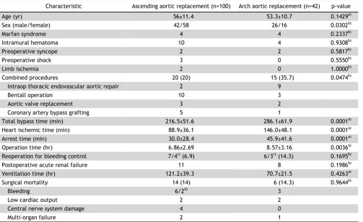
Characteristic Ascending aortic replacement (n=100) Arch aortic replacement (n=42) p-value
Age (yr) 56±11.4 53.3±10.7 0.1429
a)Sex (male/female) 42/58 26/16 0.0302
b)Marfan syndrome 4 4 0.2337
b)Intramural hematoma 10 4 0.9308
b)Preoperative syncope 2 2 0.5817
b)Preoperative shock 3 0 0.5550
b)Limb ischemia 2 0 1.0000
b)Combined procedures 20 (20) 15 (35.7) 0.0474
b)Intraop thoracic endovascular aortic repair 2 9
Bentall operation 10 3
Aortic valve replacement 3 2
Coronary artery bypass grafting 5 1
Total bypass time (min) 216.5±51.6 286.1±61.9 0.0001
a)Heart ischemic time (min) 88.9±36.1 146.0±48.1 0.0001
a)Arrest time (min) 30.0±28.4 45.9±41.6 0.0001
a)Operation time (hr) 6.86±2.69 8.57±3.16 0.0036
a)Reoperation for bleeding control 7/4
c)(6.9) 6/3
c)(14.3) 0.1695
b)Postoperative acute renal failure 11 8 0.1986
b)Ventilation time (hr) 121.2±39.3 70.7±21.5 0.4263
a)Surgical mortality 14 (14) 6 (14.3) 0.9644
b)Bleeding 6/2
d)3
Low cardiac output 2 2
Central nerve system damage 4 0
Multi-organ failure 2 1
Values are presented as mean±standard deviation, number, or number (%).
a)
By t-test.
b)By χ
2-test.
c)Cases of death.
d)Early anastomotic rupture.
replacement in prevention of long-term postoperative distal aortic aneurysms, we researched the distal aortic status after total aortic arch replacement in
acute DeBakey type I aortic dissections and com- pared the results to that of the ascending aortic re- placement group.
Methods
Between January 1996 and March 2015, 142 pa- tients of with acute DeBakey type I aortic dissection were treated surgically at Dong-A University Hospital.
Seventy percent of the cases (100 patients) under-
went ascending aortic replacements (include 13 hem-
iarch replacement cases and 1 innominate artery by-
pass case), while the remaining 30% of the cases (42
patients) underwent total aortic arch replacements,
which includes the aorta from the root to the begin-
ning of the descending aorta with the 3 arch branch-
es (Table 1). The arch replacement technique was
changed during that period. Prior to the year 2010,
21% of the cases (30 patients) were performed with
the arch-first technique under deep hypothermic cir-
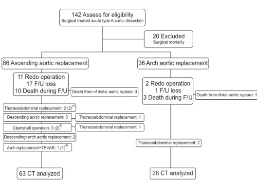
Fig. 2. Enrolled patients and F/U status. F/U, follow-up; TEVAR, tho- racic endovascular aortic repair; CT, computed tomography.
a)Operative mortality.
culatory arrest [1]. After the year 2010, 9% of the cases (12 patients) were performed with selective arch replacement with a 4-vessel branched arch graft, under moderate hypothermia and antegrade cerebral perfusion. The rate of performance of each of the two procedures is described in Fig. 1. Twenty patients of the ascending aortic replacement group and 15 patients of the total arch aortic replacement group underwent concomitantly combined procedures, which included 11 intraoperative thoracic endovas- cular aortic repairs (TEVAR), 13 Bentall operations, 5 aortic valve replacements, and 6 coronary artery by- pass grafting procedures (Table 1). Eighty-six percent of the cases (122 patients) survived these operations with a mean follow-up period of 6.9±13.3 years.
During the follow-up period, 64% of the patients (63 from the ascending aortic replacement group, 28 from the total arch aortic replacement group) were checked by serial CT scan in our out-patient clinic (Fig. 2). The follow-up CT scan allowed us to check the distal aortic pseudolumen status and diameter, the level of the largest portion of the distal remnant aorta, and the interval change between the immedi- ate postoperative period and the late postoperative period. The mean interval between the two periods was 4.9±2.9 years. The level of the dilated aorta was defined as the proximal descending aorta above the sixth intercostal level and below was the distal de-
scending aorta [1]. We categorized the aortic change of the serial CT scan into 5 classes: class A–the distal remnant aorta was remodeled to normal size and the remnant distal aorta shrank when compared to the immediate postoperative CT; class B–the pseudolu- men was totally obliterated but the size of the rem- nant distal aorta did not shrink; class C–the pseudo- lumen remained but the size of the remnant distal aorta dilatation was <40 mm in diameter; class D–
the remnant distal aorta dilatation was >40 mm but
<60 mm in diameter, which was generally the in- dicator for surgical correction; and class E–the rem- nant distal aorta was >60 mm in diameter or the CT was checked just before reoperation. Class D and E eventually resulted in an aneurysmal dilatated dis- tal remnant aorta, while class A, B, and C, a normal- ized aorta.
We analyzed the statistical differences between the ascending aortic replacement group and the total arch aortic replacement group with a χ
2-test and t-test. The survival rate was calculated with the Kaplan-Meier method of life test. The categorical dif- ference between the two groups was calculated with stratified analysis using Cochran-Mantel-Haenszel sta- tistics. The multivariate analysis was calculated with logistic regression analysis. We considered a differ- ence statistically significant with the p-value <0.05.
The statistics were calculated with the SAS ver. 9.3
Table 2. Long-term follow-up data
Variable Ascending aortic
replacement (n=86)
Arch aortic
replacement (n=36) p-value
Distal aortic reoperation 13/2
a)2 0.0001
b)Thoracoabdominal aortic replacement 4/2
c)2
Descending aortic replacement 3/1
a)Total thoracic aortic replacement 3/1
a), 3/2
c)Descending +arch aortic replacement 2
Arch replacement +intraoperative thoracic endovascular aortic repair 1/1
c)Long-term mortality 10 3 0.2809
b)Non-surgical death 5/3
d)3/1
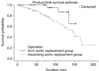
d)Mean survival duration of long-term mortality cases (mo) 76.6±47.5 123.3±10.4 0.0332
e)Values are presented as number or mean±standard deviation.
a)
Trido operation.
b)By χ
2-test.
c)Death from surgery.
d)Death from distal aortic rupture.
e)By Student t-test.
Table 3. Characteristics of patients with late aortic events
Sex/age (yr) Group Interval
a)PL
b)Redo operative procedure Result
F/33
c)Asc
d)38 ( +) Type II thoracoabdominal aortic replacement Death from redo surgery F/54 Asc 105 ( +) Type II thoracoabdominal aortic replacement Death from redo surgery
F/40 Asc 5 ( +) Descending aortic replacement Trido type IV thoracoabdominal replacement → arch aortic dilatation
M/49 Asc 88 ( +) Descending aortic replacement Distal aortic dilatation F/38 Asc 25 ( +) Descending aortic replacement Normal with false lumen
M/60 Asc 84 ( +) Clamshell operation
e)Death from redo surgery
F/56 Asc 59 ( +) Clamshell operation Death from redo surgery
M/40
c)Asc 31 ( +) Clamshell operation Trido type IV thoracoabdominal replacement → normal
M/40 Asc 77 ( +) Descending+arch aortic replacement Normal
F/52 Asc 121 ( +) Descending+arch aortic replacement Distal anastomosis pseudoaneurysm M/39 Asc 60 ( +) Arch replacement+intraoperative thoracic
endovascular aortic repair
Death from redo surgery
F/61 Asc 133 ( −) ( −) Death from arch aortic aneurysm rupture
F/67 Asc 39 ( +) ( −) Death from distal aortic rupture
F/63 Asc 170 ( +) ( −) Death from distal aortic rupture
M/43
c)Arch
f)56 ( +) Type IV thoracoabdominal aortic replacement Normal M/50 Arch 22 ( +) Type IV thoracoabdominal aortic replacement Normal
F/41
c)Arch 41 ( +) ( −) Death from abdominal aortic aneurysm rupture
F, female; M, male.
a)
Interval between initial operative day and the event day of the month.
b)Pseudolumen patency.
c)Marfan syndrome.
d)Ascending aortic replacement group.
e)Total thoracic aortic replacement with clamshell incision.
f)Arch replacement group.
(SAS Institute Inc., Cary, NC, USA).
Results
The cardiopulmonary bypass time, heart ischemic time, circulatory arrest time, and operation time of the total arch aortic replacement group were longer than those of the ascending aortic replacement group.
However, postoperative renal function and ventilator
support period showed no statistically significant dif-
ference between the two groups (Table 1). Fourteen
percent of the cases (20 patients) resulted in surgical
mortality. However, there was no statistically sig-
nificant difference between the ascending aortic re-
placement group and the total arch aortic replace-
ment group. The main cause of surgical death was
Table 4. Long-term follow-up data with CT scan
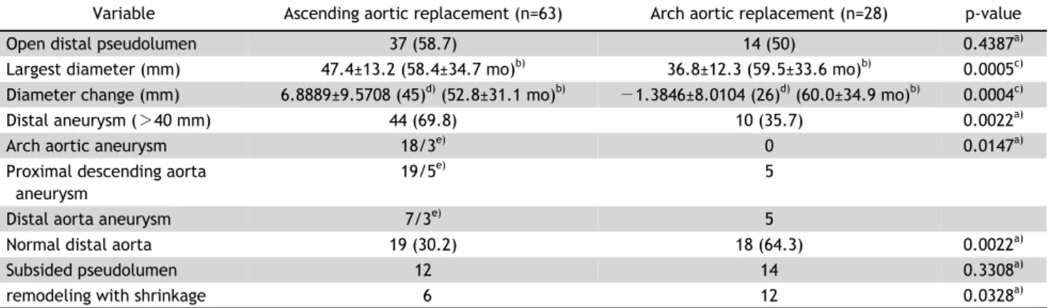
Variable Ascending aortic replacement (n=63) Arch aortic replacement (n=28) p-value
Open distal pseudolumen 37 (58.7) 14 (50) 0.4387
a)Largest diameter (mm) 47.4±13.2 (58.4±34.7 mo)
b)36.8±12.3 (59.5±33.6 mo)
b)0.0005
c)Diameter change (mm) 6.8889±9.5708 (45)
d)(52.8±31.1 mo)
b)−1.3846±8.0104 (26)
d)(60.0±34.9 mo)
b)0.0004
c)Distal aneurysm ( >40 mm) 44 (69.8) 10 (35.7) 0.0022
a)Arch aortic aneurysm 18/3
e)0 0.0147
a)Proximal descending aorta aneurysm
19/5
e)5
Distal aorta aneurysm 7/3
e)5
Normal distal aorta 19 (30.2) 18 (64.3) 0.0022
a)Subsided pseudolumen 12 14 0.3308
a)remodeling with shrinkage 6 12 0.0328
a)Values are presented as number (%) or mean±standard deviation.
CT, computed tomography.
a)
By χ
2-test.
b)Mean follow-up period.
c)By t-test.
d)No. of cases with immediately postoperative CT available.
e)Cases of large aneurysm in need of surgery (diameter >60 mm).
bleeding. There were 13 reoperations for surgical bleeding and 7 of them died of bleeding complica- tions. Two other patients in the ascending aortic re- placement group developed delayed anastomotic ru- pture. Still there was no statistically significant differ- ence between the two groups (Table 1). Among the other causes of death due to damage of the central nerve system (CNS) in the ascending replacement group included 2 cases of middle cerebral artery in- farction, a case of severe hypoxic brain damage, and a case of intracerebral hemorrhage. However, there was no mortality due to CNS damage in the total arch aortic replacement group.
1) Distal remnant aortic events
During the follow-up period, 13 patients under- went 15 cases of late reoperations with a mean age of 45.6±8.2 years and a mean interval between initial operation and reoperation of 59.3±34.6 months. Thir- teen cases were performed in the ascending aortic replacement group and 2 cases were performed in the total arch aortic replacement group (p=0.0001).
Among the 13 cases of the ascending aortic replace- ment group, 4 were thoracoabdominal aortic replace- ments (2 of them were trido thoracoabdominal aortic replacement), 3 were descending aortic replacements, 3 were total thoracic aortic replacements with clam- shell incision, 2 were descending and arch replace- ments, and 1 was arch replacement with intraope- rative TEVAR. The 5 redo operative cases resulted in surgical mortality: 2 redo thoracoabdominal aortic re-
placements, 2 total thoracic aortic replacements with clamshell incision, and 1 arch replacement with in- traoperative TEVAR (Tables 2, 3). Of the 6 patients who survived in the ascending aortic replacement group, 3 developed remnant aortic aneurysms (Table 3). All the reoperative cases in the total arch aortic replacement group survived without any distal aortic problems.
Thirteen cases of late mortality occurred; 5 redo surgical mortalities, 4 distal aortic ruptures, and 4 medical causes of death (Table 2). Ten cases were in the ascending aortic replacement group and 3 cases were in the total arch aortic replacement group. In the ascending aortic replaced group 8 cases (9.2%) resulted in late mortality due to distal aortic events but in the total arch aortic replacement group only 1 case (2.7%) developed a distal aortic event, which was an abdominal aortic aneurysm rupture; the pa- tient had Marfan syndrome and failed to follow-up (Table 3).
We reviewed the long term CT scans and identi-
fied 11 candidates for reoperation (category E). All
candidates were in the ascending aortic replacement
group and the maximal aneurysm diameter was >60
mm (Table 4). When these candidates were included
into the redo operations and the late mortalities due
to distal aortic rupture, the total distal aortic events
were statistically different between the two groups
(Fig. 3). In the ascending aortic replacement group,
there were 27 adverse distal aortic events (31.4%),
which occurred during a mean follow-up period of
Table 5. Classification of distal remnant aorta