127
R E V IE W ARTICLE
pISSN: 2384-3799 eISSN: 2466-1899 Int J Thyroidol 2016 November 9(2): 127-130 https://doi.org/10.11106/ijt.2016.9.2.127
Received May 2, 2014 / Revised July 13, 2016 / Accepted July 14, 2016
Correspondence: Sun Wook Cho, MD, Department of Internal Medicine, Seoul National University Hospital, 101 Daehak-ro, Jongno-gu, Seoul 03080, Korea
Tel: 82-2-2276-2094, Fax: 82-2-762-2292, E-mail: [email protected]
Copyright ⓒ 2016, the Korean Thyroid Association. All rights reserved.
This is an open-access article distributed under the terms of the Creative Commons Attribution Non-Commercial License (http://creative- commons.org/licenses/by-nc/4.0/), which permits unrestricted non-commercial use, distribution, and reproduction in any medium, provided the original work is properly cited.
갑상선자극호르몬이 골대사에 미치는 영향
서울대학교병원 내과
조선욱
Effects of Thyroid Stimulating Hormone on Bone Metabolism
Sun Wook Cho
Department of Internal Medicine, Seoul National University Hospital, Seoul, Korea
Bone is a dynamic tissue undergoing life-long remodeling, a process of bone resorption by osteoclast and bone formation by osteoblast, regulated by diverse hormones including estrogen. Recently, several pituitary hormones have been identified as a modulator of this process. Here, we reviewed the role of thyroid stimulating hormone signaling per se in bone metabolism.
Key Words: Thyroid stimulating hormone, Bone resorption, Bone formation, Bone remodeling
Bone Remodeling and Pituitary Hormone
성인의 뼈 조직은 일생 동안 재형성(remodeling)된 다. 이 과정은 파골세포(osteoclast)의 골흡수(bone re- sorption) 작용에 의해 시작이 되며, 조골세포(osteoblast) 의 골형성(bone formation)이 뒤따르게 된다. 그리고 이 들 파골세포와 조골세포의 연결 작용(coupling)은 골수 내 미세 환경의 여러 호르몬과 싸이토카인에 의해서 섬세하게 조절된다. 여성 호르몬인 에스트로겐은 폐경 후 여성의 골다공증에 관한 연구 결과가 축적되면서 골재형성을 조절하는 가장 대표적인 조절 호르몬으로 정립되었다.1) 그러나 최근 10여 년간, 에스트로겐 이외 에도 폐경을 전후하여 변화하는 각종 뇌하수체호르몬 들, 특히 성장호르몬(growth hormone; GH),2) 여포자극 호르몬(follicular stimulating hormone; FSH),3) 갑상선 자극호르몬(thyroid stimulating hormone; TSH),4) 유즙 분비호르몬(prolactin),5) 그리고 옥시토신(oxytocin)6)이
골 대사를 조절한다는 여러 연구가 진행되고 있다. 갑상선호르몬은 사춘기 이전 골조직의 형성과 성장 에 매우 중요한 호르몬이다. 선천성 갑상선기능저하증 환자에서 골형성 장애 및 성장 지연을 보이고, 이러한 골 표현형이 갑상선호르몬 치료 이후 호전된다는 임상 보고가 그 근거이다.7) 이러한 갑상선호르몬은 시상하 부-뇌하수체-갑상선 축의 음성 되먹이기 기전에 의해 조절을 받는다(Fig. 1). 시상하부에서 분비되는 갑상선 자극호르몬방출호르몬(thyrotropin-releasing hormone;
TRH)은 뇌하수체에서 TSH의 분비를 자극하고, TSH 는 갑상선의 TSH 수용체(TSH receptor; TSHR)에 작 용하여 갑상선호르몬인 T4 (thyroxine)와 T3 (triiodo- thyronine)의 분비를 촉진한다. 혈중 T3 농도가 증가할 경우, 시상하부와 뇌하수체의 갑상선호르몬 수용체 (thyroid hormone receptor; TR) 베타(TRβ)에 작용하 여 음성 되먹이기 기전으로 TRH와 TSH의 분비가 억 제된다.8) 골조직에는 TRα가 존재하여 T3의 작용이 일어나는 것으로 알려졌는데,9) 최근에는 골세포에 TSHR이 존재함이 밝혀져서 T3 작용과는 별도로 TSH
Sun Wook Cho
Vol. 9, No. 2, 2016 128 Fig. 1. Hypothalamus-pituitary-thyroid-bone axis. The hypo-
thalamic neurons secrete thyrotropin releasing hormone (TRH) is secreted by hypothalamic neurons and carried down to the pituitary gland where it releases thyroid stimulating hormone (TSH). TSH reaches thyroid glands and stimulates the productions of thyroxin (T4) and triiodothyronine (T3) by binding to TSH receptor (TSHR). T3 exerts its actions on bone mainly through thyroid hormone receptor α (TRα). TSH can directly act on bone by binding to TSHR. Increased levels of circulating T3 can get negative feedback loop by inhibiting TRH and TSH releases.
가 단독으로 골조직에 영향을 미친다는 보고가 있다.10)
Thyroid Hormone Actions in Bone
갑상선기능항진증과 그레이브스 환자에서 골밀도가 감소하고 고관절 골절이 증가함이 보고된 이후, 여러 세포 실험과 조직 배양 실험을 통하여 갑상선호르몬은 골흡수를 촉진시키는 작용을 가지고 있음이 정립되었 다. 이는 파골세포에 대한 갑상선호르몬의 직접 작용 에 의한 것일 뿐만 아니라, tumor necrosis factor (TNF)- α와 Interleukin-6 등 골흡수를 촉진시키는 각종 싸이 토카인이 갑상선기능항진증에서 증가되기 때문인 것 으로 보고되었다.11) 또한 갑상선호르몬은 태아기에는 insulin-like growth factor-1 (IGF-1)을 통하여 골격계의 성장을 촉진하는 작용을 함께 가지고 있어 발생기와 성장기에는 조골세포의 증식 및 분화를 촉진하여 골격 계의 발생 및 성장을 일으키고, 성체에서는 골흡수를 촉진하여 골재형성을 초래하는 이중 작용을 하는 것으 로 정립되었다.12,13)
그런데, 위와 같이 정립된 갑상선호르몬의 골격계에
대한 작용으로 해석하기에는 무리가 따른 몇몇 연구 결과가 있었다. 첫째, 선천성 갑상선기능저하증을 보이 는 PAX8 유전자 결핍 마우스와 TSHR에 hyt/hty 돌연 변이를 가지는 마우스에서 TSH의 현저한 증가에도 불 구하고 골형성 지연과 골량 감소를 보이는 점,14) 둘째, 갑상선호르몬이 정상인 무증상 갑상선기능항진증 환 자에서 빠른 골 교체율을 보인다는 점, 그리고 골량 골 절의 위험이 갑상선호르몬이 아닌 TSH와 의미 있는 상관관계를 보인다는 점이다.4,15) 따라서, 최근 10여 년 간 TSH 신호 자체가 골격계에 어떤 작용을 하지 않을 까에 대한 연구가 집중적으로 이루어졌다.
Disruption of the Normal Reciprocal Relationship between
Thyroid Hormone and TSH
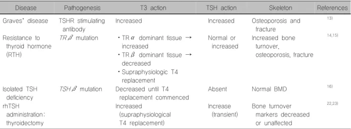
생체 내에서 TSH와 갑상선호르몬은 서로 유기적인 음성 되먹이기 작용을 하기 때문에, TSH가 갑상선호 르몬과는 별도로 어떠한 기능을 가지고 있는가를 연구 하는 것은 쉽지 않은 일이다. 따라서, 우선 이들의 상호 작용이 분리된 질환에서 골대사의 변화를 확인해 봄으 로써 TSH가 골대사에 미치는 영향을 추정해 보겠다 (Table 1). 그레이브스병에 의한 갑상선기능항진증의 경우는 갑상선호르몬이 증가되어 있으면서, TSH 수용 체에 대한 자극 항체가 형성되어 TSH의 작용도 함께 증가되어 있다. 이들 환자에서는 골다공증 및 골절이 증가되는 골 표현형을 보여, 증가된 갑상선호르몬의 역할이 주도적인 것으로 해석이 되고, 적어도 증가된 TSH 신호가 골조직에 대한 갑상선호르몬의 작용을 억 제하는 효과는 없거나 미미한 것으로 해석이 된다.16) 갑상선호르몬 내성증후군은 β-갑상선호르몬 수용체 유전자의 돌연변이에 의한 질환으로 갑상선호르몬의 작용은 α-갑상선호르몬 수용체가 우월한 조직에서는 증가, β-갑상선호르몬 수용체가 우월한 조직에서는 감소되어 있고, TSH 신호 작용은 정상이거나 증가되 어 있다. 이 경우 골연령의 지연, 골 교체율의 증가, 골 다공증 및 골절 증가의 골 표현형을 보인다.17,18) TSH 신호가 부재하는 경우를 살펴보면, TSH β 유전자의 점돌연변이로 인한 선천성 갑상선기능저하증의 증례 에서 보면 혈청 TSH는 측정되지 않고 갑상선호르몬도 낮은데, 이 경우 갑상선호르몬을 보충하면 정상 골 표 현형을 보인다.19) 이상으로, 갑상선호르몬과 TSH 신호 의 상호관계가 깨어진 증례들을 종합해 보았을 때, 골 조직에 대한 뚜렷한 TSH 단독의 작용이 있음을 밝히
TSH Signaling on Bone
129 Int J Thyroidol
Table 1. Bone phenotypes in dissociation of reciprocal relationship between thyroid hormone and TSH
Disease Pathogenesis T3 action TSH action Skeleton References
Graves’ disease TSHR stimulating antibody
Increased Increased Osteoporosis and
fracture
13)
Resistance to thyroid hormone (RTH)
TRβ mutation ㆍTRα dominant tissue → increased
Normal or increased
Increased bone turnover,
osteoporosis, fracture
14,15)
ㆍTRβ dominant tissue → decreased
ㆍSupraphysiologic T4 replacement Isolated TSH
deficiency
TSHβ mutation Decreased until T4 replacement commenced
Absent Normal BMD 16)
rhTSH
administration:
thyroidectomy
Increased
(supraphysiological T4 replacement)
Increase (transient)
Bone turnover markers decreased or unaffected
22,23)
BMI: body mass index, TSH: thyroid stimulating hormone
기는 어려웠다.
Anti-Resorptive Actions of TSH Signaling in Bone
TSH 신호 자체가 갑상선호르몬과 별도로 골재형성 과정에서 중요한 작용을 하는 것을 TSHR을 제거한 마 우스 모델에서 연구가 처음 이루어졌다. TSHR+/− 마 우스의 경우 갑상선호르몬은 정상 농도를 유지하면서 도 빠른 골 교체율을 보였으며, TSHR−/− 마우스의 경 우도 갑상선호르몬 보충 여부에 상관없이 골다공증의 표현형을 보였다.20,21) 이후 여러 마우스 후속 연구에서 TSHR의 결핍은 파골세포 활성도를 증가시켰으며, TSH 투여로 증가된 파골세포의 생존, 분화, 그리고 활 성도를 억제할 수 있음이 밝혀졌으며,22) 이는 in vitro 세포 실험에서도 증명되었다.10,23) 그리고 이러한 TSH 의 골흡수 억제 효과는 TSH가 골수 내 CD11b+ 파골 세포의 전구 세포에 직접 작용하여 TNFα 분비를 촉 진하여 이루어지는 것으로 보고되었다.22)
사람에서는 분화갑상선암 환자에서 인간 재조합 TSH (recombinant human TSH; rhTSH)를 주사한 후 골 표지자를 분석한 연구 결과가 제시되었다. 폐경 후 여성 환자에서 rhTSH 1회 주사한 경우, 골흡수의 지표 인 C-telopeptide (CTx)는 48시간 이내에 폐경 전 여성 의 정상 농도까지 억제되었다가 7일째 회복되는 양상 을 보여,24) TSH의 골흡수 억제 효과에 대한 가설을 다 시 한 번 입증하였다. 한편, Martini 등25)은 비슷한 연구 에서 rhTSH가 골형성 표지자인 type-1 procollagen N-terminal propeptide (P1NP)를 증가시킴을 보여 TSH
의 골형성을 촉진할 가능성을 시사하였다.
Anabolic Actions of TSH Signaling in Bone
간헐적인 TSH 주사가 랫트와 마우스 난소절제술 모델에서 골감소를 억제할 뿐만 아니라 골형성을 촉진 한다는 연구가 발표된 이후, TSH 신호의 골형성능에 대한 연구가 활발히 진행되고 있다. 난소절제술-랫트 모델에서는 주 3회 TSH 주사로 수술 후 28주째 골형 성이 촉진되어 에스트로겐 결핍으로 인한 골량 감소가 억제됨이 보고되었으며, 이 연구에서는 골조직의 형광 표지법을 통하여 TSH 주사 군에서 골형성 자체가 촉 진되었음을 증명하였다.26) 또한 난소절제술-마우스 모 델에서도 수술 후 28주간 2주 1회 TSH 주사요법으로 골형성이 촉진됨을 보였다.22) 그러나, TSH가 조골세포 에 직접적으로 어떤 영향을 미치는 지에 대해서는 아 직까지 논란이 많다. 최근 Baliram 등27)은 태아줄기세 포에서 TSH 신호가 Wnt 신호전달 체계를 활성화하여 조골세포로의 분화를 촉진함을 보여, 적어도 미성숙 세포의 단계에서 TSH 신호는 골형성 촉진 효과가 있 음을 보인 바 있다.
결 론
최근 연구 결과를 종합해 보면 TSH 신호 체계가 갑 상선호르몬과는 별도로 골대사 조절에 기여한다는 점 은 정립되었다. 그 기전으로 보면, 파골세포의 골흡수 작용을 억제한다는 연구 결과가 가장 많이 제시되고
Sun Wook Cho
Vol. 9, No. 2, 2016 130 있고, 조골세포에 대한 작용은 아직 논란이 있다. 또한
성장이 끝난 골에서의 작용과 성장기의 골에서의 역할 이 다를 가능성이 제시되어 보다 많은 연구가 필요할 것으로 사료된다.
중심 단어: 갑상선자극호르몬, 골흡수, 골형성, 골재 형성.
References
1) Weitzmann MN, Pacifici R. Estrogen deficiency and bone loss:
an inflammatory tale. J Clin Invest 2006;116(5):1186-94.
2) Menagh PJ, Turner RT, Jump DB, Wong CP, Lowry MB, Yakar S, et al. Growth hormone regulates the balance between bone formation and bone marrow adiposity. J Bone Miner Res 2010;25(4):757-68.
3) Ebeling PR. What is the missing hormonal factor controlling menopausal bone resorption? J Clin Endocrinol Metab 2010;
95(11):4864-6.
4) Bauer DC, Ettinger B, Nevitt MC, Stone KL, Study of Osteoporotic Fractures Research Group. Risk for fracture in women with low serum levels of thyroid-stimulating hormone.
Ann Intern Med 2001;134(7):561-8.
5) Seriwatanachai D, Thongchote K, Charoenphandhu N, Pandaranandaka J, Tudpor K, Teerapornpuntakit J, et al.
Prolactin directly enhances bone turnover by raising osteoblast- expressed receptor activator of nuclear factor kappaB ligand/
osteoprotegerin ratio. Bone 2008;42(3):535-46.
6) Tamma R, Colaianni G, Zhu LL, DiBenedetto A, Greco G, Montemurro G, et al. Oxytocin is an anabolic bone hormone.
Proc Natl Acad Sci U S A 2009;106(17):7149-54.
7) Rivkees SA, Bode HH, Crawford JD. Long-term growth in juvenile acquired hypothyroidism: the failure to achieve normal adult stature. N Engl J Med 1988;318(10):599-602.
8) Abel ED, Ahima RS, Boers ME, Elmquist JK, Wondisford FE. Critical role for thyroid hormone receptor beta2 in the regulation of paraventricular thyrotropin-releasing hormone neurons. J Clin Invest 2001;107(8):1017-23.
9) Harvey CB, O'Shea PJ, Scott AJ, Robson H, Siebler T, Shalet SM, et al. Molecular mechanisms of thyroid hormone effects on bone growth and function. Mol Genet Metab 2002;75(1):17-30.
10) Abe E, Marians RC, Yu W, Wu XB, Ando T, Li Y, et al.
TSH is a negative regulator of skeletal remodeling. Cell 2003;
115(2):151-62.
11) Kim CH, Kim HK, Shong YK, Lee KU, Kim GS. Thyroid hormone stimulates basal and interleukin (IL)-1-induced IL-6 production in human bone marrow stromal cells: a possible mediator of thyroid hormone-induced bone loss. J Endocrinol 1999;160(1):97-102.
12) Bassett JH, Nordstrom K, Boyde A, Howell PG, Kelly S, Vennstrom B, et al. Thyroid status during skeletal development determines adult bone structure and mineralization. Mol
Endocrinol 2007;21(8):1893-904.
13) Ishikawa Y, Genge BR, Wuthier RE, Wu LN. Thyroid hormone inhibits growth and stimulates terminal differentiation of epiphyseal growth plate chondrocytes. J Bone Miner Res 1998;13(9):1398-411.
14) Bassett JH, Williams AJ, Murphy E, Boyde A, Howell PG, Swinhoe R, et al. A lack of thyroid hormones rather than excess thyrotropin causes abnormal skeletal development in hypo- thyroidism. Mol Endocrinol 2008;22(2):501-12.
15) Morris MS. The association between serum thyroid-stimulating hormone in its reference range and bone status in postmenopausal American women. Bone 2007;40(4):1128-34.
16) Vestergaard P, Mosekilde L. Hyperthyroidism, bone mineral, and fracture risk--a meta-analysis. Thyroid 2003;13(6):585-93.
17) Weiss RE, Refetoff S. Effect of thyroid hormone on growth.
Lessons from the syndrome of resistance to thyroid hormone.
Endocrinol Metab Clin North Am 1996;25(3):719-30.
18) Weiss RE, Refetoff S. Treatment of resistance to thyroid hormone--primum non nocere. J Clin Endocrinol Metab 1999;84(2):401-4.
19) Papadimitriou A, Papadimitriou DT, Papadopoulou A, Nicolaidou P, Fretzayas A. Low TSH levels are not associated with osteoporosis in childhood. Eur J Endocrinol 2007;157(2):
221-3.
20) Novack DV. TSH, the bone suppressing hormone. Cell 2003;115(2):129-30.
21) Zaidi M, Sun L, Davies TF, Abe E. Low TSH triggers bone loss: fact or fiction? Thyroid 2006;16(11):1075-6.
22) Sun L, Vukicevic S, Baliram R, Yang G, Sendak R, McPherson J, et al. Intermittent recombinant TSH injections prevent ovariectomy-induced bone loss. Proc Natl Acad Sci U S A 2008;105(11):4289-94.
23) Hase H, Ando T, Eldeiry L, Brebene A, Peng Y, Liu L, et al. TNFalpha mediates the skeletal effects of thyroid-stimulating hormone. Proc Natl Acad Sci U S A 2006;103(34):12849-54.
24) Mazziotti G, Sorvillo F, Piscopo M, Cioffi M, Pilla P, Biondi B, et al. Recombinant human TSH modulates in vivo C-telopeptides of type-1 collagen and bone alkaline phosphatase, but not osteoprotegerin production in postmenopausal women monitored for differentiated thyroid carcinoma. J Bone Miner Res 2005;20(3):480-6.
25) Martini G, Gennari L, De Paola V, Pilli T, Salvadori S, Merlotti D, et al. The effects of recombinant TSH on bone turnover markers and serum osteoprotegerin and RANKL levels.
Thyroid 2008;18(4):455-60.
26) Sampath TK, Simic P, Sendak R, Draca N, Bowe AE, O'Brien S, et al. Thyroid-stimulating hormone restores bone volume, microarchitecture, and strength in aged ovariectomized rats. J Bone Miner Res 2007;22(6):849-59.
27) Baliram R, Latif R, Berkowitz J, Frid S, Colaianni G, Sun L, et al. Thyroid-stimulating hormone induces a Wnt-dependent, feed-forward loop for osteoblastogenesis in embryonic stem cell cultures. Proc Natl Acad Sci U S A 2011;108(39):16277-82.