118 Copyright © 2018 Korean Neurological Association
JCN
Open AccessWolff-Parkinson-White Syndrome in a Patient
with Mitochondrial Encephalopathy, Lactic Acidosis, and Stroke-like Episodes Syndrome Mimicking Juvenile
Myoclonic Epilepsy
Dear Editor,
Cardiac involvement in mitochondrial encephalopathy, lactic acidosis, and stroke-like episodes (MELAS) syndrome reportedly presents mainly as cardiomyopathy, but there is a paucity of reports of cardiac conduction disturbance in MELAS syndrome. Although a few studies have described Wolff-Parkinson-White (WPW) syndrome in patients with MELAS syndrome,1-3 the electroencephalography (EEG) findings in patients with concurrent ME- LAS and WPW syndromes were not described in detail. We present a case of MELAS with WPW syndrome mimicking juvenile myoclonic epilepsy (JME).
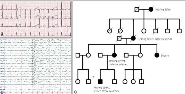
A 19-year-old man presented with recurrent episodes of loss of consciousness for the previous 3 years. His past medical history, general physical examination, and brain MRI findings were unremarkable. A laboratory examination revealed no abnormal findings ex- cept for slightly elevated lactic acid [3.2 mmol/L (reference range: 0.7–2.5 mmol/L)]. Elec- trocardiography revealed a short PR interval and a delta wave with QRS-complex widening, suggesting WPW syndrome (Fig. 1A). He was therefore initially referred to the Department of Cardiology. Since no evidence was found of atrioventricular reentrant tachycardia pre- ceded by loss of consciousness, he was referred to the Department of Neurology having not undergone any interventional procedure. The findings of a neurologic examination were unremarkable. His height was 161 cm, which is lower than the third percentile. History- taking revealed that he often experienced myoclonus, which had developed when he was 14 years old. His myoclonus occurred irrespective of exposure to light; however, all of the six generalized tonic-clonic seizures that he had experienced occurred when he was exposed to bright sunlight. He had not experienced an absence seizures. Abundant 2–3 Hz generalized spikes/polyspikes and wave discharges were found in an EEG examination (Fig. 1B). The electroclinical features led to an initial consideration of JME as a diagnosis. A detailed fami- ly history-taking revealed that his mother and maternal grandmother had been diagnosed with epilepsy, hearing defect, and diabetes. His maternal great-grandmother also had a hearing defect, and his aunt was diagnosed with epilepsy (Fig. 1C).
Considering the patient’s short stature and family history, genetic studies for identifying hereditary causes of epilepsy were performed. The mitochondrial 3243A>G mutation in the MT-TL1 gene was confirmed, and so he was diagnosed as MELAS syndrome. Further evalu- ations for identifying the involvement of other organs were performed. Electromyography, brainstem auditory evoked potentials, and an ophthalmologic examination showed no ab- normalities. Although he did not complain of any deterioration of his hearing, pure-tone au- diometry revealed moderate sensorineural hearing loss with preserved speech discrimina- Joo Hye Sung
Jung Hoon Han Hayom Kim Jung Bin Kim
Department of Neurology,
Korea University College of Medicine, Seoul, Korea
pISSN 1738-6586 / eISSN 2005-5013 / J Clin Neurol 2018;14(1):118-119 / https://doi.org/10.3988/jcn.2018.14.1.118
Received July 17, 2017 Revised August 20, 2017 Accepted August 24, 2017 Correspondence Jung Bin Kim, MD Department of Neurology,
Korea University College of Medicine, 73 Inchon-ro, Seongbuk-gu, Seoul 02841, Korea Tel +82-2-920-6277 Fax +82-2-925-2472
E-mail [email protected]
cc This is an Open Access article distributed under the terms of the Creative Commons Attribution Non-Com- mercial License (http://creativecommons.org/licenses/by-nc/4.0) which permits unrestricted non-commercial use, distribution, and reproduction in any medium, provided the original work is properly cited.
LETTER TO THE EDITOR
www.thejcn.com 119
Sung JH et al.
JCN
tion. He was receiving ubidecarenone and an antiepileptic drug (valproate). He experienced no seizure attack during the 4-month follow-up after discharge. Due to the possibility of mitochondrial toxicity of valproate, we are planning to replace this with other antiepileptic drugs.
While there are a few reports of WPW syndrome in pa- tients with MELAS syndrome,1-3 to our best knowledge EEG findings mimicking JME in patients with concurrent MELAS and WPW syndromes have not been described. Various pat- terns of epileptiform discharges can be found in patients with MELAS syndrome, although none of the findings are pathog- nomonic.4,5 Focal and multifocal spikes in association with ce- rebral lesions have been reported more frequently than gener- alized spikes in patients with MELAS syndrome.4,5 The present case implies that an EEG examination is essential in WPW syndrome with loss of consciousness without evidence of preceding atrioventricular reentrant tachycardia. In addition, it has been unclear whether WPW syndrome might be an epi- phenomenon in MELAS syndrome that mainly involves tis- sues with high energy demand. However, a genetic study should be considered when an epileptogenic condition has been confirmed in patients with WPW syndrome. Given the
electroclinical features of the present MELAS case mimicking JME, a genetic study should be considered if WPW syndrome or other symptoms suggesting MELAS are comorbid in pa- tients who are diagnosed electroclinically as JME.
Conflicts of Interest
The authors have no financial conflicts of interest.
REFERENCES
1. Sproule DM, Kaufmann P, Engelstad K, Starc TJ, Hordof AJ, De Vivo DC. Wolff-Parkinson-White syndrome in patients with MELAS. Arch Neurol 2007;64:1625-1627.
2. Lee MH, Sung YJ, Yoon JH, Kim J, Oh IY, Choi EK, et al. Wolff-Par- kinson-White syndrome in a patient with mitochondrial encephalop- athy, lactic acidosis and stroke-like episodes syndrome. Korean Circ J 2011;41:674-676.
3. Malfatti E, Laforêt P, Jardel C, Stojkovic T, Behin A, Eymard B, et al.
High risk of severe cardiac adverse events in patients with mitochon- drial m.3243A>G mutation. Neurology 2013;80:100-105.
4. Chevallier JA, Von Allmen GK, Koenig MK. Seizure semiology and EEG findings in mitochondrial diseases. Epilepsia 2014;55:707-712.
5. Lee HN, Eom S, Kim SH, Kang HC, Lee JS, Kim HD, et al. Epilepsy characteristics and clinical outcome in patients with mitochondrial en- cephalomyopathy, lactic acidosis, and stroke-like episodes (MELAS).
Pediatr Neurol 2016;64:59-65.
Hearing defect
Seizure
Pt
Hearing defect, diabetes, seizure
Hearing defect, diabetes, seizure
Hearing defect, seizure, WPW syndrome
A
B C
Fig. 1. A: Electrocardiography shows a short PR interval (96 msec) and QRS-complex widening (114 msec) with a delta wave. B: Electroencephalogra- phy shows 2–3 Hz frontal-dominant generalized polyspikes and wave discharges. C: Pedigree of the patient shows maternal inheritance traits. WPW:
Wolff-Parkinson-White.