Noninvasive Imaging of Pericardium
심막의 영상 소견
Bae Young Lee, MD*
Department of Radiology, Eunpyeong St. Mary’s Hospital, College of Medicine, The Catholic University of Korea, Seoul, Korea
The clinical manifestation of pericardial disease is similar to that of coronary artery disease and aortic disease. Therefore, a timely and accurate diagnosis is necessary. The pericardium is a 2-layered membrane that envelops the heart and great vessels, and there are numerous ana- tomic variations and pathologic conditions. Large or unusually located pericardial recesses can be easily mistaken for abnormal findings. Additionally, primary pericardial diseases resulting from infections, tumors, and injuries are possible; further, diseases can quickly spread along the pericardium. Echocardiography is generally the first imaging tool used to evaluate the pericardi- um. However, it has limited windows and poor resolution. Besides, the evaluation of postoper- ative echocardiography is sometimes limited. Currently, CT and MR imaging are useful for eval- uating pericardial diseases. Detailed knowledge of the pericardium is important for interpreting the images and clinical results.
Index terms Pericardium; Pericarditis; Pericardial Effusion; Heart Neoplasms
서론
심막(Pericardium)
심막은 심혈관계 질병이 있는 환자의 예후에 지대한 영향을 미치고 이의 평가를 위해서는 비침습적인 영상 검사가 중요한 역할을 한다(1-3). 지금까지 심막의 평가에는 심초음파가 일 차적인 검사 방법이며 중요한 역할을 해왔으나 제한된 음창, 낮은 해상도와 조직 분해능, 기 술적인 어려움 특히 폐기종, 비만, 수술 후 등의 경우 만족스러운 영상 획득에 문제가 있어 점점 CT, MR의 활용이 늘어나고 있다. CT는 영상 획득 속도와 공간 분해능이 좋아지면서 활용도 가 늘어났고 MR은 연부조직의 특성을 파악하는데 유용해서이다(1-3). 이번 종설에서는 심 막의 해부학, 생리학적 기능, 영상 획득 방법과 비교, 각 질환의 병리기전 및 영상 소견을 다 루고자 한다.
Received February 10, 2020 Revised March 13, 2020 Accepted March 20, 2020
*Corresponding author Bae Young Lee, MD Department of Radiology, Eunpyeong St. Mary’s Hospital, College of Medicine,
The Catholic University of Korea, 1021 Tongil-ro, Eunpyeong-gu, Seoul 03312, Korea.
Tel 82-2-2030-3020 Fax 82-2-2030-3026 E-mail [email protected] This is an Open Access article distributed under the terms of the Creative Commons Attribu- tion Non-Commercial License (https://creativecommons.org/
licenses/by-nc/4.0) which permits unrestricted non-commercial use, distribution, and reproduc- tion in any medium, provided the original work is properly cited.
ORCID iD Bae Young Lee https://
orcid.org/0000-0002-4125-255X
심막의 해부학 및 영상 소견
심막은 심장과 대혈관들의 시작 부분을 둘러싼 플라스크 모양으로 생긴 질긴 섬유막이며 심장 자체와 직접적으로 붙어 있는 부위는 없다(1). 내장 심막(visceral pericardium)은 심외막(epicar- dium)을 둘러싼 매우 얇은(< 1 mm) 중피(mesothelium)가 있고 그 사이에는 얇은 지방이나 다른 연결조직이 있어 서로 단단히 붙어 있으나 분리될 수도 있다. 벽층 심막(pariteal pericardium)은 바깥쪽의 질긴 섬유(fibrous) 심막과 안쪽의 얇은 중피로 구성된다. 내장 심막의 중피와 벽층 심막 의 중피 두 개가 합쳐져 장액(serous) 심막을 형성하며 내부에 15~50 mL의 장액(serous fluid)이 있다(1-3).
바깥의 섬유 심막은 주로 콜라겐 섬유로 구성돼 있고 중간중간에 짧은 탄력섬유(elastic fibrils) 가 섞인 구조이다. 섬유 심막은 위로는 대혈관들의 바깥막(adeventitia) 및 깊은 목 근막(deep cer- vical fascia)으로 연결되고 아래로는 횡격막의 중심널 힘줄(central tendon)과 연결된다. 앞으로 는 복장뼈(sternum)와 연결되고 왼쪽 4~6 늑골의 연골에 직접적으로 붙어 있을 수 있다. 앙 옆으로 는 종격동 흉막과 붙어 있고 뒤로는 주요 기관지, 식도, 하행 대동맥 등이 연해 있다. 횡경막 신경은 폐문 앞에서 섬유 심막과 종격동 흉막 사이에 위치한다(1-3).
복부의 지방과 마찬가지로 심외막 지방(epicardial fat)도 비만 시 증가하며 이는 관상동맥 죽상 경화증에 직접적으로 영향을 미친다. 지방 분포는 불균일하며 보통 심장의 오른쪽이 왼쪽보다 3~4배 더 많은 경향을 보인다. 이 지방층은 관상동맥, 정맥, 림프, 신경 등을 포함한다(1-3).
심막은 해부학적으론 0.8~1 mm 정도의 두께이나 영상 소견에서는 좀 더 두껍게 보인다. 얆은 부분이 CT에선 0.7~1.2 mm, MR에선 1.2~1.7 mm 정도로 측정되는데 이는 심막 내부의 구분되지 는 않아도 정상적으로 있는 심막액 때문으로 생각된다(Fig. 1). 경흉부 심초음파는 심막 두께 측정 에 유용하지 않으며 경식도 심초음파는 CT와 비슷한 소견을 보인다(1-4).
내측의 심막이 대동맥과 폐동맥을 둘러싸고 대정맥과 폐정맥을 둘러싸면서 가로 굴(transverse sinus) 및 빗굴(oblique sinus)을 구성하고 추가로 몇 개의 작은 오목(recess)들을 구성하게 된다.
이는 심막액이 늘어날 때 받아 주는 예비 공간의 역할을 한다(4, 5). 가로 굴은 대동맥 및 폐동맥 뒤 와 심방들과 상대정맥 앞에 위치하며 또한 좌심방 천장 위에 위치하게 된다. 이는 상행대동맥 오른 쪽으로 올라가 상대정맥 사이에서 위 대동맥 오목(superior aortic recess)을 형성하고 아래로 대 Fig. 1. Imaging of normal pericardium.
A-C. The pericardium (arrows) is thicker on a 3-mm slice thickness (A) compared to a 1-mm slice thickness (B) on CT. The right pericardial fat is more abundant on the right side than on the left. MRI (C) shows a dark signal rim of the pericardium.
A B C
동맥판까지 이어지며 여기서 생기는 상행대동맥과 우심방 사이의 공간을 아래 대동맥 오목(inferi- or aortic recess)이라고 한다(4, 5). 옆으로는 좌우 폐동맥을 따라 생기는 폐 오목(pulmonary re- cess)이 있으며 상대정맥의 뒤와 오른쪽 바깥쪽에 있는 대정맥 뒤 오목(postcaval recess)이 있다.
빗굴은 U자를 거꾸로 한 모습으로 좌심방 뒤에 위치하며 폐정맥들과 하대정맥에 의해 경계지어지 고 기관 분기부(carina)와 바로 붙어 있다(Fig. 2). 위 대동맥 오목, 왼쪽 오른쪽 폐 오목, 빗굴 같은 큰 오목은 약 29~45%에서 잘 보이고, 11~20%에서는 오른쪽, 왼쪽 폐 정맥 오목, 위 대동맥 오목, 대정맥 뒤 오목 같은 작은 오목들이 잘 보이는데 이런 경우 임파절 비대 같은 이상 소견들과 잘 구 분하여야 한다(Fig. 3) (4, 5).
심막의 기능 및 심실 상호의존
심막과 심막액은 윤활제 역할을 해서 마찰을 최소화하고 외부로부터 심장 감염을 막는 장벽의 Fig. 2. Transverse sinus, oblique sinus, and recesses of the pericardium.
Transverse sinus: 1. anterior portion of the superior aortic recess, 2. posterior portion of the superior aortic recess, 3. inferior aortic recess, 4. left pulmonic recess, 5. right pulmonic recess. Oblique sinus: 6. oblique si- nus. Pericardial sinus proper: 7. postcaval recess, 8. left pulmonary venous recess, 9. right pulmonary ve- nous recess.
역할도 하며 심장 형태를 보존하고 심방과 심실 간의 상호작용을 돕는 역할을 한다. 비교적 비탄력 적인 심막은 심실 확대(ventricular dilatation)를 최소화하고 심막액과 표면 활성제(surfactant) 를 통해 심장을 보호하며 종격동에서 심장의 이탈을 막는 역할을 한다(1-3). 특히 벽이 얇은 우심방 우심실에서는 정상 이완 압력(normal diastolic pressure)의 50% 이상이 심막의 영향을 받는다 (3). 이러한 비탄력적인 심막은 우심실 좌심실이 따로 움직이지 않도록 하며 이를 심실 커플링 (ventricular coupling) 내지는 심실 상호의존(ventricular interdependence)이라고 한다(1-3).
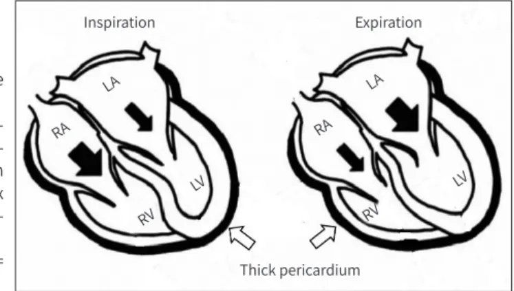
정상적으로는 좌심실의 이완기 압력이 우심실 보다 약간 높으므로 심실 중격이 오른쪽 심장으로 약간 불거진 모양을 보이나 병적인 경우 즉 우심실 부피나 압력이 증가하거나 심장막 내 압력이 증 가하는 경우 중격이 납작하거나 반대로 불룩할 수 있다(3, 6). 심낭(pericardial sac)은 약간의 음압 (negative pressure)을 가지며 주변 흉부의 장기들과 해부학적으로 구분되나 기능적으로는 상호 작용을 하게 된다(1). 정상인 경우 흡기 시 흉곽 내 압력이 떨어지고 이는 전신 정맥 환류(systemic venous return)와 우심실 충만(right ventricle filling)을 증가시키는 반면 좌심실 충만은 별 영향 을 받지 않는다. 호기 시에는 반대로 흉강 내 압력이 증가하고 폐정맥 환류(pulmonary venous return)와 좌심실 충만(left ventricle filling)을 증가시키고 전신 정맥 환류 및 우심실 충만을 감소 시킨다(3, 6). 교착성 심막염같이 심막에 이상이 있는 경우 호흡 시 흉곽 내 압력변화가 심장 내로 전해지는게 방해받아 심실 충만에 문제가 생기게 된다(Fig. 4) (Supplementary Video 1 in the online-only Data Supplement, Courtesy of Jung Im Jung from the Catholic University of Korea) (1-3, 6).
심막의 영상 획득 방법
심초음파가 일차적인 검사 방법이며 CT, MR은 그 후 더 자세한 평가를 위해 시행하는 것이 보통 Fig. 3. Large superior pericardial recess (arrows) of the pericardium in axial and coronal images.
A, B. This recess is continuous with other recesses and can be confused with pathologic lesions such as a mediastinal mass or lymphadenopathy.
A
B
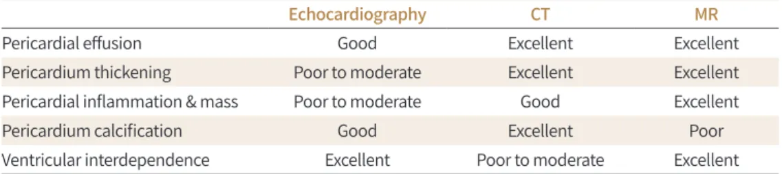
이다(1-3). 심초음파는 사용하기 쉽고 병상 옆에서도 시행 가능하며 해부와 기능적인 면을 동시에 볼 수 있는 장점이 있어 널리 쓰이나 조직 분해능이 떨어지고 시술자에 따른 차이가 크며 음창이 제한되는 경우가 많다(1, 3). 특히 비만, 만성 폐쇄성 폐질환, 심장 주변 수술 후 같은 경우 초음파 음창이 심히 제한되는 경우를 보인다(3). CT는 실제 해부학적인 해상도나 심장 주변의 평가, 수술 계획 수립, 심막 석회화 평가 등에 있어 우월하며 검사 시간도 수초에 불과하다. 다만 방사선 피폭 이 있고 조영제를 사용하며 숨을 참아야 하는 관계로 호흡과 관련된 검사를 하기 불편하다는 것이 단점이다. 촬영 조건은 고해상도의 얆은 두께로 분석한 영상이 필요하며 심전도 동기화된(ECG- gated) 영상을 얻어야 심장운동으로 인한 인공물을 피할 수 있다(1, 3). 심막 두께는 영상 획득 절 편 두께와 관련이 있는데 이는 절편 두께가 얇으면 더 얇아 보이고 두꺼우면 더 두툼하게 보이므 로 이를 감안해 판단하여야 한다(Fig. 1) (1). MR은 기능적인 평가와 해부학적 평가가 둘 다 용이 한 것이 장점이며 조직 분해능이 좋아 연부조직 평가에 우월해 염증 등의 평가에 아주 유용하다.
그러나 비용이 비싸고 검사 시간이 많이 걸리며 석회화 평가 능력이 떨어진다는 것이 단점이다(1- 3). 각 영상기기의 장점과 영상 소견별 기능은 다음과 같이 정리하였다(Tables 1, 2). 최근 가이드라
Expiration
RA RA
RV RV
LA LA
LV LV
Thick pericardium Inspiration
Fig. 4. Ventricular interdependence with constrictive pericarditis.
Changes in blood flow and ventricu- lar septum according to the respira- tion cycle. Interventricular septum with pericardium constriction convex toward the LV on inspiration and to- ward the RV on expiration.
LA = left atrium, LV = left ventricle, RA = right atrium, RV = right ventricle
Table 1. Strengths and Limitations of Echocardiography, CT, and MR
Modality Strengths Limitations
Echocardiography First line
Readily available, portable Low cost
No radiation
Respiro-phasic assessment
Limited acoustic window (obesity, postoperative) Narrow field of view
Operator dependent Low signal to noise ratio Limited tissue characterization
CT Improved anatomic delineation
Extracardiac disease Preoperative planning Pericardial calcifications Fast: a few seconds
Radiation Iodinated contrast Functional?
Imaging acquisition affected by arrhythmias and ectopy Breath-hold
Hemodynamically stable
MR Improved anatomic delineation
Superior tissue characterization
Ability to assess for pericardial inflammation Functional
High cost Time consuming Calcification not well seen
Gadolinium contrast: contraindication in renal impairment Hemodynamically stable
인에 의하면 심막 질환은 T1과 T2 영상, T1 mapping, 좌심실과 우심실 동영상(cine imaging), 지 연 조연증강 영상이 적절한 검사이며 혈류영상(velocity encoding gradient echo image, VENC) 이나 관류영상(perfusion imaging)은 그 효과가 확실치 않으며 심부하 검사는 적절치 않은 검사 라고 하였다(7). PET-CT는 종양이나 감염의 경우 강한 fluorodeoxyglucose (이하 FDG) 섭취를 보이나 감염이나 종양이 아닌 염증의 경우, FDG 섭취가 약간 증가하거나 거의 증가하지 않아 감 별에 도움이 된다(Fig. 5) (8).
심막의 질환
심막액
비정상적인 심막액의 원인으로는 심부전, 신부전, 간부전, 염증, 감염, 종양, 심근염, 의인성, 결합 조직질환, 특발성(idiopathic) 등으로 매우 다양하다. 대체적으로 한국을 비롯한 동양에서는 결핵 을 비롯한 감염 원인이 많으며 서양에서는 특발성 원인인 경우가 많다(8-10). 영상 검사를 하는 이 유로는 심막액의 유무 확인, 정도 파악, 심막액의 성질과, 심장 막의 염증 유무 확인, 천자 유도 등 Fig. 5. Usability of PET-CT in evaluating tumor invasion into the pericardium.
A, B. Invasion of the pericardium by thymic carcinoma (arrows) is strongly suggested in the CT scan (A), but there is no definite increased uptake of the pericardium in the PET-CT (B). No tumor invasion is observed during surgery.
A B
Table 2. Comparison of Imaging Modalities in the Evaluation of Pericardium
Echocardiography CT MR
Pericardial effusion Good Excellent Excellent
Pericardium thickening Poor to moderate Excellent Excellent
Pericardial inflammation & mass Poor to moderate Good Excellent
Pericardium calcification Good Excellent Poor
Ventricular interdependence Excellent Poor to moderate Excellent
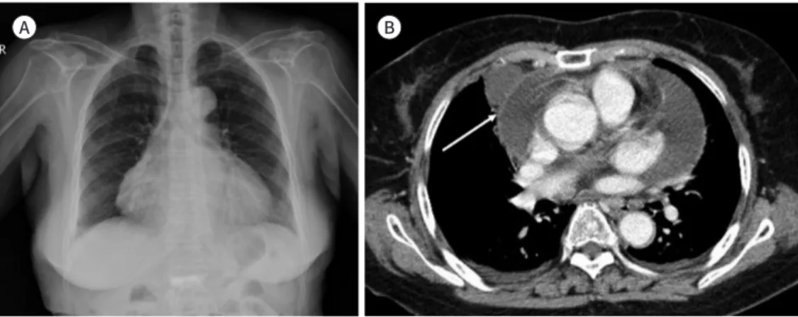
이다. 흉부 방사선 사진에서는 삼출액이 300 mL 이상 고여야 보이며 양쪽으로 대칭적으로 커져 심 장이 가죽 물주머니 모양을 보인다(Fig. 6) (8, 9).
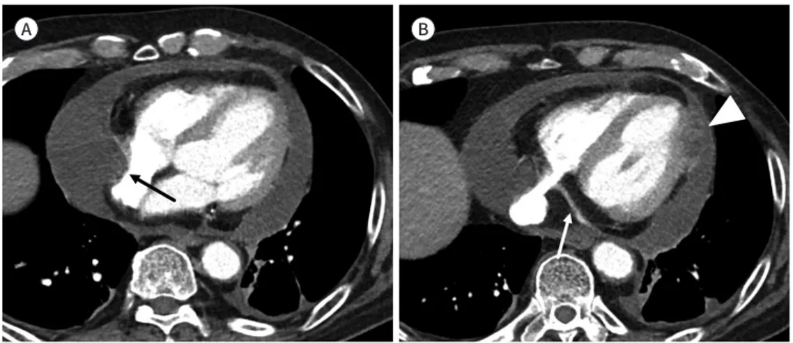
심초음파가 일차적인 검사이긴 하나 CT나 MR으로 더 추가 검사하는 경우가 많은데 그 이유로 는 두 방법이 심막액의 위치 및 분포를 파악하는데 더 유용해서이다. 대체로 심막의 두께가 4 mm 이상이면 비정상으로 간주한다(11, 12). 그러나 정확한 심막액 양은 확인하기 어려운데 각종 오목 에 고이는 것은 평가가 어렵기 때문이다. 그러나 우심실 앞에서 5 mm 이상의 심막 공간이 있으면 중등도의 심막액(100~500 mL)이 있는 것으로 생각할 수 있다(11, 12). CT나 MR 영상으로 심막액 의 성질을 추정해 볼 수 있는데, CT에서 물보다 높은 Hounsfield units (이하 HU), 즉 25 HU 이상 의 밀도를 보인 경우 혈액심막(hemopericardium), 악성 종양, 화농성 삼출액, 갑상선 기능저하 로 인한 심막액 등을 의심해 볼 수 있다(Fig. 7) (11, 12). 또한 물보다 낮은 밀도를 보이는 경우 유 미심막증(chylopericardium)을 의심할 수 있다(1). MR 영상에서는 단백질과 세포가 많은 삼출액 (exudate)의 경우 T1 신호강도가 올라가고 T2 신호강도가 떨어지는 소견을 보이며 혈액심장막의 경우 T1에서 높은 신호강도를 보이고 시네(cine) 영상에서 불균질한 낮은 신호강도를 보인다.
Fig. 7. Hemopericardium.
A, B. High-density hemopericardium (arrows) is seen around the heart and aorta in axial and coronal images.
Aorta dissection (arrowhead) is also seen and might be the cause of hemopericardium.
A B
Fig. 6. Pericardial effusion with tuberculosis.
A. Chest radiograph shows a leather water bag-shaped heart contour with enlarging contour on both sides.
B. CT axial image shows a large pericardial effusion with septation (arrow).
A B
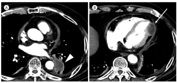
심장 압전(Cardiac Tamponade)
심막 공간에 액체, 혈액, 농, 공기 등이 차서 심막 주변의 압력이 올라가 심방과 심실을 압박하여 이완기 충전(diastolic filling)에 문제가 생기는 생명을 위협하는 응급상태이다. 심박출량 감소, 저 혈압, 빈맥 등이 오면서 심장성 쇼크가 올 수 있다(10). 원인은 다양한데 외상, 염증, 대동맥 박리, 종양, 심근 경색, 심장 수술 등이다. 심막액이 늘어나는 초기에는 각종 오목들의 여유 공간이 있어 압력 변화가 크지는 않지만 여유 공간 이상 늘어나면 급격하게 압력이 증가한다. 또한 심막 자체 가 잘 늘어나지 않는 조직이라서 심막액 전체 양도 중요하지만 심막액이 늘어나는 속도도 중요하 다. 빠르게 늘어난다면 소량이 늘어나도 바로 문제가 생기지만 천천히 늘어난다면 압력이 천천히 올라가 양이 많이 늘어도 문제가 천천히 생길 수 있다(10). 영상 소견으로는 우심실의 자유벽(free wall)의 허탈이 이완기에서 보이며 이는 심막 주변의 압력이 심실의 압력을 능가하는 것을 의미한 다. 다른 소견으로 심초음파에서는 수축기 초반에 우심방 압박, 승모판 및 삼첨판 혈액 유입의 호 흡에 따른 변화(흡기 시 삼첨판 혈류 증가, 승모판 혈류 감소) 등을 볼 수 있고, CT, MR에서는 납작 한 심장(flattened heart), 눌린 관상정맥동, 휘어진 중격(septal bowing), 상대정맥 및 하대정맥 직경 확대, 홀정맥 및 하대정맥으로 조영제 역류 등이 정지된 영상에서 잘 보일 수 있다(Fig. 8) (1- 3, 10-12).
심막 염증
심막 염증의 원인은 다양하나 급성 심막염은 원인을 모르는 경우가 80~85% 정도로 아마도 바이러 스 감염에 의한 것으로 추정하고 있다. 결핵에 의한 심막염은 개발도상국에서 흔하게 보고되며, hu- man immunodeficiency virus 감염 환자들에게서 칵테일 치료 전에는 심막액 빈도가 5~46%로 보 고되었는데, 심장 압전의 경우 결핵이 가장 흔한 원인이었다(Fig. 6) (13-16). 그 외 침습적인 심장 치 료 즉 수술, 스텐트 시술, 심박동기 삽입술 등이 심막 염증의 흔한 원인이 될 수 있다. 심근경색 후에 약 10% 정도에서는 급성으로 심막 염증이 생길 수 있으며 이는 경색과 연관된 염증 반응의 확산이
Fig. 8. Cardiac tamponade.
A. There is a large pericardial effusion, which is compressing the right ventricle (arrow).
B. A relatively narrow coronary sinus (arrow) and metastasis of the pericardium (arrowhead) are seen. Com- pression of the right ventricle and narrow coronary sinus are a sign of cardiac tamponade.
A B
원인으로 생각된다(13-16). 심막염은 흉부 방사선 치료의 가장 흔한 심장 부작용 중의 하나이며 어떤 부검 연구에서는 70%에서 심막 손상이 있다고 한다. 방사선 치료 후에 바로 나타나는 경우는 드물 고 임상 양상은 비정형적인 흉통, 발열, 심전도 변화 등이다. 보통은 영상 소견 없이 임상적으로 진단 이 되고 휴식과 염증 치료로 좋아진다. 영상 소견으로는 심막염, 심막 삼출액, 교착성 심막염 등의 다 양한 소견을 보일 수 있고, 심막이 조영증강될 수 있다. 심막 삼출은 방사선 치료 후 수개월 수년 후 에 생길 수 있으며 증상이 없는 경우 보통 저절로 좋아지나 급격히 증가하는 경우 치료를 해야 한다 (Fig. 9) (17).
교착성 심막염(Constrictive Pericarditis)
심막이 두꺼워지면서 탄력을 잃어 이완 기능장애가 오는 질환으로 우심실 부전에서 오는 증상이 나타나게 된다. 제한성 심근증과 비슷한 증상을 보이나 이 심근증은 교착성 심막염과 다르게 호흡 에 따른 변화는 보이지 않고 교착성 심막염은 심막절제술로서 교정이 가능하다는 것이 차이점이 다(18, 19).
원인은 급성 심막염과 크게 다르지는 않으나 급성 심막염 없이 생기는 경우도 있다. 그러나 급성 심막염에서 만성 심막염으로 진행되는 경우는 극히 드물며 반복되는 심막염이 있는 환자들도 교 착성 심막염까지 진행되는 경우는 드물다(18-20). 결핵이 흔한 원인이기는 하나 요즘은 특발성(id- iopathic)이 흔한 원인이며 최근에는 심장 수술이나 방사선 치료 후에 많이 생기는 경향이 있다(18).
교착성 심낭염의 병태생리를 좀 더 자세히 살펴보면, 심막이 탄력을 잃으면서 심낭 내의 부피가 고정되고 이완기의 기능장애가 오게 된다. 정상적으로는 흡기 시 흉곽 내 압력이 떨어져 전신 정맥 환류(systemic venous return)와 우심실 충만(right ventricle filling)이 증가하고 호기 시에는 반대 로 흉강 내 압력이 증가하면서 폐정맥 환류(pulmonary venous return)와 좌심실 충만(left ventri- cle filling)이 증가하고 전신 정맥 환류 및 우심실 충만이 감소한다(18-20). 교착성 심막염같이 심막 의 탄력이 떨어진 경우 호흡 시 흉곽 내 압력변화가 심장 내로 전해지는데 방해받고 양 심실이 제한 된 같은 공간을 사용하면서 정상보다 상대적으로 흡기 시 우심실 혈액 유입 증가가 늘고 호기 시에 는 우심실 혈류 유입은 더 감소하고 좌심실 혈류 유입은 더 증가하게 되면서 흡기 시 심실 중격이
Fig. 9. Pericardial effusion after radia- tion treatment for breast cancer.
Thick skin and increased density of the subcutaneous tissue (arrowhead) due to radiation and pericardial effu- sion (arrow).
오른쪽으로 움직이는 즉, 중격이 바운싱(bouncing) 하는 모습을 보이게 된다(Fig. 4) (18-20).
교착성 심막염은 혈류역학적인 진단이라서 영상 소견만으로는 진단할 수는 없다(21, 22). 심막의 비후가 대표적인 영상 소견이나 실제로 약 삼분의 일 정도에서는 심막 비후가 없으며 심막의 석회 화도 수술한 환자를 대상으로 했을 때 23%에서만 보였다는 보고도 있다(21). 즉 심막의 비후나 석 회화가 없더라도 교착성 심막염을 배제할 수는 없고 반대로 심막 석회화가 있다 하더라도 다 교착 성 심막염이다라고 할 수는 없다. 물론 석회화는 만성을 나타내는 대표적인 소견으로 CT로는 68%, 흉부 X-선 사진으로는 49%에서 보이며 흔하게 침착되는 부위는 양쪽 심실의 기저부, 우심실 유출로, 우심방, 심방심실고랑(atrioventricular groove)이다(21). 그 외에 심실 유입이 안 되어 생 기는 부가적인 변화로 대정맥이나 간정맥 확장, 관상 정맥동 및 심장의 확대, 흉수, 복수, 튜브 같은 심실의 모습, 납작한 우심실 자유벽 등이 보일 수 있다(Fig. 10) (21, 22).
전형적인 교착성 심막염과 다른 비전형적인 교착성 심막염도 있는데 정상 심막을 가진 교착성 심막염과 일시적 교착성 심막염이다. 교착성 심막염으로 수술한 환자의 약 18%에서는 심막 두께 가 정상이라는 보고가 있었으며 일시적인 교착성 심막염의 경우 교착성 심막염의 약 16% 정도에 서 2~3개월 후 병변이 없어졌다는 보고가 있었다(19). 가역적인 교착성 심막염과 비가역적 교착성 심막염으로 나누는 경우도 있는데 이는 가역적인 경우 심장막의 염증이 내과적인 치료에 반응해 Fig. 10. Constrictive pericarditis with pericardial calcifications.
A. Chest radiograph shows thin curvilinear calcification (arrow) along the inferior border of the heart shadow with pleural effusion.
B, C. CT images show thick pericardium with calcifications (arrows), enlarged atria, and tubular ventricles.
A B C
Fig. 11. Transient fluorodeoxyglucose-PET uptake in constrictive pericarditis.
A, B. Strong uptake of the pericardium (arrows) is seen in the initial study (A), and this uptake disappears after appropriate medical treatment (B) (Courtesy of Sung-A Chang, Samsung Medical Center).
A B
서 좋아지는 것을 의미하고 비가역적인 경우에는 심막절제술로 치료하는 것을 의미한다(18, 23, 24). MR에서 심막의 조영증강이 있으면 좀 더 가역적이라고 한다(23). PET-CT에서는 심막의 18F- FDG 섭취가 높으면 가역적일 가능성이 크다는 보고가 있다(Fig. 11) (24).
심막외 지방 괴사(Epicardial Fat Necrosis)
급성 흉통의 원인 중 하나로 원인은 잘 모른다. 영상 소견은 심장 주변에 지방음영의 종괴로 보 이며 석회화는 간혹 보인다. 보통은 저절로 좋아지는 경우가 많다(Fig. 12) (2, 25).
심막 종양
일차 심막 종양은 드물다. 양성으로는 심막낭종(pericardial cyst)이 가장 흔하고 악성으로는 중 피종(mesothelioma)이 가장 흔하며 다른 일차 악성 종양으로 육종(sarcoma), 임파종, 원시성 신 경 외배엽 종양(primitive neuroectodermal tumor) 등이 있다. 증상이나 임상 양상은 심막 삼출 액, 심막 염증, 주변 장기 침범 정도 등에 따라 결정된다(26).
심막낭종은 중피세포 낭종으로서 중피세포층과 연결조직으로 구성된 벽으로 형성 된다. 가장 흔한 부위는 오른쪽 심장가로막각(cardiophernic angle)이며 CT 영상에선 경계가 좋은 조영증강 Fig. 12. Epicardial fat pad necrosis.
A, B. Axial (A) and oblique (B) chest CT images show a fat attenuation mass with internal stranding in the epicardial fat pad (arrows).
A B
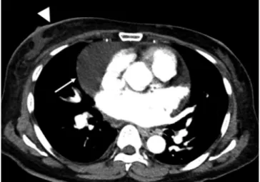
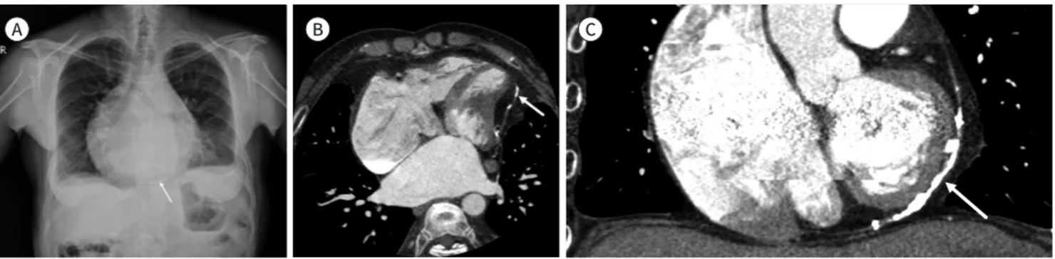
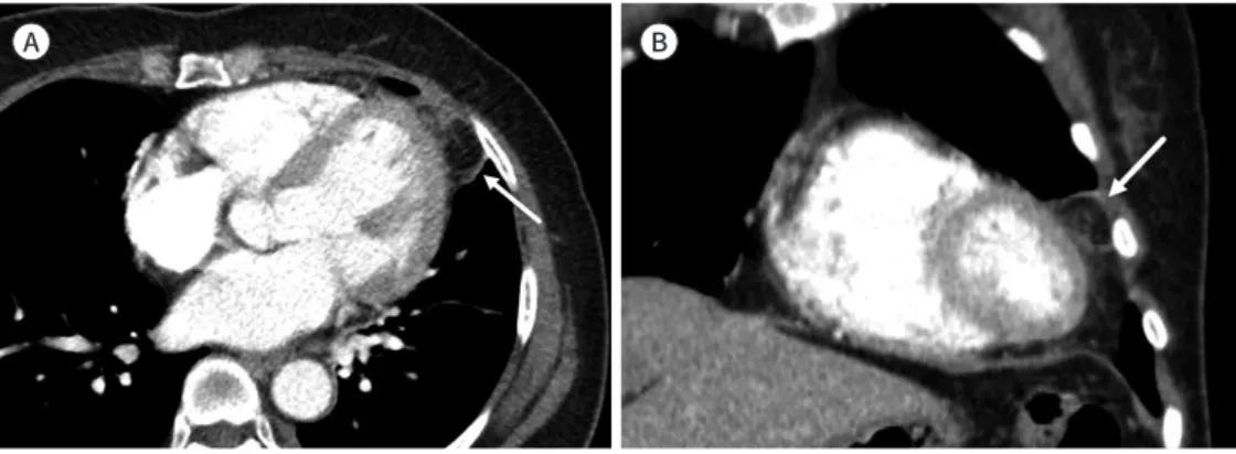
Fig. 13. Pericardial cyst.
A, B. Bulging contour (arrowhead) of the right cardiac border in the chest radiograph (A) and a well-defined homogenous cyst (arrows) along the right side of the heart in the axial and coronal CT images (B and C).
A B C
이 안 되는 액체 음영을 보이고 MR에선 액체의 성분에 따라 다르기는 하나 대체로 T1 저신호, T2 고신호강도를 보인다(Fig. 13) (26). 심막 중피종은 일차 심막암의 50% 정도를 차지하기는 하나 그 빈도는 매우 낮아 0.0022% 이하이다(2). 석면과의 연관성은 확실하지 않으나 약 삼분의 일에서 석 면 노출 병력이 있다. 예후는 극히 불량하여 극히 일부만 12개월 이상 생존한다(2). 이차 암의 심막 전이는 일차에 비해 훨씬 흔하며 임파선 전이로는 유방암, 임프종 등이 흔하고 혈류 전이는 흑생 종, 신장암, 직접 침범으로는 폐암이 대표적이다(Fig. 14) (2, 26).
결론
심막 질환은 생명을 위협하는 관상동맥이나 대동맥 질환들과 증상이 비슷한 경우가 많아 빠른 감별이 중요하다. 심막의 내부의 심장과 대혈관을 둘러싸며 특히 오목(recess)은 모양이 다양하여 질환으로 오인될 수도 있다. 그러므로 심막의 해부학적 구조를 숙지하고 병변의 다양한 영상 소견 을 익히는 것이 심막과 이를 둘러싼 장기들의 질환을 이해하고 평가하는데 필수적이다.
심막을 평가하는데 있어서 전통적으로 심초음파가 일차적인 검사 방법이나 그 해상도가 떨어지 고 주변의 폐나 뼈에 의해 음창이 제한되고 수술 후에는 평가가 힘든 경우가 많아 CT나 MR에 의 한 영상평가가 점점 늘어나고 유용한 경우가 많아 이러한 영상 소견에 익숙해지는 것이 해당 분야 의 의료진에 큰 도움이 될 것이다.
Supplementary Video Legend
Video 1. Septal bouncing due to this paradoxical septal motion of the interventricular septum is ob- served.
Supplementary Materials
The online-only Data Supplement is available with this article at http://dx.doi.org/10.3348/
jksr.2020.81.2.337.
Fig. 14. Pericardial metastasis.
A, B. Lung cancer (arrowhead) and pericardial metastasis (arrow) in the axial images of the chest CT are seen with high-density pericardial effusion.
A B
Conflicts of Interest
The author has no potential conflicts of interest to disclose.
REFERENCES
1. Bogaert J, Francone M. Pericardial disease: value of CT and MR imaging. Radiology 2013;267:340-356 2. Kligerman S. Imaging of pericardial disease. Radiol Clin North Am 2019;57:179-199
3. Klein AL, Abbara S, Agler DA, Appleton CP, Asher CR, Hoit B, et al. American Society of Echocardiography clini- cal recommendations for multimodality cardiovascular imaging of patients with pericardial disease: en- dorsed by the Society for Cardiovascular Magnetic Resonance and Society of Cardiovascular Computed To- mography. J Am Soc Echocardiogr 2013;26:965-1012.e15
4. Kodama F, Fultz PJ, Wandtke JC. Comparing thin-section and thick-section CT of pericardial sinuses and re- cesses. AJR Am J Roentgenol 2003;181:1101-1108
5. Z·urada A, Ustymowicz A, Loukas M, Michalak M, Czyz·ewska D, Gielecki J. Computerized tomography of the transverse pericardial sinus: normal or pathologic? Clin Anat 2017;30:61-70
6. Kim DH, Choi SI, Chun EJ, Choi SH, Park JH. The cardiac MR images and causes of paradoxical septal motion.
J Korean Soc Radiol 2010;62:427-434
7. Jo Y, Kim J, Park CH, Lee JW, Hur JH, Yang DH, et al. Guideline for cardiovascular magnetic resonance imag- ing from the Korean society of cardiovascular imaging-Part 1: standardized protocol. Korean J Radiol 2019;
20:1313-1333
8. James OG, Christensen JD, Wong TZ, Borges-Neto S, Koweek LM. Utility of FDG PET/CT in inflammatory car- diovascular disease. Radiographics 2011;31:1271-1286
9. Korean Society of Thoracic Radiology. Thoracic radiology. Chapter 17. 1st ed. Seoul: Koonja Publisher 2009 10. Jung HO. Pericardial effusion and pericardiocentesis: role of echocardiography. Korean Circ J 2012;42:725-
734
11. Bogaert J, Francone M. Cardiovascular magnetic resonance in pericardial diseases. J Cardiovasc Magn Reson 2009;11:14
12. Yared K, Baggish AL, Picard MH, Hoffmann U, Hung J. Multimodality imaging of pericardial diseases. JACC Cardiovasc Imaging 2010;3:650-660
13. Troughton RW, Asher CR, Klein AL. Pericarditis. Lancet 2004;363:717-727
14. Imazio M, Trinchero R. Myopericarditis: etiology, management, and prognosis. Int J Cardiol 2008;127:17-26 15. Restrepo CS, Diethelm L, Lemos JA, Velásquez E, Ovella TA, Martinez S, et al. Cardiovascular complications of
human immunodeficiency virus infection. Radiographics 2006;26:213-231
16. Yevenes S, Broncano J, Gutierrez FR. CT and MR update of pericardial diseases: a systematic review. Curr Car- diol Rep 2016;4:52
17. Walker CM, Saldaña DA, Gladish GW, Dicks DL, Kicska G, Mitsumori LM, et al. Cardiac complications of onco- logic therapy. Radiographics 2013;33:1801-1815
18. Sohn DW. Constrictive pericarditis as a never ending story: what’s new? Korean Circ J 2012;42:143-150 19. Peebles CR, Shambrook JS, Harden SP. Pericardial disease--anatomy and function. Br J Radiol 2011;84:
S324-S337
20. Alajaji W, Xu B, Sripariwuth A, Menon V, Kumar A, Schleicher M, et al. Noninvasive multimodality imaging for the diagnosis of constrictive pericarditis. Circ Cardiovasc Imaging 2018;11:e007878
21. Bogaert J, Meyns B, Dymarkowski S, Sinnaeve P, Meuris B. Calcified constrictive pericarditis: prevalence, dis- tribution patterns, and relationship to the myocardium. JACC Cardiovasc Imaging 2016;9;1013-1014 22. Hanneman K, Thavendiranathan P, Nguyen ET, Moshonov H, Paul NS, Wintersperger BJ, et al. Cardiovascular
CT in the diagnosis of pericardial constriction: predictive value of inferior vena cava cross-sectional area. J Cardiovasc Comput Tomogr 2014;8:149-157
23. Feng D, Glockner J, Kim K, Martinez M, Syed IS, Araoz P, et al. Cardiac magnetic resonance imaging pericar- dial late gadolinium enhancement and elevated inflammatory markers can predict the reversibility of con- strictive pericarditis after antiinflammatory medical therapy: a pilot study. Circulation 2011;124:1830-1837 24. Chang SA, Oh JK. Constrictive pericarditis: a medical or surgical disease? J Cardiovasc Imaging 2019;27:178-
186
25. Lee BY, Song KS. Calcified chronic pericardial fat necrosis in localized lipomatosis of pericardium. AJR Am J
Roentgenol 2007;188:W21-W24
26. Restrepo CS, Vargas D, Ocazionez D, Martínez-Jiménez S, Betancourt Cuellar SL, Gutierrez FR. Primary peri- cardial tumors. Radiographics 2013;33:1613-1630
심막의 영상 소견
이배영*
심막 질환은 응급질환인 관상동맥, 대동맥 질환과 증상이 비슷하여 빠른 감별이 중요하다. 심 막은 두겹으로 된 막으로서 다양한 해부학적 변이와 질환이 있다. 크거나 비전형적인 위치를 보이는 심막의 오목(recess)은 이상 소견으로 오인될 수도 있다. 심막에는 감염이나 종양 같 은 질환이 생기며 또한 주변에서 생기는 이러한 질환의 전파경로가 될 수 있다. 심막을 평가 하는데 있어 심초음파가 일차적인 검사방법이나 해상도가 떨어지고 주변의 폐나 뼈에 의한 음창이 제한될 수 있으며 수술 후 평가가 힘든 경우가 많다. 이때 CT나 MR에 의한 영상평가 가 유용한 경우가 많아 검사가 점점 늘어나고 있다. 심막의 형태와 질환에 대한 지식 및 영상 소견에 대한 이해는 환자 치료에 중요하다.
가톨릭대학교 의과대학 은평성모병원 영상의학과