급성 호흡기 증상으로 중환자실에 입원한 소아에서 호흡기 바이러스 감염의 임상적 특징
신연심1, 강동수1, 이건송1, 김재경2, 정은희3
단국대학교 의과대학 1소아청소년과학교실 · 2진단검사의학과교실, 3국립중앙의료원 소아청소년과
Clinical characteristics of respiratory virus infection in children admitted to an intensive care unit
Youn Shim Shin1, Dong Soo Kang1, Kun Song Lee1, Jae Kyoung Kim2, Eun Hee Chung3
Departments of 1Pediatrics and 2Laboratory Medicine, Dankook University College of Medicine, Cheonan; 3Department of Pediatrics, National Medical Center, Seoul, Korea
Purpose: To investigate the clinical characteristics and the risk factors for mechanical ventilator treatment and incidence of compli- cations in children admitted to an intensive care unit (ICU) with detected respiratory viruses.
Methods: Eighty-two patients who were detected respiratory viruses by multiplex real-time polymerase chain reaction from naso- pharyngeal aspirates were enrolled among the 123 children admitted to ICU with acute respiratory manifestations during the study period from January 2006 to December 2012.
Results: Detection rate of respiratory viruses were 66% and 13 patients (16%) had two viruses isolated. The most common respira- tory virus isolated was respiratory syncytial virus (RSV) (35%) followed by rhinovirus (19%), adenovirus (13%), parainfluenza virus (11%), influenza virus (11%), human metapneumovirus (6%), and human coronavirus (5%). Pneumonia (70%) was the most com- mon clinical diagnosis. The mean age of patient with RSV infection was the youngest and with influenza virus infection was the old- est among other viruses infection (mean± standard deviation, 5.9± 10.1 months vs. 51.0± 26.1 months; P= 0.01). Forty Patients (49%) who had the underlying diseases were not associated with incidence of mechanical ventilator treatment and complications.
Bacterial coinfection with respiratory virus was the significant risk factor of mechanical ventilator care and incidence of complica- tions (odds ratio [OR], 50.003; 95% confidence interval [CI], 3.955–632.144; P= 0.003, and OR, 15,569; 95% CI, 1.803–134.452;
P= 0.013).
Conclusion: The significant morbidity of pediatric patient admitted to ICU with respiratory virus infection (RVI) was associated with bacterial coinfection. Furthermore, multicenter study should be performed to investigate the epidemiology of RVI in pediatric pa- tients admitted to ICU in domestic. (Allergy Asthma Respir Dis 2013;1:370-376)
Keywords: Respiratory virus; Intensive care units; Child
서 론
소아에서 급성 호흡기 감염은 가장 흔한 질환이나 소수에서는 중환자실 입원이 필요한 경우도 발생한다.1) 급성 호흡기 감염으로 인한 호흡부전의 경우 미숙아 혹은 심폐질환 및 면역부전 등의 위 험인자가 있는 환아에서 주로 발생하지만 건강한 영유아에서도 발
생한다.2,3) 과거에 비해 백신의 보급과 위생 환경이 좋아지면서 세균 감염의 빈도가 감소하여 호흡기 바이러스가 급성 호흡기 감염의 주 요 원인이 되었다.4) 진단 검사 방법의 발전으로 최근에는 호흡기 감 염의 원인 바이러스 검출이 매우 용이하여 임상적으로 널리 사용 되고 있으며5) 과거에는 세균성 감염이 아닌 경우 감기 바이러스에 의한 호흡기 증상이라고 설명하였던 것에 비해 respiratory syncy- Allergy Asthma Respir Dis 1(4):370-376, December 2013 http://dx.doi.org/10.4168/aard.2013.1.4.370 ORIGINAL ARTICLE
Correspondence to: Kun Song Lee
Department of Pediatrics, Dankook University Hospital, Dankook University College of Medicine, 201 Manghyang-ro, Dongnam-gu, Cheonan 330-715, Korea
Tel: +82-41-550-3968, Fax: +82-41-550-3949, E-mail: pdlks@dankook.ac.kr Received: May 31, 2013 Revised: August 9, 2013 Accepted: August 10, 2013
© 2013 The Korean Academy of Pediatric Allergy and Respiratory Disease The Korean Academy of Asthma, Allergy and Clinical Immunology This is an Open Access article distributed under the terms of the Creative Commons Attribution Non-Commercial License
tial virus (RSV), adenovirus (ADV), human rhinovirus (hRV), human metapneumovirus (hMPV) 등에 의한 바이러스 감염임을 알 수 있게 되었다. 분자진단법인 multiplex real-time polymerase chain reaction (PCR) 방법을 이용하여 소아에서 호흡기 바이러스 감염의 임상 양상 및 각각의 바이러스 감염의 빈도, 특성 등에 대한 연구가 많이 보고되었으며 지금도 많은 연구가 진행 중이다.2,4-7) 그 러나 중환자실에 입원한 소아 환자를 대상으로 호흡기 바이러스 감염의 역학 및 임상적 특징에 대한 연구는 매우 적은 실정이다. 이 에 저자들은 단일 병원에서 급성 호흡기 증상으로 중환자실에 입 원한 소아 환자를 대상으로 호흡기 바이러스 감염의 종류와 임상 증상 그리고 인공호흡기 치료 및 치료 중 사망했거나 치료 후 만성 폐질환이 발생한 합병증 발생에 원인이 된 위험인자들을 분석하여 추후 급성 중증 호흡기 바이러스 감염에 대한 역학적 자료로 제시 하고자 한다.
대상 및 방법
1. 대상
2006년 1월부터 2012년 12월까지 급성 호흡기 증상으로 단국대 학교 병원 응급실을 방문하였거나 외래진료실을 통해 입원하여 시 행한 분당 호흡 수가 60회 이상의 빈호흡과 함께 동맥혈 가스분석 에서 고탄산혈증(pCO2≥60 mmHg)이 관찰되거나 연령을 기준 으로 빈호흡을 보이고 5 L/min의 산소를 마스크를 통해 투여에도 불구하고 산소포화도가 92% 이상 상승하지 않아 단국대학교병원 중환자실에 입원하여 치료 받았던 123명 중 호흡기 바이러스 감염 이 확인된 82명의 소아 환자를 대상으로 하였다.
2. 검체 채취 및 호흡기 바이러스 검출
급성 호흡기 증상으로 응급실을 통해 입원하였거나 외래에서 입 원한 환아에서 입원 24시간 이내에 8 French 무균 카테터를 점액 추 출기에 연결시킨 후 외비공을 통해 약 5–7 cm 정도 삽입시켜 음압 60–80 mmHg하에 흡입하여 비인두액을 채취하였다. 채취한 검체 는 가능한 빨리 검사실로 의뢰하였다. 지연된 경우는 DNA 추출 전 까지 4°C에 보관하였고, 추출한 DNA는 검사 실시 전까지 –70°C에 보관하였다. 비인두액에서 RNA를 분리하여 cDNA로 재구성한 후 호흡기 감염을 일으키는 11가지 종류의 RNA 바이러스; RSV type A, B, influenza virus (IFV) type A, B, parainfluenza virus (PIV) type 1, 2, 3, hRV A, coronavirus (CoV) 229E/NL63, OC43, hMPV 와 1가지 종류의 DNA 바이러스; ADV를 검사할 수 있게 제작된 Seeplex Respiratory virus detection kit-1 (Seegene, Seoul, Korea) 를 사용하여 증폭을 수행하였다. PCR은 PTC 200 PCR system (MJ Research, Watertown, MA, USA)을 사용하여 진행하였고, 94°C에 서 30초, 60°C에서 1.5분, 72°C에서 1.5분의 과정을 40회 반복하였
고, 마지막으로 72°C에서 10분간 진행하였다. 증폭된 PCR 산물은 ethidium bromide를 포함하는 2% agarose gel에서 100–150 V로 30 분간 전기 영동한 후 결과를 판독하였다.
3. 의무 기록 고찰
대상 환아의 입원 시 연령, 성별, 입원 시 혈액검사 소견, 미숙 아 및 선천성 기형 및 증후군, 천식 등의 위험인자에 대한 분석과 인공호흡기 치료, 치료 중 사망 했거나 혹은 치료 후 만성 폐질환 등의 합병증 발생에 대한 유무, 폐렴(이학적 검사에서 라음이 청 진되고, 영상의학적 소견에서 명확한 폐의 침윤이 존재한 경우) 및 모세기관지염의 유무 등을 의무기록지를 통해 후향적으로 분 석하였다. 본 연구는 환자에 대한 후향적 의무기록 분석에 대해 단국대학교병원 임상윤리위원회 심의를 통과하였다(No. 2013- 04-011).
4. 통계 분석
PASW ver. 18 (IBM Co., Armonk, NY, USA)을 사용하여 통계 분석을 하였다. 바이러스 특성에 대한 연속변수에 대한 분석은 Kruskal-Wallis검정을 이용하였고, 범주형 변수는 chi-square test 를 이용하였다. 인공호흡기 치료에 대한 위험인자와 합병증 발생의 위험인자에 대한 다변량 분석을 시행하였고 인공호흡기 치료에 대 한 분석의 경우는 성별, 입원 시 나이 및 체중, 입원 경로, 기저질환 의 유무, 바이러스 중복 감염, 무호흡, 바이러스와 세균 감염의 중 복 등의 독립변수에 대해 분석을 하였고 합병증 발생 분석의 경우 는 인공호흡기 치료에 대한 분석의 독립변수에 기저질환의 유무를 제외하고 인공호흡기 치료 여부를 추가하였고 중환자실 치료 기간 과 입원 시 체중, 나이를 보정하여 logistic regression을 이용하여 분석하였다. P 값이 0.05 미만인 경우 통계학적으로 유의한 것으로 판단하였다.
결 과
1. 환자의 특성
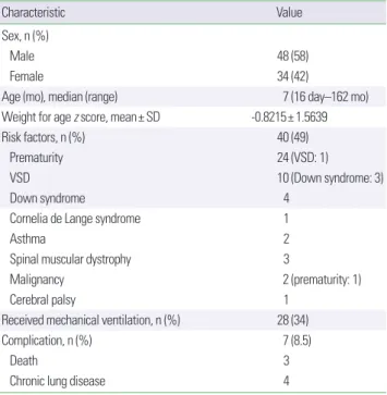
급성 호흡기 증상으로 중환자실에 입원한 총 123명 중 82명 (66%)에서 바이러스가 검출 되었으며 남아는 48명, 여아 34명으로 남녀 비는 1.4:1이었다. 연령 분포는 중앙값이 7개월(범위, 16일–162 개월)로 12개월 이하가 52명(63%)이었다. 미숙아, 심실중격결손, 다 운증후군, 천식, 근육질환, 뇌성마비, 다운증후군을 포함한 선천성 증후군 등의 위험인자 중 한가지라도 가지고 있던 환아는 82명 중 40명(49%)이었다. 중환자실에 입원 중 인공호흡기 치료를 받은 환 아는 82명 중 28명(34%)이었으며, 치료 중 또는 치료 후 사망 또는 만성 폐질환 등의 합병증이 발생한 환자는 80명중 7명(8.5%)이었 다(Table 1).
2. 호흡기 바이러스 감염의 양상 1) 바이러스 감염의 빈도
바이러스 감염이 확인된 82명 중 13명(16%)은 바이러스 중복 감 염이 확인되었으며 중복 감염된 바이러스를 포함하여 검출된 바이 러스의 전체 빈도는 RSV, 34회(35%); hRV, 18회(19%); ADV, 12회 (13%); PIV, 10회(11%); IFV, 10회(11%); hMPV, 6회(6%); CoV, 5회 (5%)였다(Fig. 1).
2) 월별 바이러스 감염의 양상
12월에 호흡기 바이러스 감염이 확인된 환자가 20명(21%)으로 가장 많았으며 11월에 18명(19%), 5월에 11(12%)명이었으며 8월에 2명(2%)으로 가장 적었다. RSV 감염은 9월부터 시작되어 11월에
가장 많았고, PIV 감염은 3월부터 시작되어 6월에 가장 많았다 (Fig. 2).
3) 바이러스 감염에 따른 임상 진단
호흡기 바이러스 감염에 의해 폐렴으로 진단된 경우가 57예 (70%)로 가장 많았으며 세기관지염은 12예(15%), 크룹은 2예(2%),
Table 1. The characteristics of patients
Characteristic Value
Sex, n (%)
Male 48 (58)
Female 34 (42)
Age (mo), median (range) 7 (16 day–162 mo)
Weight for age z score, mean± SD -0.8215± 1.5639
Risk factors, n (%) 40 (49)
Prematurity 24 (VSD: 1)
VSD 10 (Down syndrome: 3)
Down syndrome 4
Cornelia de Lange syndrome 1
Asthma 2
Spinal muscular dystrophy 3
Malignancy 2 (prematurity: 1)
Cerebral palsy 1
Received mechanical ventilation, n (%) 28 (34)
Complication, n (%) 7 (8.5)
Death 3
Chronic lung disease 4
SD, standard deviation; VSD, ventricular septal defect.
Fig. 1. Incidence of respiratory viruses detected in pediatric patients admitted to intensive care unit with acute respiratory manifestations. RSV, respiratory syncytial virus; hRV, human rhinovirus; ADV, adenovirus; PIV, parainfluenza virus;
IFV, influenza virus; hMPV, human metapneumovirus; CoV, coronavirus.
40 35 30 25 20 15 10 5 0
RSV hRV ADV PIV IFV hMPV CoV
Number of detection
Fig. 2. Monthly distribution of respiratory viruses detected. CoV, coronavirus; hMPV, human metapneumovirus; hRV, human rhinovirus; IFV, influenza virus; ADV, adeno- virus; PIV, parainfluenza virus; RSV, respiratory syncytial virus.
CoV hMPV hRV IFV ADV PIV RSV 25
20
15
10
5
0 1 2 3 4 5 6 7 8 9 10 11 12
Months
Number of patients
11 1
2 3
4
3
8 13
2 2 1
2 2 2 3 3 1 1
1 2
11 1 2
2 1
1 1 2
1 2 2 1 2
1 2
11 1 3
1 2 1
11 1
1
천식 악화는 2예(2%)였다. 환아 중 입원 시 시행한 혈액배양 혹은 흉수배양검사에서 세균이 배양되거나, 호흡기 바이러스검사와 같 이 시행한 비인두액 PCR 검사에서 양성으로 확인된 세균성 폐렴 은 총 9예였다. 비인두액 PCR 검사와 혈청학적 검사에서 모두 양성 이 확인된 미코플라스마 2예, 비인두액 검사에서 양성이 확인된 백 일해 1예가 있었으며 혈액배양검사에서 확인된 경우는 폐렴연쇄구 균 2예와 메치실린 내성 황색 포도상구균 1예, group B 연쇄상구균 1예였다. 그 외에 기관지 흡입액 배양 및 PCR 검사에서 확인된 결핵 1예, 쥐폐포자충 1예가 있었다.
4) 바이러스 감염에 의한 임상적 차이
바이러스 감염에 대한 임상적 차이를 알아보기 위해 임상 양상 에 영향을 줄 수 있는 위험인자를 가지고 있는 환아 40명을 제외한 42명에서 중복 감염이 된 다섯명을 포함하여 47건의 바이러스 감 염을 대상으로 입원 시 연령, white blood cell, C-reactive protein,
산소 포화도, 중환자실에서의 입원 기간 등의 연속 변수에 대한 분 석과 폐렴 및 모세기관지염 발생, 인공호흡기 치료의 빈도에 대한 범주형 변수에 대해 교차분석을 시행하였다. 연속변수에 대한 평 균값 분석에서는 RSV 감염의 평균 연령이 5.9개월로 가장 어렸고, 인플루엔자 감염의 평균 연령은 51개월로 가장 높게 나타났으며 통 계적으로 유의함을 보였으나(P= 0.01) 이외에 연속 변수는 바이러 스에 따른 평균값에 차이가 없었으며 바이러스 감염에 따른 폐렴 과 모세기관지염 및 인공 호흡기 치료의 빈도는 통계적으로 유의함 을 보이지 않았다(Table 2).
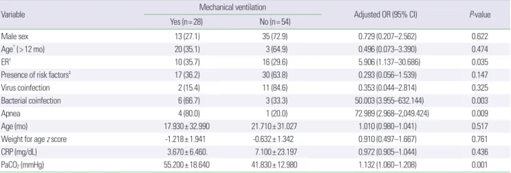
3. 인공호흡기 치료의 위험인자에 대한 다변량 분석
인공호흡기 치료와 연관된 위험인자에 대한 다변량 분석에서는 응급실에서 중환자실로 입원한 경우, 호흡기 바이러스와 세균성 폐렴이 동반된 경우, 무호흡, 입원 시 동맥혈 이산화탄소 분압이 통 계적으로 의미는 위험인자였다(Table 3).
Table 2. The clinical characteristics and diseases association of respiratory viruses infection
Characteristic RSV (n= 20) ADV (n= 8) hRV (n= 6) IFV (n= 5) hMPV (n= 4) PIV (n= 2) CoV (n= 2) P-value
Age (mo) 5.9± 10.1 8.4± 6.7 15.2± 12.4 51.0± 26.1 13.0± 7.8 6.0± 7.0 42.5± 58.6 0.010*
WBC (/mm3) 13,402± 7,909 14,341± 9,006 10,575± 3,921 14,972± 8,471 13,875± 10,716 17,355± 12,013 11,810± 1,173 0.869*
CRP (mg/dL) 9.6± 34.8 1.0± 1.4 5.8± 11.9 11.7± 18.0 4.0± 4.8 1.6± 1.3 6.7± 8.0 0.250*
SPO2 (%) 88.3± 11.9 80.5± 32.7 87.8± 9.3 92.6± 10.6 95.8± 1.0 90.6± 10.6 64.6± 44.6 0.404*
Duration of ICU care (day) 5.6± 5.6 3.4± 1.2 5.7± 3.0 5.6± 4.8 4.0± 0.8 4.5± 2.1 4.5± 2.1 0.765*
Pneumonia 16 (41.0) 7 (17.9) 5 (12.8) 4 (10.3) 3 (7.7) 2 (5.1) 2 (5.1) 1.000†
Bronchiolitis 4 (50.0) 1 (12.5) 1 (12.5) 1 (12.5) 1 (12.5) 0 (0) 0 (0) 1.000†
Ventilator care 5 (25.0) 1 (12.5) 3 (50.0) 2 (40.0) 0 (0) 1 (50.0) 1 (50.0) 0.563†
Values are presented as mean± standard deviation or number (%).
RSV, respiratory syncytial virus; ADV, adenovirus; hRV, human rhinovirus; IFV, influenza virus; hMPV, human metapneumovirus; PIV, parainfluenza virus; CoV, coronavirus; WBC, white blood cell; CRP, C-reactive protein; ICU, intensive care unit.
*Kruskal-Wallis test. †Fisher exact test.
Table 3. Evaluating risk factors on the treatment of mechanical ventilation
Variable Mechanical ventilation
Adjusted OR (95% CI) P-value
Yes (n= 28) No (n= 54)
Male sex 13 (27.1) 35 (72.9) 0.729 (0.207–2.562) 0.622
Age* (> 12 mo) 20 (35.1) 3 (64.9) 0.496 (0.073–3.390) 0.474
ER† 10 (35.7) 16 (29.6) 5.906 (1.137–30.686) 0.035
Presence of risk factors‡ 17 (36.2) 30 (63.8) 0.293 (0.056–1.539) 0.147
Virus coinfection 2 (15.4) 11 (84.6) 0.353 (0.044–2.814) 0.325
Bacterial coinfection 6 (66.7) 3 (33.3) 50.003 (3.955–632.144) 0.003
Apnea 4 (80.0) 1 (20.0) 72.989 (2.968–2,049.424) 0.009
Age (mo) 17.930± 32.990 21.710± 31.027 1.010 (0.980–1.041) 0.517
Weight for age z score -1.218± 1.941 -0.632± 1.342 0.910 (0.497–1.667) 0.761
CRP (mg/dL) 3.670± 6.460. 7.100± 23.197 0.972 (0.905–1.044) 0.436
PaCO2 (mmHg) 55.200± 18.640 41.830± 12.980 1.132 (1.060–1.208) 0.001
Values are presented as number (%) or mean± standard deviation.
OR, odds ratio; CI, confidence interval; ER, emergency room; CRP, C-reactive protein.
*Age at admission. †Admission route to intensive care unit. ‡Prematurity, ventricular septal defect, Down syndrome, Cornelia de Lange syndrome, asthma, spinal muscular dys- trophy, malignancy, cerebral palsy.
4. 합병증과 연관된 위험인자에 대한 다변량 분석
치료 중 전원으로 인해 합병증의 유무를 알 수 없었던 2명을 제 외하고 80명에 대한 합병증 발생의 위험인자에 대한 다변량 분석에 서는 세균의 중복 감염만이 사망, 만성 폐질환 등의 합병증 발생에 위험인자로 통계적으로 유의함을 보였다(Table 4).
고 찰
본 연구에서 급성 호흡기 증상으로 중환자실에 입원한 소아에서 각각의 호흡기 바이러스 감염의 특성은 입원 시 평균 연령을 제외 하고 혈액검사와 인공호흡기 치료의 빈도 및 진단에는 의미 있는 차이는 없었다. 인공호흡기 치료의 위험인자로는 응급실을 통한 입 원, 바이러스 감염 이외에도 세균 감염이 합병된 경우, 무호흡, 혈중 이산화탄소 농도가 높은 경우에 기계환기의 위험도가 증가하였다.
합병증 발생의 위험인자로는 바이러스 감염과 세균 감염이 동반되 어 있는 경우 의미 있는 위험인자라는 결론을 얻었다.
PCR 방법을 통해 급성 호흡기 증상의 원인 바이러스를 규명하 고 이를 토대로 소아 호흡기 바이러스 감염의 역학 및 임상 양상에 대한 많은 논문이 국외뿐만 아니라 국내에서도 다수 보고되어 바 이러스 감염의 계절 분포, 임상적 특징 등에 대해 유익한 참고 자료 로 사용되고 있다.2,5,8-14) 국외에서 중환자실에 입원한 소아 환자에 대한 호흡기 바이러스 감염에 대한 연구가 소수 있으나 국내에서는 Ko 등15)이 급성 호흡곤란증후군으로 중환자실에 입원한 환아에서 원인 바이러스 감염원에 대한 보고가 있을 뿐이다.1,16-18) Ghani 등1) 의 연구에서는 호흡기 증상이 있던 709명의 소아 환자 가운데 62.9%의 환자에서 호흡기 바이러스가 검출되었고, hRV가 가장 많 이 검출 되었으며 RSV, ADV, IFV A, PIV 순서로 검출 빈도를 보였 다. 본 연구에서는 호흡기 바이러스 검출률이 66%였으며 RSV 가 장 높은 빈도를 보였고, hRV, ADV 등의 순서로 Ghani 등1)의 연구 와 비슷한 결과를 보였다. Ghani 등1)은 계절에 따른 호흡기 바이러 스 검출 빈도와 소아중환자실에 검출된 호흡기 바이러스 검출 빈 도는 일치한다고 하여 병원 내 감염이 아니라면 계절에 유행하고 있는 호흡기 바이러스 감염이 급성 호흡기 증상으로 중환자실에 입 원한 환자에서 호흡기 증상의 원인 바이러스임을 유추할 수 있다
고 하였으나 본 연구에서는 계절에 따른 호흡기 바이러스 감염의 빈도에 대한 조사가 없어 시점에 유행하고 있는 바이러스 감염과 중환자실에 입원한 환자에서의 원인 바이러스 감염원을 비교할 수 없었다.
Ghani 등1)은 중환자실에 입원한 환자의 경우 67%가 선천성 질 환을 포함한 선행 질환이 있는 것으로 보고하여 기저질환이 있는 경우 중환자실에 입원할 위험률이 높다는 일반적인 예측을 증명할 수 있었고 본 연구에서는 82명의 환아 중 40명(49%)이 미숙아, 선천 성 증후군, 심실중격결손, 근육질환, 뇌성마비, 천식 등의 질환을 가 지고 있어 Ghani 등1)의 연구에 비해 기저질환이 있는 환아의 비율 은 낮았으나 입원 환자의 절반을 차지하고 있어 본 연구에서도 기 저질환을 가지고 있는 환아의 경우 심한 급성 호흡기 증상을 나타 낼 위험성이 있다고 할 수 있겠다.
호흡기 바이러스 감염에 있어 임상적 양상만으로 각각의 바이러 스를 구별할 수 있는 특징적인 소견은 없으나,19,20) 2009년 전세계적 으로 유행하였던 H1N1 신종 인플루엔자의 경우 빈호흡을 동반한 폐렴과 급성 호흡곤란증후군이 발생하는 임상적 특징이 있으며 RSV의 경우는 어린 영유아에서의 호흡기 감염과 연관이 있고, 아 데노바이러스의 경우는 다른 바이러스에 비해 치사율이 더 높다 는 연구가 있다.10,21) 바이러스 검출 수가 각각 다르고, 대상 바이러 스의 수가 적다는 한계점이 있으나 본 연구에서는 진단 시 환자의 연령만이 유의하게 차이가 있었으며 각각의 바이러스에 따른 폐렴 및 모세기관지염의 발생, 혈액학적 소견, 인공호흡기 사용 및 합병 증 발생 등은 유의한 차이가 없어 증상을 통한 바이러스 감염원을 유추할 수는 없었다. 그러나 RSV 감염이 인플루엔자 바이러스 감 염 환아에 비해 유의하게 어린 연령에서 발생하여 연령은 원인 바 이러스를 예측할 수 있는 중요 인자가 될 수 있을 것으로 생각된다.
호흡기 바이러스 감염 시 두 가지 이상의 바이러스에 감염되는 중복 감염은 본 연구에서 16%의 빈도를 보였으며 기계환기요법이 나 합병증 발생과는 무관하였다. 다른 연구에서도 15% 정도의 빈 도를 보이며 나쁜 예후와는 연관성이 없으나 바이러스 중복 감염 이 있을 때 호흡기 증상으로 일반 병실에 입원한 경우 보다 소아중 환자실에 입원하게 되는 경우가 많고, 기저질환을 가지고 있거나 미숙아의 경우 중복 감염률이 더 높다는 보고를 하였다.1,9) 그러나 Table 4. The logistic regression analysis on risk factors of developing complications
Factor No. of patients* (%) Adjusted OR† 95% CI P-value
Admission route: ER 2 (28) 1.341 0.145–12.383 0.796
Virus coinfection 1 (14) 4.305 0.275–67.393 0.298
Male 4 (57) 0.892 0.100–7.962 0.928
Ventilator treatment 5 (71) 5.365 0.547–52.631 0.149
Bacterial coinfection 6 (85) 15.569 1.803–134.452 0.013
OR, odds ratio; CI, confidence interval; ER, emergency room.
*Total number of patients developed complications was seven. †Adjusted for age, z score, duration of intensive care unit care
본 연구에서는 일반 병실 환자에 대한 대조군 연구를 시행하지 못 하였고 대상 환자 수가 적어 통계적으로 바이러스에 중복 감염이 된 경우 기저질환이 있는 것과의 연관성은 통계적으로 유의함을 보 이지 않았다.
호흡기 바이러스 감염 후 급성 호흡곤란으로 중환자실에 입원하 는 경우, 이 중 3%–5%의 환자는 24시간 이내에 기계환기요법이 필 요해진다고 보고된 바 있다.22) Straliotto 등18)의 연구에서는 소아중 환자실에 입원한 261명 중 25명(9.6%)에서 기계환기요법을 받았으 며 본 연구에서는 82명 중 28명(34%)으로 타 연구에 비해 높은 빈 도를 보였다. 이는 다른 연구들에 비해 병원 내 감염 환자가 없고 지 역에서 감염이 되어 급성 호흡곤란 증상으로 내원하여 응급실 및 병실 등에서 치료가 이루어 지지 않은 상태에서 증상이 악화되어 기도 삽관의 비율이 타 연구에 비해 높았을 것으로 추측된다.
기계환기요법을 시행하게 되는 위험인자 분석에서 세균과의 중 복 감염, 응급실에서 중환자실로 입원한 경우, 무호흡 등이 통계적 으로 유의하게 의미가 있었다. Ghani 등1)의 연구에서도 세균과의 중복 감염이 있을 경우 기계환기요법을 시행할 확률이 더 높고, 기 계환기요법을 보다 오랫동안 유지하게 되어 중환자실에서의 치료 기간이 길어졌음을 보고하여 본 연구에서와 같이 세균과의 중복 감염은 기계환기요법의 위험인자임을 알 수 있었다. 본 연구에서는 82명 중 9명(11%)에서 입원 시 시행한 검사에서 세균 감염과 호흡 기 바이러스의 중복 감염이 확인되었으며 미코플라즈마와 폐구균 이 각각 2명으로 다른 세균에 비해 검출 빈도가 높았다. 세균 감염 이 동반되었던 환아 9명 중 과거 병력이 없던 환아는 3명으로, 6명 은 미숙아가 3명, 급성 림프구성 백혈병이 1명, 선천성 심장질환이 1 명, 선천성 증후군(DeLange syndrome)이 1명으로 동반질환이 있 는 환아에서 세균성 감염의 비율이 높음을 알 수 있었으나 대상자 수의 부족으로 통계적 유의성을 확인하지는 못하였다.
Straliotto 등18)과 Hon 등10)의 연구에서는 아데노바이러스 감염 시 사망률이 높다는 보고를 하였으나 본 연구에서는 치료 도중 사 망하거나 만성 폐질환 등의 합병증이 발생한 경우 각각의 바이러스 에 따른 차이가 없었고 합병증 발생의 위험인자에 대한 다변량 분 석에서 세균과의 중복 감염만이 통계적으로 의미 있는 위험인자로 확인되어 급성 호흡기 감염에 의한 급성 호흡부전 환자에서는 호 흡기 바이러스 검사 외에도 혈액배양 및 흉막액 배양 등에 대한 검 사를 적극적으로 시행하여 세균성 감염의 유무를 확인해야 할 것 으로 생각된다.
본 연구는 중환자실에 입원한 환자만을 대상으로 하여 일반 병 실 환자의 대조군이 없다는 점과 단일 기관에서의 연구로 대상 환 자가 적어 연구의 결과를 일반적으로 적용할 수 없고 통계적 힘이 작은 한계점이 있다. 그러나 국내에서 아직까지 보고가 많지 않았 던 호흡기 바이러스 감염으로 인해 중환자실에 입원한 소아 환자 의 역학 및 임상적 특성에 대한 연구로 추후 참고자료로서 의의가
있을 것으로 생각된다. 추후 소아 중환자실에 급성 호흡기 증상으 로 입원한 환자를 대상으로 다기관의 연구가 시행되어 국내의 소아 중증 호흡기 바이러스 감염의 특징과 나쁜 예후와 연관된 위험인 자 등에 대한 보다 광범위한 연구가 필요할 것으로 생각된다.
REFERENCES
1. Ghani AS, Morrow BM, Hardie DR, Argent AC. An investigation into the prevalence and outcome of patients admitted to a pediatric intensive care unit with viral respiratory tract infections in Cape Town, South Af- rica. Pediatr Crit Care Med 2012;13:e275-81.
2. Kwon JH, Chung YH, Lee NY, Chung EH, Ahn KM, Lee SI. An Epide- miological study of acute viral lower respiratory tract infections in hospi- talized children from 2002 to 2006 in Seoul, Korea. Pediatr Allergy Respir Dis 2008;18:26-36.
3. Diniz EM, Vieira RA, Ceccon ME, Ishida MA, Vaz FA. Incidence of re- spiratory viruses in preterm infants submitted to mechanical ventilation.
Rev Inst Med Trop Sao Paulo 2005;47:37-44.
4. Lim G, Park TS, Suh JT, Lee HJ. Comparison of R-mix virus culture and multiplex reverse transcriptase-PCR for the rapid detection of respiratory viruses. Korean J Lab Med 2010;30:289-94.
5. Lim JS, Woo SI, Kwon HI, Baek YH, Choi YK, Hahn YS. Clinical charac- teristics of acute lower respiratory tract infections due to 13 respiratory viruses detected by multiplex PCR in children. Korean J Pediatr 2010;
53:373-9.
6. Kim HY, Kim KM, Kim SH, Son SK, Park HJ. Clinical manifestations of respiratory viruses in hospitalized children with acute viral lower respira- tory tract infections from 2010 to 2011 in Busan and Gyeongsangnam- do, Korea. Pediatr Allergy Respir Dis 2012;22:265-72.
7. Aramburo A, van Schaik S, Louie J, Boston E, Messenger S, Wright C, et al. Role of real-time reverse transcription polymerase chain reaction for detection of respiratory viruses in critically ill children with respiratory disease: Is it time for a change in algorithm? Pediatr Crit Care Med 2011;
12:e160-5.
8. Roh EJ, Chang YP, Kim JK, Rheem IS, Park KS, Chung EH. Clinical sig- nificance of codetection of the causative agents for acute respiratory tract infection in hospitalized children. Korean J Pediatr 2009;52:661-6.
9. Richard N, Komurian-Pradel F, Javouhey E, Perret M, Rajoharison A, Bagnaud A, et al. The impact of dual viral infection in infants admitted to a pediatric intensive care unit associated with severe bronchiolitis. Pedi- atr Infect Dis J 2008;27:213-7.
10. Hon KL, Leung E, Tang J, Chow CM, Leung TF, Cheung KL, et al. Pre- morbid factors and outcome associated with respiratory virus infections in a pediatric intensive care unit. Pediatr Pulmonol 2008;43:275-80.
11. Crowcroft NS, Zambon M, Harrison TG, Mok Q, Heath P, Miller E. Re- spiratory syncytial virus infection in infants admitted to paediatric inten- sive care units in London, and in their families. Eur J Pediatr 2008;167:
395-9.
12. Lee SJ, Shin EW, Park EY, Oh PS, Kim KN, Yoon HS, et al. Epidemiology and clincal analysis of acute viral respiratory tract infections in children (September, 1998-May, 2003). Korean J Pediatr 2005;48:266-75.
13. Han MY, Lee BL, Seo WH, Ahn SH, Kim SJ, Hwang SJ, et al. Epidemiol- ogy of respiratory syncytial virus lower respiratory tract infection and analysis of risk factors in severe infections from 1995 to 2004. Pediatr Al- lergy Respir Dis 2005;15:150-9.
14. Kim MR, Lee HR, Lee GM. Epidemiology of acute viral respiratory tract
infections in Korean children. J Infect 2000;41:152-8.
15. Ko JM, Ha EJ, Lee EH, Lee SY, Kim HB, Hong SJ, et al. Clinical outcome and prognostic factors of acute respiratory distress syndrome in children.
Korean J Pediatr 2005;48:599-605.
16. Farias JA, Fernandez A, Monteverde E, Flores JC, Baltodano A, Mencha- ca A, et al. Mechanical ventilation in pediatric intensive care units during the season for acute lower respiratory infection: a multicenter study. Pe- diatr Crit Care Med 2012;13:158-64.
17. van de Pol AC, Wolfs TF, Jansen NJ, van Loon AM, Rossen JW. Diagnos- tic value of real-time polymerase chain reaction to detect viruses in young children admitted to the paediatric intensive care unit with lower respiratory tract infection. Crit Care 2006;10:R61.
18. Straliotto SM, Siqueira MM, Machado V, Maia TM. Respiratory viruses in the pediatric intensive care unit: prevalence and clinical aspects. Mem
Inst Oswaldo Cruz 2004;99:883-7.
19. Kim NK, Choi JY. Respiratory syncytial virus prevention in children with congenital heart disease: who and how? Korean J Pediatr 2011;54:197- 20. Laurent C, Dugue AE, Brouard J, Nimal D, Dina J, Parienti JJ, et al. Viral 200.
epidemiology and severity of respiratory infections in infants in 2009: a prospective study. Pediatr Infect Dis J 2012;31:827-31.
21. Choi BG, Lee DW, Kim YH, Hyun MC, Lee HJ. Clinical aspects of pneu- monia with tachypnea in pediatric patients with influenza H1N1. Pediatr Allergy Respir Dis 2010;20:114-21.
22. Ventre KM, Wolf GK, Arnold JH. Pediatric respiratory diseases: 2011 update for the Rogers' textbook of pediatric intensive care. Pediatr Crit Care Med 2011;12:325-38.