INTRODUCTION
Ankylosing spondylitis (AS) is a chronic inflammatory arthritis with an unknown etiology and occurs most frequently in young men. AS primarily affects the spine and the sacroiliac joints and can create new bone growth leading to pain and impaired mobility; AS may also affect other organs in the body1-3). AS may affect hip joints and can cause painful restricted range of motion (ROM) in early stages of the condition, progressing to flexion contracture and complete stiffness depending on disease progression4-7). Total hip arthroplasty (THA) can be considered for improvements in pain of the affected hip, correction of deformities and ROM
Long-term Outcomes of Cemented Total Hip Arthroplasty in Patients with Ankylosing
Spondylitis at a Minimum Follow-up of 10 Years
Soo Jae Yim, MD, PhD, Yong Bok Park, MD, Junyoung Kim, MD, Sin Hyung Park, MD, PhD Department of Orthopedic Surgery, Soonchunhyang University Bucheon Hospital,
Soonchunhyang University College of Medicine, Bucheon, Korea
Purpose: This study was performed to evaluate long-term clinical and radiologic outcomes of cemented total hip arthroplasty in patients with ankylosing spondylitis.
Materials and Methods: A retrospective study of 12 patients (16 cases) diagnosed with ankylosing spondylitis undergoing total hip arthroplasty with cemented femoral stem from November 2002 to January 2006 with a minimum follow up of 10 years. Clinical outcomes were assessed using Harris Hip Scores and measures of pain and range of motion. Radiologic outcomes were assessed with serial plain X-ray. Fixation and stability of implant, enthesopathy of ischium and development of heterotopic ossification were also evaluated.
Results: Mean Harris Hip Scores significantly improved from pre-operative levels (58 points; range, 39-81 points) to post-operative (92 points; range, 68-100 points). Mean flexion contracture levels decreased from pre- operative (13。) to post-operative (5。), and mean post-operative range of motion improved 106。compared to pre- operative levels. No newly developed osteolysis lesions or implant loosening were observed in last follow up X- rays. One heterotopic ossification and one greater trochanter fracture were observed. Greater trochanter fracture was treated conservatively, and was resulted in bony union. No patients underwent revisions.
Conclusion: This study revealed positive long-term clinical and radiologic outcomes following total hip arthroplasty with cemented femoral stems in patients with ankylosing spondylitis patients.
Key Words: Ankylosing spondylitis, Total hip arthroplasty, Cemented femoral stem
ORIGINAL ARTICLE
Hip Pelvis 30(3): 175-181, 2018 http://dx.doi.org/10.5371/hp.2018.30.3.175
Submitted:February 1, 2018 1st revision:May 14, 2018 2nd revision:May 29, 2018 Final acceptance:June 6, 2018 Address reprint request to
Sin Hyung Park, MD, PhD
(https://orcid.org/0000-0003-1804-0986)
Department of Orthopedic Surgery, Soonchunhyang University Bucheon Hospital, 170 Jomaru-ro, Wonmi-gu, Bucheon 14584, Korea
TEL:+82-32-621-6706 FAX:+82-32-621-6950 E-mail:[email protected]
This is an Open Access article distributed under the terms of the Creative Commons Attribution Non-Commercial License (http://creativecommons.
org/licenses/by-nc/4.0) which permits unrestricted non-commercial use, distribution, and reproduction in any medium, provided the original work is properly cited.
of joints3,8). A wide range of attempts have been made to manage hip involvement in patients with AS, including cemented implants and recently used cementless components.
However, unlike individuals with common forms of arthritis, AS patients have severe osteoporosis, reduced rates of bone union and deformity of the proximal femur that can cause technical difficulties during surgery9).
Many reports have documented surgical efficacy of THA for patients with AS with hip involvement. On the contrary, only few studies have reported the long-term outcomes of THA using cemented hip implants in Korea. The authors of this study aimed to present the long-term results of cemented THA in patients with AS.
MATERIALS AND METHODS
This study retrospectively reviewed 17 patients (23 cases) who underwent cemented THA from November 2002 to January 2006 after being diagnosed with AS in our hospital.
Of these, a total of 12 patients (16 cases) were included in the study, excluding 2 who were not followed up for a minimum of 10 years and 3 who died during the follow-up period.
The mean duration of follow-up was 12.5 years (range, 10-14.3 years) and all 12 patients were male. The mean age at the time of surgery was 53.6 years old (range, 33-74 years), and the mean body mass index was 22.4 kg/m2 (range, 20-24.6 kg/m2). Based on the modified New York criteria10), all patients were positive for HLA-B27 antigens.
This study was performed after gaining IRB approval from Soonchunhyang University Bucheon Hospital (IRB No.
2018-02-009).
1. Surgical Methods and Postoperative Management
All operations were performed by a single surgeon and the surgical procedures were carried out using a transgluteal approach. Secur-Fit acetabular cups (Stryker, Kalamazoo, MI, USA) and Exeter femoral stems (Howmedica, Benoist- Girard, France) were used in all cases and bone cement was used to fix prosthetic components. Ceramic-on-PE bearing was used in 4 cases and ceramic-on-ceramic bearing was used in 12 cases.
In cementing technique, loose cancellous bone was first removed after intramedullary reaming, and brushing and pulsatile irrigation of the femoral canal were conducted. The distal femoral canal was blocked using an intramedullary plug and low viscosity cement (Simplex; Howmedica,
Rutherford, NJ, USA) was used to fill the femoral canal retrogradely with a vent tube and a cement gun. When the medullary cavity was filled, the proximal femur was sealed and cement was pressurized by increasing intramedullary pressure with continuous insertion of bone cement. The Exeter femoral stem with a centralizer was inserted. Complete removal of osteophytes was performed in the acetabulum, and decortication was done using a reamer. To ensure a congruent fit, trial with a cup that is the same size as the reamer was fit into the acetabulum and fixed with screws leaving no space on acetabular margin. Sitting was allowed in most patients on the 2nd and 3rd postoperative days, and standing was begun within the 1st postoperative week.
Partial weight bearing was done for two months after surgery and full weight bearing was allowed by gradually increasing weight load.
2. Evaluation Methods
Clinical assessment was done based on pre- and post- operative: i) hip pain, ii) ROM, and iii) Harris Hip Score (HHS). Hip ROM was compared by measuring flexion contracture, further flexion, abduction, adduction and internal and external hip rotation. Scores were categorized as follows:
90 or higher (excellent), 80 to 89 (good), 70 to 79 (fair) and less than 70 (poor).
Radiologic assessments were made by evaluating fixation and stability, enthesopathy of the ischium and development of complications including heterotopic ossification based on the anteroposterior and lateral hip radiographs taken preoperatively, at 2 weeks, 3 months, 6 months, and then annually after surgery. To identify proximal femur geometry, pre-operative radiographs were evaluated according to the Dorr classification. Cement mantle was determined in radiographs taken immediately after surgery according to criteria of Barrack et al11). Cement mantle around the femoral component was divided into 7 Gruen zones on AP view
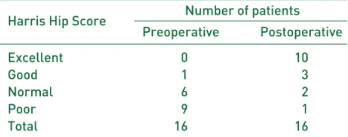
Table 1. Preoperative and Postoperative Clinical Scores Using Harris Hip Scoring System
Harris Hip Score Number of patients Preoperative Postoperative
Excellent 00 10
Good 01 03
Normal 06 02
Poor 09 01
Total 16 16
and lateral view each according to the Gruen method, and all 14 zones were examined12). Radiolucency was defined as a radiolucent line greater than 2 mm thick at cement-bone interface. To identify osteolysis, loosening of the femoral stem component was categorized as definite, probable or possible according to criteria of Harris et al13). The degree of heterotopic bone formation was determined using the classifications of Brooker et al14).
RESULTS
1. Clinical Results
Mean HHS showed significant improvement from preoperative 58 points (range, 39-81 points) to postoperative 92 points (range, 68-100 points) (P<0.001).
As shown in Table 1, clinical outcomes at final follow- up were excellent (n=10), good (n=3), fair (n=2) and poor (n=1). As shown in Table 2, mean postoperative flexion contracture values decreased from preoperative levels (from 13。to 5。; P<0.001); mean postoperative further flexion values increased from preoperative levels (from 58。to 107。; P<0.001); mean postoperative abduction and adduction values increased from preoperative levels (from 21。to 56。; P<0.001), and mean range of postoperative external and internal rotation values increased from preoperative levels (from 21。
to 44。; P<0.001) Mean ROM improved by 106。(101。
[preoperative] to 207。[postoperative]; P<0.001).
2. Radiological Results
Femoral morphologies observed from immediate preoperative radiographs according to the Dorr classification of proximal femur geometry were as follows: type A (n=1), type B (n=12), and type C (n=3). Cement mantle grades of the femoral stem using radiographs taken immediately following surgery are as follows: A (n=10) and B (n=6). No newly developed osteolytic lesions or prosthetic loosening were detected on last follow up X-rays (Fig. 1). There were no cases with enthesopathy of ischium, and 1 case of heterotopic ossification of Brooker grade I (Fig. 2). A case with greater trochanter fracture was treated conservatively, and bony union was obtained. No patients received revision surgery (Fig. 3).
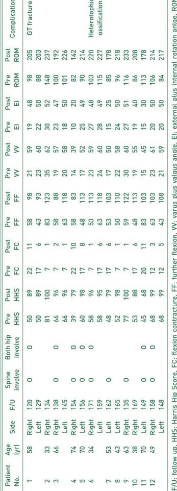
Table 2.Data for All Patients with Ankylosing Spondylitis PatientAge SideF/USpineBoth hipPrePostPrePostPrePostPrePostPrePostPrePost Complication No.(yr)involveinvolveHHSHHSFCFCFFFFVVVVEIEIROMROM 0158Right120OO5008922115809821591948098205GT fracture Left1295008917064309323602250088203 0233Right1348110007018312335623052148237 0366Right138OO6609607025808819572347100192 Left1456609607016311820581850101226 0474Right154OO3907922105808314391020082142 0570Left156OO6009817084811317522549090214 0634Right171OO5809607015311323592748103220Heterotophic Left1595809517066311824602849115227ossification 0753Left162O4807917065310317501525085178 0843Left1655209807015011022582450096218 0963Right135O7710007015912230602751116233 1038Right1695308817064811319551940086208 1170Left149OO4506820118310315451530113178 1249Right158OO6809912036310323612050106214 Left1486809912054310821592050084217 F/U: follow up, HHS: Harris Hip Score, FC: flexion contracture, FF: further flexion, VV: varus plus valgus angle, EI: external plus internal rotation anlge, ROM: range of motion, GT: greater trochanter.
DISCUSSION
This study achieved stable fixation and positive clinical results without loosening of the femoral component after performing THA using cement femoral implants in patients with AS at long-term follow up.
When Kim et al.15)performed THA in 19 patients (31 cases) with AS, mean HHS improved from 50 points (preoperatively) to 87 points (at final follow up) and mean flexion contracture decreased from 11。(preoperatively) to 3。(postoperatively). Yoo et al.16)performed uncemented
and cemented THA in 28 patients (45 cases) with AS and HHS was excellent (n=37), good (n=7), fair (n=1) and poor (n=3). Few domestic studies have been conducted on THA in AS patients and, in particular, no studies have been done over a long-term period of 10 years or more. The current study achieved satisfactory results of THA in 12 patients (16 cases) with AS at longer than 10-year follow-up. In our study, mean HHS improved from preoperative levels (58 points) to postoperative (92 points) and postoperative ROM improved by 106。compared to preoperative levels.
Sochart and Porter3) documented long-term clinical
F
Fiigg.. 11.. Pre-operative X-ray (AA, BB), post-operative X-ray (CC, DD) post-operative 10 years X-ray (EE, FF) of cemented total hip arthroplasty in 58 years old male patient with ankylosing spondylitis.
A C E
B D F
results of Charnley low-friction arthroplasty in 24 patients with AS with a mean follow-up of 22.7 years. Pain, hip function, and ROM improved and 20-year and 30-year survival rate were 91% and 83% in Kaplan-Meier survival analyses, repsectively.
Lehtimäki et al.17)reported long-term outcomes of THA in 76 cases with AS with hip involvement during 8 to 28 years of follow-up. The survival rates were 80%, 66%, and 62% at 10, 14, and 20 years, respectively.
The most expected improvement after THA is mainly pain relief in patients with AS. Brinker et al.18)noted that pain was relieved completely or by more than 90% after THA in 18 of 20 patients with AS. Walker and Sledge19) observed complete pain remission in 28 out of 29 patients (96.5%). We also observed good clinical results in this study (as 14 of 16 cases [87.5%] had pain relief). Increases in ROM differed slightly by study. Kilgus et al.20)reported that ROM improved by 109。(67。[preoperative] to 176。
[postoperative]). Bhan et al.9)suggested that ROM improved (from 0。[preoperative] to 156.2。[postoperative]) after cementless THA in patients with complete ankylosis of the hip joint as a result of AS. Improvement in ROM appears to be affected by the preoperative degree of stiffness, heterotopic ossification and other conditions.
The types of femoral implant available for AS patients are cemented or cementless components. When performing THA in patients with AS, the choice of femoral implant is a primary concern. The durability of hip implants is important
because AS commonly occurs at a young age and heavier loads can be placed on the hip joint due to bony union of the spine9,21). Moreover, accurate fixation may be challenging with the use of cementless implants because of possible anatomical deformity in the proximal femoral region.
Typically, femoral anteversion increases with externally rotated proximal femur in patients with AS, and it is difficult to press-fit a femoral stem in the cortex. Bony union may be delayed due to changes in bone metabolism and the use of cementless prosthesis may fail due to severe osteoporosis.
Cemented femoral components may not require bony union, be flexible to anatomical change in the proximal femur, and obtain favorable long-term results with good-quality cementing based on classification of cementation according to Barrack et al11). In particular, since decreases in physical activity and weight loss with disease progression may reduce physiological stress in patients with AS, the durability of well-fixed implants at the initial surgery is important.
Sochart and Porter3)achieved good surgical outcomes at long-term follow-up after Charnley low-friction arthroplasty with cement in 43 cases. The authors of this study initially preferred cemented femoral implants, and gained satisfactory results using cemented stem-type component. Saglam et al.22)described that although both cemented and uncemented components can be used, cemented THA was performed in patients with AS with Dorr type-C femurs.
F
Fiigg.. 22.. Post-operative X-ray anteroposterior (AP) view (AA), post-operative 14 years X-ray AP view (BB) of 74 years old male patients with ankylosing spondylitis who underwent total hip arthroplasty. Heterotopic ossification (grade I in Barrack classification) occurred (black arrow).
A B
F
Fiigg.. 33.. Post-operative X-ray anteroposterior (AP) view (AA), post-operative 10 years X-ray AP view (BB) of 66 years old male patient with ankylosing spondylitis who underwent total hip arthroplasty. Greater trochanter avulsion fracture occurred at operation time, and it was displaced upward at last follow up (black arrows).
A B
Heterotopic ossification is one of the most common complications in patients with AS. This complication may decrease postoperative hip ROM14,23,24). The incidence rate of heterotopic ossification following THA ranges from 3% to 90%25-27). Surgical removal can be considered as a treatment option in heterotopic ossification, but the recurrence rate is high28-30). Many studies suggest that the incidence of heterotopic ossification in patients with AS after THA is higher than that of patients with arthritis after THA31,32), but some studies have contradicted this outcome33). Thilak et al.34)performed THA in 24 patients (47 cases) with AS, and of these, heterotopic ossification occurred in 7 cases (14.9%). Saglam et al.22)carried out THA in 32 patients (59 cases) with AS from 1997 to 2012, and of these, heterotopic ossification occurred in 14 cases (13.3%). In this study, 12 patients (16 cases) with AS underwent THA and heterotopic ossification occurred in 1 case (6.3%)–a relatively lower rate compared to previous studies. Though several approaches have been recommended to prevent heterotopic ossification after THA, the use of diphosphonate/indomethacin and low-dose radiation therapy is commonly implemented. In our study, daily administration of 75 mg indomethacin for the first postoperative month was used for pain control and prevention of heterotopic ossification and good results were obtained.
In the current study, greater trochanter fracture occurred in 1 case; bone union was obtained with conservative treatment. This fracture was not seen intraoperatively or in plain X-ray done immediately after surgery. Migration (roughly 1 cm) of greater trochanter bone fragment was found in plain X-rays at the 6th postoperative month, but did not progress further.
When performing THA due to severe ankylosis in patients with AS, periprosthetic fracture may occur because exposure or dislocation is uneasy. To prevent this type of complication, several studies performed osteotomy of the greater trochanter3,4,17). Although we were not able to find literature on greater trochanter fracture, there was a case where wire fixation was also done to manage intraoperative periprosthetic fractures. Brinker et al.18)conducted cementless THA in 12 patients (20 cases) with AS, and bony union was achieved despite a non-displaced fracture around the femoral calcar intrapoperatively. Saglam et al.22)performed THA in 61 patients (105 cases) with AS, and periprosthetic fracture occurred in 2 cases during surgery and were treated with plate and cable.
There are several limitations to note in the present study.
First, this study was limited by the relatively small sample
size. Long-term follow-up of AS patients was challenging as the survival rate declined since these patients often have problems in other parts of the body including upper and lower limbs and respiratory tract. Second, more systematic measurement of clinical parameters was not done because only ROM and HHS were used for the evaluation of clinical and radiological results. Third, this study was unable to compare our findings with the results of cementless THA in patients with AS.
CONCLUSION
This study achieved stable fixation and favorable clinical results without loosening of the femoral component after performing THA using cement femoral component in patients with AS at long-term follow up.
CONFLICT OF INTEREST
The authors declare that there is no potential conflict of interest relevant to this article.
REFERENCES
01. Cooksey R, Brophy S, Husain MJ, Irvine E, Davies H, Siebert S. The information needs of people living with ankylosing spondylitis: a questionnaire survey. BMC Musculoskelet Disord. 2012;13:243.
02. Vander Cruyssen B, Muñoz-Gomariz E, Font P, et al. Hip involvement in ankylosing spondylitis: epidemiology and risk factors associated with hip replacement surgery. Rheumatology (Oxford). 2010;49:73-81.
03. Sochart DH, Porter ML. Long-term results of total hip replacement in young patients who had ankylosing spondylitis.
Eighteen to thirty-year results with survivorship analysis. J Bone Joint Surg Am. 1997;79:1181-9.
04. Joshi AB, Markovic L, Hardinge K, Murphy JC. Total hip arthroplasty in ankylosing spondylitis: an analysis of 181 hips. J Arthroplasty. 2002;17:427-33.
05. Ibn Yacoub Y, Amine B, Laatiris A, Hajjaj-Hassouni N. Gender and disease features in Moroccan patients with ankylosing spondylitis. Clin Rheumatol. 2012;31:293-7.
06. Bangjian H, Peijian T, Ju L. Bilateral synchronous total hip arthroplasty for ankylosed hips. Int Orthop. 2012;36:697-701.
07. Hamdi W, Alaya Z, Ghannouchi MM, Haouel M, Kchir MM. Associated risk factors with worse functional prognosis and hip replacement surgery in ankylosing spondylitis. Joint Bone Spine. 2012;79:94-6.
08. Goodman SM, Zhu R, Figgie MP, Huang WT, Mandl LA.
Short-term total hip replacement outcomes in ankylosing spondylitis. J Clin Rheumatol. 2014;20:363-8.
09. Bhan S, Eachempati KK, Malhotra R. Primary cementless total hip arthroplasty for bony ankylosis in patients with ankylosing spondylitis. J Arthroplasty. 2008;23:859-66.
10. Moll JM, Wright V. New York clinical criteria for ankylosing spondylitis. A statistical evaluation. Ann Rheum Dis. 1973;
32:354-63.
11. Barrack RL, Mulroy RD Jr, Harris WH. Improved cementing techniques and femoral component loosening in young patients with hip arthroplasty. A 12-year radiographic review. J Bone Joint Surg Br. 1992;74:385-9.
12. Gruen TA, McNeice GM, Amstutz HC. “Modes of failure”
of cemented stem-type femoral components: a radiographic analysis of loosening. Clin Orthop Relat Res. 1979;141:17-27.
13. Harris WH, McCarthy JC Jr, O’Neill DA. Femoral component loosening using contemporary techniques of femoral cement fixation. J Bone Joint Surg Am. 1982;64:1063-7.
14. Brooker AF, Bowerman JW, Robinson RA, Riley LH Jr.
Ectopic ossification following total hip replacement. Incidence and a method of classification. J Bone Joint Surg Am.
1973;55:1629-32.
15. Kim YM, Kim HJ, Kang SB, Choi ES, Lee SM. Total hip replacement arthroplasty in ankylosing spondylitis. J Korean Orthop Assoc. 1996;31:469-76.
16. Yoo MC, Chung DW, Kim JJ, Lee HK. Total hip replacement in the ankylosing spondylitis. J Korean Rheum Assoc. 1994;
1:23-32.
17. Lehtimäki MY, Lehto MU, Kautiainen H, Lehtinen K, Hämäläinen MM. Charnley total hip arthroplasty in ankylosing spondylitis: survivorship analysis of 76 patients followed for 8-28 years. Acta Orthop Scand. 2001;72:233-6.
18. Brinker MR, Rosenberg AG, Kull L, Cox DD. Primary noncemented total hip arthroplasty in patients with ankylosing spondylitis. Clinical and radiographic results at an average follow-up period of 6 years. J Arthroplasty. 1996;11:802-12.
19. Walker LG, Sledge CB. Total hip arthroplasty in ankylosing spondylitis. Clin Orthop Relat Res. 1991;(262):198-204.
20. Kilgus DJ, Namba RS, Gorek JE, Cracchiolo A 3rd, Amstutz HC. Total hip replacement for patients who have ankylosing spondylitis. The importance of the formation of heterotopic bone and of the durability of fixation of cemented components.
J Bone Joint Surg Am. 1990;72:834-9.
21. Bisla RS, Ranawat CS, Inglis AE. Total hip replacement in patients with ankylosing spondylitis with involvement of the hip. J Bone Joint Surg Am. 1976;58:233-8.
22. Saglam Y, Ozturk I, Cakmak MF, Ozdemir M, Yazicioglu O.
Total hip arthroplasty in patients with ankylosing spondylitis:
Midterm radiologic and functional results. Acta Orthop Traumatol Turc. 2016;50:443-7.
23. DeLee J, Ferrari A, Charnley J. Ectopic bone formation following low friction arthroplasty of the hip. Clin Orthop Relat Res. 1976;
(121):53-9.
24. Sundaram NA, Murphy JC. Heterotopic bone formation following total hip arthroplasty in ankylosing spondylitis. Clin Orthop Relat Res. 1986;(207):223-6.
25. Rosendahl S, Christoffersen JK, Norgaard M. Para-articular ossification following hip replacement. 70 arthroplasties ad modum Moore using McFarland’s approach. Acta Orthop Scand. 1977;48:400-4.
26. Lazansky MG. Complications revisited. The debit side of total hip replacement. Clin Orthop Relat Res. 1973;(95):96-103.
27. Padgett DE, Holley KG, Cummings M, et al. The efficacy of 500 CentiGray radiation in the prevention of heterotopic ossification after total hip arthroplasty: a prospective, randomized, pilot study. J Arthroplasty. 2003;18:677-86.
28. Warren SB, Brooker AF Jr. Excision of heterotopic bone followed by irradiation after total hip arthroplasty. J Bone Joint Surg Am. 1992;74:201-10.
29. Chao ST, Joyce MJ, Suh JH. Treatment of heterotopic ossification. Orthopedics. 2007;30:457-64; quiz 65-6.
30. Pape HC, Marsh S, Morley JR, Krettek C, Giannoudis PV.
Current concepts in the development of heterotopic ossification.
J Bone Joint Surg Br. 2004;86:783-7.
31. Healy WL, Lo TC, Covall DJ, Pfeifer BA, Wasilewski SA.
Single-dose radiation therapy for prevention of heterotopic ossification after total hip arthroplasty. J Arthroplasty.
1990;5:369-75.
32. Pellegrini VD Jr, Konski AA, Gastel JA, Rubin P, Evarts CM. Prevention of heterotopic ossification with irradiation after total hip arthroplasty. Radiation therapy with a single dose of eight hundred centigray administered to a limited field. J Bone Joint Surg Am. 1992;74:186-200.
33. Ahrengart L. Periarticular heterotopic ossification after total hip arthroplasty. Risk factors and consequences. Clin Orthop Relat Res. 1991;(263):49-58.
34. Thilak J, Panakkal JJ, Kim TY, Goodman SM, Lee SS, Salvati EA. Risk factors of heterotopic ossification following total hip arthroplasty in patients with ankylosing spondylitis.
J Arthroplasty. 2015;30:2304-7.