Received on February 2, 2012. Revised on February 15, 2012. Accepted on February 17, 2012.
CC This is an open access article distributed under the terms of the Creative Commons Attribution Non-Commercial License (http://creativecommons.org/licenses/by-nc/3.0) which permits unrestricted non-commercial use, distribu- tion, and reproduction in any medium, provided the original work is properly cited.
*Corresponding Author. Tel: 82-53-420-4835; Fax: 82-53-256-1566; E-mail: ksuk@knu.ac.kr Keywords: Neuroinflammation, Astrocyte, Microglia, Glial activation, Pain
Glia as a Link between Neuroinflammation and Neuropathic Pain
Mithilesh Kumar Jha, Sangmin Jeon and Kyoungho Suk*
Department of Pharmacology, Brain Science & Engineering Institute, Kyungpook National University School of Medicine, Daegu 700-422, Korea
Contemporary studies illustrate that peripheral injuries acti- vate glial components of the peripheral and central cellular circuitry. The subsequent release of glial stressors or activat- ing signals contributes to neuropathic pain and neuroinflam- mation. Recent studies document the importance of glia in the development and persistence of neuropathic pain and neuroinflammation as a connecting link, thereby focusing at- tention on the glial pathology as the general underlying fac- tor in essentially all age-related neurodegenerative diseases.
There is wide agreement that excessive glial activation is a key process in nervous system disorders involving the re- lease of strong pro-inflammatory cytokines, which can trig- ger worsening of multiple disease states. This review will briefly discuss the recent findings that have shed light on the molecular and cellular mechanisms of glia as a connecting link between neuropathic pain and neuroinflammation.
[Immune Network 2012;12(2):41-47]
INTRODUCTION Glia
Glia refers to a diverse set of specialized cell types that are found both in the peripheral nervous system (Schwann cells, satellite glia, perineural glia) and in the central nervous sys- tem (CNS) (astrocytes, oligodendrocytes, microglia, and peri- vascular glia) (1). Glial cells constitute 70% of the total cell population in the brain and spinal cord. Glial cells can be subdivided into two primary categories: microglia, comprising 5% to 10% of the glial population, and macroglia, which in-
clude astrocytes and oligodendrocytes (2). Glial cells that were, up to several years ago, considered the forgotten brain cells, or neglected stepchildren of neuroscience, now act as orchestrators in the tetrapartite synapse, and control of their activation state is crucial to the integrity of CNS function.
Recent technical advances and increased interest in elucidat- ing the role of non-neuronal cells of the CNS in both the physiological and pathophysiologial processes have cata- pulted these cells into the forefront of neuroscience research.
Glial cells do not conduct nerve impulses, and they provide structural support for the brain, assisting in nervous system development, repair and maintenance, supplying nutrients and biosynthetic products to neurons, imparting metabolic functions to neurons, destroying and removing injured and dead neurons and, finally, regulating the neuronal micro- environment. The well known quote the neuron is the struc- tural and functional unit of the brain has been challenged by a wealth of findings which have, to the contrary, in- troduced the neuronal-glial complex as the structural and functional unit of the brain. Glia regulate brain vasculature and the blood-brain barrier, modulating ischemia and migraines. Moreover, they are important in the repair of neu- rons after injury and also contribute to neuropathology in neurodegenerative diseases. Microglia can either protect or damage neurons depending on where and how they are activated. Microglia are chronically engaged in repairing mi- nor insults and that clinical diseases are observed only when these repair efforts fail. Fully activated microglia are detri- mental to neurons, but other stages in the sequence of re-
active states may improve neuronal survival by releasing neu- rotrophic factors or by removing excess glutamate from the extracellular space (3). Glial cells, which include oligoden- drocytes, astrocytes, and microglia, have been found to play key roles in neuroinflammation and neuropathic pain. Given that less is known about the involvement of oligodendrocytes, this paper will focus primarily on the role of astrocytes and microglial cells in neuroinflammation and neuropathic pain.
In view of the neuroinflammation and neuropathic pain proc- esses, activation of glial cells in the spinal dorsal horn, espe- cially microglia and astroglia, plays a predominant role.
Furthermore, astrocytes and microglia are known to play a role in the development, spread, and potentiation of neuro- pathic pain (4-12).
Neuroinflammation
Neuroinflammation is a normal and necessary process. In the acute phase after injury, neuroinflammation is tightly con- trolled. In its chronic phase or when directed against normal tissue (in an autoimmune response), neuroinflammation is detrimental when it manifests itself as multiple sclerosis (MS), amyotrophic lateral sclerosis (ALS), various types of dementia, Huntington s disease, or other diseases (13-16). A wide range of neurodegenerative diseases, including those affecting the CNS, such as Alzheimer s disease (AD), Parkinson s disease (PD), ALS, and MS, are associated with chronic inflammation (17-21). Although inflammation may not be the initiating fac- tor, emerging evidence in animal models suggests that sus- tained inflammatory responses involving microglia and as- trocytes contribute to disease progression (22). The activation of perivascular microglia and endothelial cells (lining the ca- pillary bed), astrocytes (making up the blood-brain barrier), parenchymal microglia and astrocytes by the means of stress- es, leads to the subsequent production of cytokines, cellular adhesion molecules, chemokines, and the expression of sur- face antigens that enhance a CNS immune cascade. If left un- checked, this neuroimmune activation can lead to the traffick- ing of leukocytes into the perceived area of injury as a mech- anism for neuroprotection. Therefore, neuroinflammation can be defined as the infiltration of immune cells into the site of injury in response to damage to the peripheral or CNS (23).
Neuropathic pain
Neuropathic pain, initiated or caused by primary lesions or dysfunction in the CNS (brain and spinal cord) or the periph- eral nervous system (nerves outside the brain and spinal
cord), having occurred following viral infection, trauma, cer- tain medications, or metabolic insults, and many diseases such as MS and stroke, is especially problematic because of its severity, chronicity, and resistance to simple analgesics (24). The possible mechanisms of neuropathic pain could be classified as: a) chemical excitation of non-nociceptors, b) re- cruitment of nerves outside the site of injury, c) excitotoxicity, d) excess sodium channels, e) ectopic discharge, f) central sensitization maintained by peripheral input, and g) sym- pathetic involvement.
THE ROLE OF GLIA IN NEUROINFLAMMATION The role of microglia in initiating or promoting inflammatory processes in the CNS by facilitating the recruitment of periph- eral immune cells has been well documented (25-27). During the neuroinflammatory process, microglial cells release proin- flammatory mediators such as cytokines, matrix metal- loproteinases (MMP), reactive oxygen species (ROS), and ni- tric oxide (NO). For a long time, glial cells have been consid- ered to merely support the neuronal environment. However, the innovative research in the field of neuroscience has strongly propelled glial cells as new players in neuroinflam- mation and neuropathic pain. Neuroinflammation is a charac- teristic feature of both acute and chronic CNS disorders and is a process that results primarily from the presence of chroni- cally activated glial cells (astrocytes and microglia) in the brain, and is a common feature of several neurodegenerative conditions. Activated glia release a variety of neuroexcitatory substances that potentiate neurotransmission, especially pro- inflammatory cytokines. Blocking glial activity may be a novel way of controlling neuropathic pain. Neuroinflammation in- duces a complex and dynamic change in glial cell pheno- types. One of the first cell types to respond are microglial cells, which retract their processes and migrate towards the site of injury, where they release proinflammatory cytokines such as IL-1β, TNF-α, and IL-6 (28-30).
THE ROLE OF GLIA IN NEUROPATHIC PAIN
Neuropathic pain is a debilitating condition that affects mil- lions of individuals worldwide. It is now thought that solely considering neuronal activity provides an incomplete under- standing of the creation and maintenance of chronic neuro- pathic pain (31). Glia have recently emerged as key contrib- utors to pathological and chronic pain mechanisms and are
emerging as a new target for drug development (32,33).
Spinal cord glial activation seems to be a common underlying mechanism that leads to pathological pain in a number of pain syndromes with dramatically different aetiologies (for ex- ample, diabetic neuropathy, chemotherapy-induced neuro- pathy, peripheral nerve inflammation and trauma, and spinal cord inflammation) (34). Upon activation, both the astrocytes and microglia respond to and release a number of signaling molecules which have protective and/or pathological func- tions. These include, among others, the classic immune sig- nals: cytokines and chemokines. The role of glia in the CNS is intimately integrated with the functions of the other players in the tetrapartite synapse composed of pre- and post- syn- aptic neurons, astrocytes, and microglial cells. In trauma or disease states, the spinal glia become activated and the dorsal horn neurons become hyperexcitable, contributing to sensi- tized neuronal-glial circuits. The maladaptive spinal circuits directly affect synaptic excitability, including the activation of intracellular downstream cascades, that results in enhanced evoked and spontaneous activity in dorsal horn neurons lead- ing to the development of abnormal pain syndromes (35).
INTERLINKING INFLAMMATION AND NEUROPATHIC PAIN
Inflammation is a component of the body s wisdom. Inflam- mation is the body s foremost response to an injury or disease. Webster defines inflammation as redness, swelling and fever in a local area of the body, often with pain and disturbed function, in reaction to an infection or to a physical or chemical injury. Symptoms of inflammation can range from mild aches to sharp, wrenching pain that may take one s breath away. Inflammation in any part of our body is coupled with pain, due to the release of inflammatory media- tors such as prostaglandin E2 (PGE2), the proinflammatory cy- tokines TNF-α and IL-1β, and nerve growth factor (NGF).
These mediators, produced by non-neural cells or immune cells, can stimulate nociceptor terminals in the peripheral tis- sue to increase pain sensitivity (36,37). Inflammation also oc- curs in the CNS after brain trauma, brain infection, and in neurodegenerative diseases, such as AD, PD, and MS. This so-called neuroinflammation is characterized by the activation of glial cells (especially microglia and astrocytes) in the CNS and is an important contributor to the development of neuro- degeneration by releasing inflammatory mediators from glial cells (38,39). This glia-mediated neuroinflammation also plays
an important role in pain control under pathological condi- tions.
Although multiple conditions may generate neuropathic pain, a common underlying mechanism is the presence of in- flammation at the site of the damaged or affected nerve(s).
This inflammatory response initiates a cascade of events re- sulting in increased local perfusion, increased capillary per- meability, and the concentration and activation of innate im- mune cells at the site of tissue injury, irritation, or infection.
Immunoactive substances, such as cytokines, neurotrophic factors, and chemokines, released at the site of injury have local actions and can initiate a systemic immune response.
The resultant neuroinflammatory environment can cause the activation of microglia and astrocytes located in the spinal cord and brain, which appear to play a prominent role in nociception (40).
Current research suggests that microglia are involved in the early development of neuropathic pain, whereas astrocytes function to sustain neuropathic pain (12,41-43). The activa- tion of spinal cord glia is both necessary and sometimes even sufficient for the development of persistent pain states asso- ciated with various etiologies, including diabetic neuropathy, chemotherapy-induced neuropathy, peripheral nerve in- flammation and trauma, and spinal cord inflammation (44-46).
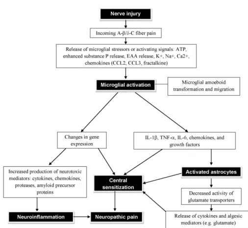
Both spinal astrocytes and microglia activate mitogen-activat- ed protein kinases (MAPKs) to induce the synthesis and re- lease of proinflammatory cytokines, such as IL-1β, IL-6, TNF- α, PGE2, and NO (47,48). There is ample evidence that both astrocyte and microglia activation lead to pro-inflammatory responses with pathological effects, such as neuronal hyper- excitability, neurotoxicity and chronic inflammation. Fig. 1 il- lustrates the consequential neuroinflammation and neuro- pathic pain subsequent to the neuronal injury via the glial cell (microglial and astrocytic) activation.
GLIA IN FORMALIN TEST & COMPLETE FREUND’S ADJUVANT (CFA)-INDUCED INFLAMMATORY PAIN MODEL
Animal models of inflammatory pain have been widely used to study the mechanisms of tissue injury that induced persis- tent pain. The formalin test is the most predictive of the mod- els for acute pain, it is predominantly used on rats and mice and involves moderate, continuous pain generated by injured tissue. In this way it differs from most traditional tests of noci- ception which rely on brief stimuli of threshold intensity.
Figure 1. Diagram showing the neuroinflammation and neuropathic pain subsequent to the neuronal injury and glial cell (microglial and astrocytic) activation. Following a peripheral injury, the synaptic projection of pain sensing neuron within the spinal cord releases ATP. Nearby microglial cells are drawn to the source of ATP and undergo morphological changes as they approach the source and become activated. Fully activated microglial cells are localized around the pain sensing neuron and begin to interact with the neurons at a molecular level, releasing various neuroinflammatory agents. These neuroinflammatory agents activate astrocytes. Upon activation, the astrocytes undergo hypertrophy and increased production of neuroinflammatory agents are secreted into the synaptic cleft. Astrocyte activation in conjugation with microglial activation significantly depolarizes the neuron increasing its sensitivity and potentiating the neuroinflammation and neuropathic pain states. EAA, excitatory amino acids.
Formalin injected beneath the footpad of a rat, mouse, or cat produces two phases of nocifensive behavior, characterized by the licking and flinching of the paw, that are separated by a short period of quiescence in which there is no apparent pain behavior (49,50). The first or acute phase typically oc- curs in the first 5 min; the second starts from 15 min and lasts about 40∼60 min after injection. The early phase seems to be caused predominantly by the direct activation of both the low-threshold mechanoreceptive and the nociceptive pri- mary afferent fibers due to the peripheral stimulus (51). The late phase, also called the tonic phase, appears to be depend- ent on the combination of an inflammatory reaction in the peripheral tissue and functional changes in the dorsal horn of the spinal cord, popularly called central processing. These functional changes seem to be initiated by the C-fiber barrage
during the early phase (52).
Complete Freund s adjuvant (CFA)-induced inflammatory pain is very commonly used as a model for chronic in- flammatory pain. CFA contains heat-killed or inactivated and dried mycobacteria, which is a primary agent responsible for stimulating antibody production, but has also been attributed to a number of undesirable side effects (53). This assay has a good track record for predicting the effectiveness of com- pounds as analgesic or anti-hyperalgesic agents as well as de- fining the mechanism behind inflammatory pain. CFA is in- jected into one hind-paw of the animal. The CFA injection in the footpad produces localized inflammation and persistent pain (54,55). Thermal hyperalgesia or mechanical allodynia associated with the inflammation are assessed by determining the hind-paw withdrawal latency. After the CFA injection into
the footpad, cutaneous inflammation appears in minutes to hours and peaks within 5∼8 hours. The average time of on- set is 2∼6 hours and persists for approximately 1∼2 weeks.
The edema peaks around 24 hours after injection (56). For the induction of hind paw inflammation, mice receive (ipl.) injection of 10μl of CFA (diluted with PBS, 2 mg/ml Mycobacterium tuberculosis). The CFA produces dose-de- pendent inflammatory responses. Behavior testing in CFA-in- jected mice is done by mechanical sensitivity testing using von Frey hairs and heat sensitivity testing.
Peripheral formalin injection induces two stages of microglial activation: p38 activation in spinal microglia plays key roles in central pain modulation in formalin test for both the early acute phase and the late secondary long-term pain state (57). These unique properties of spinal microglial activation in an animal model of pain will potentially help to further understand the contributions of spinal microglia to acute and chronic pain states. Both qualitative and quantitative analyses for the com- parison of the effects of the peripheral CFA and formalin in- jection on spinal microglia activation showed signs of micro- glia activation on the ipsilateral side of the lumbar dorsal horn on days 3, 7, and 14 after the formalin injection was introduced. However, significant microglia morphological al- teration was not found in the CFA model. At the injection site in the paw, the CFA injection induced considerably more inflammation than the formalin injection. Although spinal mi- croglia might have been activated morphologically in in- flammatory pain models, spinal microglia activation was not closely correlated with peripheral inflammation (58). Glial acti- vation is a common feature of many diseases of the CNS (59).
In the spinal cord, astrocytes are activated following periph- eral inflammation or the nerve injury and may manifest as increased expression of astrocytic markers such as glial fibril- lary acidic protein (GFAP) (60,61). The peripheral injection of CFA increased the mRNA and protein expression of the astrocytic marker GFAP in the bilateral anterior cingulate cor- tex (ACC). The inhibition of astroglial function by an as- troglial toxin blocked the place-avoidance behavior, but not the paw withdrawal threshold, suggesting the involvement of astrocytes in the ACC as the affective component of pain.
Modulating the function of astrocytes in the ACC may provide a new strategy for the prevention of chronic pain-induced emotional disturbances.
CONCLUSION AND FUTURE PERSPECTIVES
Neurons are not the only cell type in the nervous system;
∼90% of the cells in our brain are glia. Glia, which until recently were thought to be passive support cells for the neu- rons, are now considered to be, not only the link between neuroinflammation and neuropathic pain, but also an im- portant link between the immune and nervous systems under inflammatory and traumatic conditions. Microglia, not born in the nervous system but formed by the transformation of certain white blood cells called macrophages or their pre- cursors, monocytes, are now thought to be part of the im- mune system, defending the brain against infection and injury. Microglia are the macrophages of the brain and are the first responders to CNS injury, but exactly which signal triggers microglial reactivity is not fully understood. The acti- vating signals may include changes in neuronal transmission or the appearance of NO or proinflammatory cytokines.
Substantiation indicates that central glial activation depends on nerve inputs from the site of injury and the release of chemical mediators. Glia, being the secret player in the neu- roinflammation and neuropathic pain, could be exploited to provide new research avenues for therapeutic pain control.
Taking into account both the protective and the pathological effects of activated glia will be a key to the development of effective therapeutics. Although knowledge of the develop- ment and differentiation of glial cells has significantly in- creased in recent years, there are still many unanswered questions regarding the consequential neuroinflammation and neuropathic pain subsequent to the neuronal injury via glial cell (microglial and astrocytic) activation.
Therapies directed at activated glia hold promise for new approaches to intractable pain. To expedite the goal of devel- oping new diagnostic tools and new therapies for intractable pain, it is important to allow the cross-fertilization of ideas to occur between preclinical and clinical researchers. It is high time that neuroscience research focused on the charac- terization of the glial phenotypes in the circumstances of in- flammation, nerve injury and their correlation with pain behavior. It can lead to the development of new therapeutic strategies by targeting both spinal glial cells and bone mar- row-derived macrophages for effective pain relief. Finally, it can be concluded that glial cells act as orchestrators in the tetrapartite synapse and control of their activation state is cru- cial to the integrity of CNS function.
ACKNOWLEDGEMENTS
This work was supported by the National Research Founda- tion (NRF) grants funded by the Ministry of Education, Science and Technology (MEST) of Korean government (2011-0028240). This study was also supported by a grant of the Korea Health technology R&D Project, Ministry of Health
& Welfare, Republic of Korea (A111345).
CONFLICTS OF INTEREST
The authors have no financial conflict of interest.
REFERENCES
1. Kriegstein A, Alvarez-Buylla A: The glial nature of embryonic and adult neural stem cells. Annu Rev Neurosci 32;149-184, 2009.
2. Moalem G, Tracey DJ: Immune and inflammatory mecha- nisms in neuropathic pain. Brain Res Rev 51;240-264, 2006.
3. Aamodt S: Focus on glia and disease. Nat Neurosci 10;1349, 2007.
4. Watkins LR, Milligan ED, Maier SF: Glial activation: a driving force for pathological pain. Trends Neurosci 24;450-455, 2001 5. McMahon SB, Cafferty WB, Marchand F: Immune and glial
cell factors as pain mediators and modulators. Exp Neurol 192;444-462, 2005.
6. Coull JA, Beggs S, Boudreau D, Boivin D, Tsuda M, Inoue K, Gravel C, Salter MW, De Koninck Y: BDNF from microglia causes the shift in neuronal anion gradient underlying neuro- pathic pain. Nature 438;1017-1021, 2005.
7. Garrison CJ, Dougherty PM, Kajander KC, Carlton SM:
Staining of glial fibrillary acidic protein (GFAP) in lumbar spi- nal cord increases following a sciatic nerve constriction injury. Brain Res 565;1-7, 1991.
8. Meller ST, Dykstra C, Grzybycki D, Murphy S, Gebhart GF:
The possible role of glia in nociceptive processing and hyper- algesia in the spinal cord of the rat. Neuropharmacology 33;1471-1478, 1994.
9. Colburn RW, DeLeo JA, Rickman AJ, Yeager MP, Kwon P, Hickey WF: Dissociation of microglial activation and neuro- pathic pain behaviors following peripheral nerve injury in the rat. J Neuroimmunol 79;163-175, 1997.
10. Milligan ED, Mehmert KK, Hinde JL, Harvey LO, Martin D, Tracey KJ, Maier SF, Watkins LR: Thermal hyperalgesia and mechanical allodynia produced by intrathecal administration of the human immunodeficiency virus-1 (HIV-1) envelope glycoprotein, gp120. Brain Res 861;105-116, 2000.
11. Chacur M, Milligan ED, Gazda LS, Armstrong C, Wang H, Tracey KJ, Maier SF, Watkins LR: A new model of sciatic in- flammatory neuritis (SIN): induction of unilateral and bilateral mechanical allodynia following acute unilateral peri-sciatic immune activation in rats. Pain 94;231-244, 2001.
12. Raghavendra V, Tanga FY, DeLeo JA: Complete Freunds ad-
juvant-induced peripheral inflammation evokes glial activa- tion and proinflammatory cytokine expression in the CNS.
Eur J Neurosci 20;467-473, 2004.
13. Möller T: Neuroinflammation in Huntington's disease. J Neural Transm 117;1001-1008, 2010.
14. Björkqvist M, Wild EJ, Thiele J, Silvestroni A, Andre R, Lahiri N, Raibon E, Lee RV, Benn CL, Soulet D, Magnusson A, Woodman B, Landles C, Pouladi MA, Hayden MR, Khalili- Shirazi A, Lowdell MW, Brundin P, Bates GP, Leavitt BR, Möller T, Tabrizi SJ: A novel pathogenic pathway of immune activation detectable before clinical onset in Huntington's disease. J Exp Med 205;1869-1877, 2008.
15. Garden GA, Möller T: Microglia biology in health and disease.
J Neuroimmune Pharmacol 1;127-137, 2006.
16. Weydt P, Möller T: Neuroinflammation in the pathogenesis of amyotrophic lateral sclerosis. Neuroreport 16;527-531, 2005.
17. Block ML, Hong JS: Microglia and inflammation-mediated neurodegeneration: multiple triggers with a common mecha- nism. Prog Neurobiol 76;77-98, 2005.
18. Mrak RE, Griffin WS: Interleukin-1, neuroinflammation, and Alzheimer's disease. Neurobiol Aging 22;903-908, 2001.
19. Phillis JW, Horrocks LA, Farooqui AA: Cyclooxygenases, lip- oxygenases, and epoxygenases in CNS: their role and in- volvement in neurological disorders. Brain Res Rev 52;201- 243, 2006.
20. Sriram K, O'Callaghan JP: Divergent roles for tumor necrosis factor-alpha in the brain. J Neuroimmune Pharmacol 2;140-153, 2007.
21. Ubogu EE, Cossoy MB, Ransohoff RM: The expression and function of chemokines involved in CNS inflammation.
Trends Pharmacol Sci 27;48-55, 2006.
22. Weed DL: The merger of bioethics and epidemiology. J Clin Epidemiol 44 Suppl 1;15S-22S, 1991.
23. Baptista MJ, Cookson MR, Miller DW: Parkin and alpha-synu- clein: opponent actions in the pathogenesis of Parkinson's disease. Neuroscientist 10;63-72, 2004.
24. Gilron I, Watson CP, Cahill CM, Moulin DE: Neuropathic pain: a practical guide for the clinician. CMAJ 175;265-275, 2006.
25. Kielian T: Microglia and chemokines in infectious diseases of the nervous system: views and reviews. Front Biosci 9;732- 750, 2004.
26. Ransohoff RM, Glabinski A, Tani M: Chemokines in im- mune-mediated inflammation of the central nervous system.
Cytokine Growth Factor Rev 7;35-46, 1996.
27. Cartier L, Hartley O, Dubois-Dauphin M, Krause KH: Chemo- kine receptors in the central nervous system: role in brain inflammation and neurodegenerative diseases. Brain Res Brain Res Rev 48;16-42, 2005.
28. Kreutzberg GW: Microglia: a sensor for pathological events in the CNS. Trends Neurosci 19;312-318, 1996.
29. Clayton DF, George JM: Synucleins in synaptic plasticity and neurodegenerative disorders. J Neurosci Res 58;120-129, 1999.
30. Becher B, Prat A, Antel JP: Brain-immune connection: im- muno-regulatory properties of CNS-resident cells. Glia 29;293- 304, 2000.
31. Scholz J, Woolf CJ: The neuropathic pain triad: neurons, im- mune cells and glia. Nat Neurosci 10;1361-1368, 2007.
32. Halassa MM, Fellin T, Haydon PG: The tripartite synapse:
roles for gliotransmission in health and disease. Trends Mol Med 13;54-63, 2007.
33. Pocock JM, Kettenmann H: Neurotransmitter receptors on microglia. Trends Neurosci 30;527-535, 2007.
34. Watkins LR, Wieseler-Frank J, Milligan ED, Johnston I, Maier SF: Chapter 22 Contribution of glia to pain processing in health and disease. Handb Clin Neurol 81;309-323, 2006.
35. Gwak YS, Kang J, Unabia GC, Hulsebosch CE: Spatial and temporal activation of spinal glial cells: role of gliopathy in central neuropathic pain following spinal cord injury in rats.
Exp Neurol 234;362-372, 2012.
36. Julius D, Basbaum AI: Molecular mechanisms of nociception.
Nature 413;203-210, 2001.
37. Scholz J, Woolf CJ: Can we conquer pain? Nat Neurosci 5 Suppl;1062-1067, 2002.
38. Block ML, Zecca L, Hong JS: Microglia-mediated neuro- toxicity: uncovering the molecular mechanisms. Nat Rev Neurosci 8;57-69, 2007.
39. Lobsiger CS, Cleveland DW: Glial cells as intrinsic compo- nents of non-cell-autonomous neurodegenerative disease. Nat Neurosci 10;1355-1360, 2007.
40. Vallejo R, Tilley DM, Vogel L, Benyamin R: The role of glia and the immune system in the development and maintenance of neuropathic pain. Pain Pract 10;167-184, 2010.
41. Colburn RW, Rickman AJ, DeLeo JA: The effect of site and type of nerve injury on spinal glial activation and neuropathic pain behavior. Exp Neurol 157;289-304, 1999.
42. Raghavendra V, Tanga F, DeLeo JA: Inhibition of microglial activation attenuates the development but not existing hyper- sensitivity in a rat model of neuropathy. J Pharmacol Exp Ther 306;624-630, 2003.
43. Ledeboer A, Sloane EM, Milligan ED, Frank MG, Mahony JH, Maier SF, Watkins LR: Minocycline attenuates mechanical allo- dynia and proinflammatory cytokine expression in rat models of pain facilitation. Pain 115;71-83, 2005.
44. DeLeo JA, Winkelstein BA: Physiology of chronic spinal pain syndromes: from animal models to biomechanics. Spine (Phila Pa 1976) 27;2526-2537, 2002.
45. Watkins LR, Hutchinson MR, Milligan ED, Maier SF:
"Listening" and "talking" to neurons: implications of immune activation for pain control and increasing the efficacy of opioids. Brain Res Rev 56;48-69, 2007.
46. Wieseler-Frank J, Maier SF, Watkins LR: Glial activation and pathological pain. Neurochem Int 45;389-395, 2004.
47. Ji RR, Suter MR: p38 MAPK, microglial signaling, and neuro-
pathic pain. Mol Pain 3;33, 2007.
48. Zhuang ZY, Gerner P, Woolf CJ, Ji RR: ERK is sequentially activated in neurons, microglia, and astrocytes by spinal nerve ligation and contributes to mechanical allodynia in this neuropathic pain model. Pain 114;149-159, 2005.
49. Dubuisson D, Dennis SG: The formalin test: a quantitative study of the analgesic effects of morphine, meperidine, and brain stem stimulation in rats and cats. Pain 4;161-174, 1977.
50. Abbott FV, Franklin KB, Westbrook RF: The formalin test:
scoring properties of the first and second phases of the pain response in rats. Pain 60;91-102, 1995.
51. Puig S, Sorkin LS: Formalin-evoked activity in identified pri- mary afferent fibers: systemic lidocaine suppresses phase-2 activity. Pain 64;345-355, 1996.
52. Tjølsen A, Berge OG, Hunskaar S, Rosland JH, Hole K: The formalin test: an evaluation of the method. Pain 51;5-17, 1992.
53. Colpaert FC: Evidence that adjuvant arthritis in the rat is asso- ciated with chronic pain. Pain 28;201-222, 1987.
54. Millan MJ, Członkowski A, Morris B, Stein C, Arendt R, Huber A, Höllt V, Herz A: Inflammation of the hind limb as a model of unilateral, localized pain: influence on multiple opioid sys- tems in the spinal cord of the rat. Pain 35;299-312, 1988.
55. Iadarola MJ, Brady LS, Draisci G, Dubner R: Enhancement of dynorphin gene expression in spinal cord following ex- perimental inflammation: stimulus specificity, behavioral pa- rameters and opioid receptor binding. Pain 35;313-326, 1988.
56. Lao L, Zhang RX, Zhang G, Wang X, Berman BM, Ren K:
A parametric study of electroacupuncture on persistent hyper- algesia and Fos protein expression in rats. Brain Res 1020;18-29, 2004.
57. Li K, Lin T, Cao Y, Light AR, Fu KY: Peripheral formalin injury induces 2 stages of microglial activation in the spinal cord.
J Pain 11;1056-1065, 2010.
58. Lin T, Li K, Zhang FY, Zhang ZK, Light AR, Fu KY:
Dissociation of spinal microglia morphological activation and peripheral inflammation in inflammatory pain models. J Neuroimmunol 192;40-48, 2007.
59. Ren K, Dubner R: Neuron-glia crosstalk gets serious: role in pain hypersensitivity. Curr Opin Anaesthesiol 21;570-579, 2008.
60. Gao YJ, Ji RR: Targeting astrocyte signaling for chronic pain.
Neurotherapeutics 7;482-493, 2010.
61. Watkins LR, Hutchinson MR, Ledeboer A, Wieseler-Frank J, Milligan ED, Maier SF: Norman Cousins Lecture. Glia as the
"bad guys": implications for improving clinical pain control and the clinical utility of opioids. Brain Behav Immun 21;131- 146, 2007.