대뇌공기색전증은 대뇌동맥이 공기유입에 의하여 폐색된 경 우를 말하며, 발작이나 신경학적 장애를 일으키고, 심한 경우 사망에 이를 수 있는 심각한 질환이다. 수술이나 중심정맥 카 테터, Swan-Gantz 카테터의 거치 및 제거, 혈관 조영술, 폐 세침 생검, 흉관 삽입 등의 침습적 시술과 연관된 합병증으로 보고되고 있으며, 잠수병을 제외하면 침습적 시술과 연관되지 않은 대뇌공기색전증은 거의 보고된 바가 없다(1-3). 저자들 은 좌측 폐실질 파괴를 보이는 폐결핵 환자에서 배변 후 발생 한 대뇌공기색전증을 경험하였기에 이를 보고하고자 한다.
증례 보고
56세 남자가 대량 각혈을 주소로 호흡기내과에 입원하였다.
환자는 폐결핵으로 진단, 치료 받았으나 좌폐야 실질의 파괴가 진행되었고, 반복적인 각혈을 경험한 적이 있다. 입원 후 대량 각혈을 치료하기 위하여 기관 동맥과 좌 내유동맥 색전술을 시 행 받았고, 당시 기관동맥-폐정맥루가 발달하여 있는 것을 확 인하였다(Fig. 1). 색전술 시행 후 기침과 각혈은 중단되었으 며, 혈액검사와 말초혈관의 산소 포화도는 정상화되었고 기침 은 호전되었다. 대량 각혈에 대한 집중 관찰 및 치료를 위해 중 환자실로 전실되었으나, 기도 삽관이나 기계식 환기 등의 추가 적인 침습적 시술은 시행되지 않았다.
환자는 정상적인 배변 활동을 보였으며, 전날 밤에도 정상 배변을 하였다. 다음 날, 입원 5일째 오후 침상에서 대변기를 이용하여 소량 배변한 직후, 갑자기 의식저하를 보여, 약 10분 뒤 뇌전산화단층촬영을 시행하였다. 기면 의식상태가 지속되
며, 좌측 근력저하와 얼굴 감각 이상을 보였다. 배변 전, 일정 시간 동안 호흡기 증상이나 흉통 등의 증상은 보이지 않았으 며, 기존에 유지하던 정맥관 주사 외에 추가적인 약물 주입이 나 혈액 채취 등의 침습적 술기는 시행된 적이 없었다. 신경학 적 증상 발생 10분 후 응급으로 뇌전산화단층촬영을 시행하였 고, 우측 전두엽의 고랑을 따라 공기 음영으로 생각되는 원형 과 관상형의 저음영 병변을 확인하였다. 뇌전산화단층촬영 3 시간 후 뇌확산강조영상을 촬영하였으나, 뇌전산화단층촬영에 서 보이던 공기 음영 분포보다 현저히 적은 부위인 우측 전두 엽 회백질에서만 급성 경색을 시사하는 확산제한이 관찰되었 다. 대뇌공기색전증 진단 하에 100% 산소를 공급하고 아스피 린을 처방하였다. 추적 관찰을 위해 2일 후 시행한 뇌전산화단 층촬영과 뇌확산강조영상에서 공기음영은 소실되어 보이지 않 았고, 공기 음영이 있던 범위와 일치하는 우측 전두엽 고랑을 따라 CT 상 뇌실질의 저음영와 부종이 관찰되고, 뇌확산강조 영상에서 확산 제한이 보여 급성 경색이 진행되고 있음을 확인 하였다(Fig. 2).
고 찰
대뇌공기색전증은 대부분 침습적 시술 과정 또는 직후 급작 스럽게 발생하는 신경학적 장애와 함께 뇌영상 검사상 관찰되 는 공기음영을 통해 진단한다. 뇌전산화단층촬영에서 공기 색 전은 혈관의 주행 경로를 따라 원형 또는 관상형의 저음영 병 변으로 보이며, 공기 색전으로 손상된 뇌조직은 수분 함유량이 많아지면서 다른 주변 조직보다 저음영으로 관찰된다. 하지만 공기색전은 빠른 시간 안에 혈액 내로 용해되거나 순환을 통해 재분포되므로 특징적인 혈관 내 저음영은 증상 발현 초기에만
─ 307 ─ 대한영상의학회지 2010;63:307-310
폐결핵 환자에서 배변 후 발생한 대뇌공기색전증: 증례 보고1
오지영∙박동우∙함창곡∙박충기∙이승로∙이영준
대뇌공기색전증은 대부분 폐 세침 생검, 흉관 삽입, 중심정맥관 거치 및 제거, 수술 등과 같은 침습적 시술과 연관된 합병증으로 시술 직후 나타나는 경우가 대부분이며, 유입된 공기의 양에 따라 증상의 정도가 다양하게 나타나는 것으로 되어 있다. 잠수병을 제외하면, 침습적 시술과 연관되지 않은 대뇌공기색전증은 매우 드물다. 저자들은 최근 폐결핵 환자에서 배변 후 발생한 대뇌공기색전증이 뇌전산화단층촬영과 확산자기공명영상을 통해 진단된 1예를 경험하여 보고 하고자 한다.
1한양대학교 의과대학 영상의학과
이 논문은 2009년 10월 16일 접수하여 2010년 7월 25일에 채택되었음.
─ 308 ─
오지영 외: 폐결핵 환자에서 배변 후 발생한 대뇌공기색전증
A B
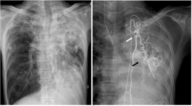
Fig. 1. A. Chest X-ray shows advanced pulmonary tuberculosis.
B. A left internal mammary angiography on the day of admission reveals extensive hypervascularity in the left lung with an arteri- ovenous fistula from the left internal mammary artery (white arrow) to the pulmonary vein (black arrow).
A B
C D
Fig. 2. A. Non-contrast axial CT demonstrates curvilinear low attenua- tion lesions in the sulci of the right frontal lobe (black arrows), suggestive of air embolism, 10 minutes after on- set of the stroke.
B. Diffusion-weighted MR image ob- tained 3 hour after onset of the stroke shows no abnormal high signal inten- sity at right frontal lobe.
C. 2 days after the stroke, the air bub- bles have disappeared and the cere- bral sulcus and cortex (white arrows) becomes effaced and swollen at the same area on the CT scan.
D. Diffusion-weighted MR image shows progression of the cortical high- intensity indicating acute infarction in the same area.
나타나고, 증상이 경미한 경우에는 병변의 발견이 어려울 수 있다(4).
대뇌공기색전증에 의한 다양한 신경학적 증상은 공기 색전 의 크기에 따라 다른 병태 생리학적 기전으로 나타난다. 크기 가 큰 공기 색전은 말초 동맥 혈류의 폐색과 이에 따른 경색에 의한 것으로 생각하며, 작은 공기 색전은 혈류에 포함되어 있 던 공기에 의해 손상된 혈관내피세포의 혈전염증반응이 증상 의 발현에 관여하는 것으로 생각되고 있다(4, 5).
혈관 내에 공기 색전이 존재하는 위치에 따라 정맥계 또는 동맥계 색전으로 나눌 수 있으며, 정맥 색전이 체순환 정맥으 로 공기가 유입되는 것과 달리 동맥 색전은 폐정맥이나 체동맥 으로 직접 유입된다고 알려져 있다. 특히 대뇌동맥공기색전증 은 세 가지 경로로 설명할 수 있는데 첫째로 폐정맥 손상에 의 해 공기가 직접 유입되는 경우이며, 둘째로 체정맥으로 유입된 후 우심방에 모인 공기가 난원공개존증이 있을 때 우심방 압력 이 상승하는 조건에서 우좌 단락을 일으켜 체순환 동맥계로 유 출되는 것을 들 수가 있다. 셋째로 양압 환기, 발살바 (Valsalva) 법, 특히 기침 등으로 기도 내압을 증가시켜, 일시 적인 기관지-폐정맥루가 형성되어 유입되는 경우이다(4-7).
본 환자에서는 폐결핵으로 파괴되어 있던 폐실질에 기관동 맥-폐정맥루가 발달되어 있었고, 반복적인 대량 각혈의 병력으 로 미루어 보아 기관지-폐정맥루가 형성되어 있었을 것으로 생 각한다. 배변 시 유도된 일시적인 흉강 내압 상승으로 인해 기 관지-폐정맥루 가 일시적으로 열리면서 공기가 유입되어 체순
환을 통해 대뇌공기색전이 발생한 것으로 생각한다.
잠수병을 제외하면 침습적 시술과 연관되지 않은 대뇌공기 색전증은 거의 보고된 바가 없다. 저자들은 좌측 폐실질 파괴 를 보이는 폐결핵 환자에서 배변 후 발생한 대뇌공기색전증을 경험하였기에 이를 보고하고자 한다.
참 고 문 헌
1. Kau T, Rabitsch E, Celedin S, Habernig SM, Weber JR, Hausegger KA. When coughing can cause stroke-a case-based update on cere- bral air embolism complicating biopsy of the lung. Cardiovasc Intervent Radiol 2008;31:848-853
2. Yamashita Y, Mukaida H, Hirabayashi N, Takiyama W. Cerebral air embolism after intrathoracic anti-cancer drug administration.
Thorac Surg 2006;82:1121-1123
3. Hsi DH, Thompson TN, Fruchter A, Collins MS, Lieberg OU, Boepple H. Simultaneous coronary and cerebral air embolism after CT-guided core needle biopsy of the lung. Tex Heart Inst J 2008;
35:472-474
4. Muth CM, Shank ES. Gas embolism. N Engl J Med 2000;342:476- 482
5. Helps SC, Parsons DW, Reilly PL, Gorman DF. The effect of gas emboli on rabbit cerebral blood flow. Stroke 1990;21:94-99 6. Murphy BP, Harford FJ, Cramer FS. Cerebral air embolism result-
ing from invasive medical procedures. Treatment with hyperbaric oxygen. Ann Surg 1985;201:242-245
7. Ashizawa K. Possible airflow around the needle in lung biopsy.
AJR Am J Roentgenol 2005;185:553
─ 309 ─ 대한영상의학회지 2010;63:307-310
─ 310 ─
오지영 외: 폐결핵 환자에서 배변 후 발생한 대뇌공기색전증
J Korean Soc Radiol 2010;63:307-310
Address reprint requests to : Dong Woo Park, M.D., Department of Radiology, College of Medicine, Hanyang University, Guri Hospital, 249-1, Gyomun-dong, Guri-si, Gyeonggi-do 471-701, Korea.
Tel. 82-31-560-2543 Fax. 82-31-560-2551 E-mail: [email protected]
A Cerebral Air Embolism That Developed Following Defecation in a Patient with Extensive Pulmonary Tuberculosis: A Case Report1
Ji Young Oh, M.D., Dong Woo Park, M.D., Chang Kok Hahm, M.D., Choong Ki Park, M.D., Seung Ro Lee, M.D., Youngjun Lee, M.D.
Department of Radiology, College of Medicine, Hanyang University
Cerebral air embolisms generally result from invasive procedures such as a percutaneous needle biopsy, chest tube insertion, central venous catheter access or removal, operations and so on. Likewise, they are most- ly iatrogenically induced and present various degrees of severity depending on the number of air bubbles.
With the exception of divers, the occurrence of a cerebral air embolism in the absence of invasive procedures is very rare. We report a case of a cerebral air embolism that developed following defecation and was detected by CT in a patient with extensive pulmonary tuberculosis.
Index words :Embolism, Air Intracranial Embolism Tuberculosis, Pulmonary
Tomography Scanner, X-Ray Computed Diffusion Magnetic Resonance Imaging