서 론
대퇴 삽입물의 회전 정렬은 성공적인 인공 슬관절 전치환술을 시
행함에 있어 중요한 요소이다. 특히 측정된 절제기술(measured resection technique)에서 대퇴 회전정렬의 기준 축으로는 대퇴 후 과간 축(posterior condylar axis, PCA), 대퇴 상과간 축(transepi- condylar axis, TEA), Whiteside 등이 제시한 대퇴 전후간 축(an- teroposterior axis, AP축) 등이 사용되어 왔으며,1,2) TEA에 평행하 도록 하거나,1,3) 혹은 PCA에 3o 외회전하도록 하거나,4) AP축에 수 직이 되도록 위치시켜 정렬을 맞추는 방법이 이용된다.2)
인공 슬관절 전시환술 시 사용되는 대퇴 삽입물 기기들은 대부
Copyright © 2017 by The Korean Orthopaedic Association
“This is an Open Access article distributed under the terms of the Creative Commons Attribution Non-Commercial License (http://creativecommons.org/licenses/by-nc/4.0/) which permits unrestricted non-commercial use, distribution, and reproduction in any medium, provided the original work is properly cited.”
The Journal of the Korean Orthopaedic Association Volume 52 Number 3 2017 Received September 2, 2016 Revised October 24, 2016 Accepted October 26, 2016 Correspondence to: Yong-Geun Park, M.D.
Department of Orthopaedic Surgery, Jeju National University Hospital, Jeju National University School of Medicine, 15 Aran 13-gil, Jeju 63241, Korea
TEL: +82-64-717-2710 FAX: +82-64-717-1131 E-mail: [email protected]
한국의 여성 골관절염 환자에서의 대퇴골의 해부학적 회전 정렬축에 대한 컴퓨터 이미지 프로그램을 이용한 분석
김경민 • 김상림 • 배종환 • 박용근
제주대학교 의학전문대학원 제주대학교병원 정형외과학교실
Rotational Alignment of the Distal Femur in Korean Female Patients with Osteoarthritis Using a Computer Image Program
Gyeong Min Kim, M.D., Sang-Rim Kim, M.D., Ph.D., Jong Hwan Bae, M.D., and Yong-Geun Park, M.D.
Department Orthopaedic Surgery, Jeju National University Hospital, Jeju National University School of Medicine, Jeju, Korea
Purpose: The purpose of this study was to propose a method to measurement of the exact anatomical alignment from the femur using a
reference axis on computed tomography (CT) images and compare the difference of alignment axis between healthy young females and female patients with osteoarthritis of knee.Materials and Methods: A total of 218 female patients with osteoarthritis of the knee joint (OA group), who underwent total knee
arthroplasty, between January 2013 and December 2014, were enrolled in this study. The control group included 50 female patients with healthy knee joint. Each study subjects took a CT scan of their knee, and a series of axial CT images of the distal femur were overlapped using the image program. Angles were measured among the anteroposterior (AP) axis, posterior condylar axis (PCA), anatomical transepicondylar axis (aTEA), and surgical transepicondylar axis (sTEA). The differences of rotation angle between the normal and osteoarthritic knee were evaluated.Results: The mean AP-PCA angle in the OA group was 92.9°±1.70°, whereas that in the control group was 96.3°±1.87° (p<0.01). The
mean AP-aTEA angle was 84.5°±2.59°, and 90.8°±1.12° respectively (p<0.01). The mean AP-sTEA angle in the OA group was 88.7°±1.98°, whereas that in the control group was 95.1°±1.27° (p<0.01). The mean aTEA-PCA angle in the OA group was 8.4°±2.84°, while control group was 5.5°±2.00° (p<0.01). The mean sTEA-PCA angle in the OA group was 4.3°±1.17°, whereas that in the control group was 1.2°±2.10° (p=0.917).Conclusion: We measured the exact relationship between the rotational axes of the distal femur by overlapping the axial images of a
CT scan. The OA group revealed a more internally rotated AP axis compared with aTEA and an increased angle of aTEA-PCA than control group.Key words: knee, osteoarthritis, alignment, total knee arthroplasty
분 PCA에 대하여 3o 외회전하도록 되어 있다. PCA에 의한 회전 정렬은 후과의 퇴행성 변화로 인한 해부학적 변이에 영향을 받을 수밖에 없고, 실제 수술 시에도 대퇴 내측과 외측 후과의 마모 정 도가 다른 경우가 흔하다. 퇴행성 관절염에 의한 회전 정렬축의 변화에 대한 연구들이 있으나 대퇴 삽입물의 정확한 회전 정렬을 위한 이상적인 해부학적 축과 삽입물 외회전의 정도에는 여러 문 헌에서 이견이 많다.5-11)
원위 대퇴골의 회전 정렬축에 관한 연구는 대부분 컴퓨터 단 층촬영(computed tomography, CT) 또는 자기공명영상(magnetic resonance imaging, MRI)을 이용하여 주로 보고되어 왔다.8-15) 영상 이미지를 이용한 대부분의 연구는 2차원인 단일 축상 이미지로 분석을 하기 때문에 정확한 회전 정렬축을 지정하기에는 한계가 있다. 3차원의 원위 대퇴골의 여러 축상 이미지에서 적절한 내/외 측 상과 기준과 후상과 기준선이 나타나는 하나의 축상 이미지를 찾아내기가 어렵고, 대퇴 활차구(trochlear groove)의 최저점의 연 결선인 Whiteside line을 단일 축상 이미지에서 정확히 지정하는 것도 힘들다.
본 연구에서 저자들은 3차원적인 원위 대퇴골의 CT 축상 이미 지들을 이미지 프로그램을 통하여 정확한 회전 정렬축을 나타내 기 위한 방법을 제시하고 이 방법을 통하여 퇴행성 관절염이 심 한 한국 여성에서 대조군과 비교하여 회전 정렬축 간의 차이를 분석하고자 한다.
대상 및 방법
2013년 1월부터 2014년 12월까지 Kellgren-Lawrence grade III 또 는 IV에 해당하는 퇴행성 골관절으로 인공 슬관절 전치환술을 시 행한 여성 환자 218명(osteoarthritis, OA군)을 대상으로 하였다. 동 일 관절 수술의 병력, 외상 및 류마티스 관절염 등에 의한 2차 골 관절염 있는 경우는 제외하였다. 대상 환자 모두 수술 전에 슬관 절 CT 및 방사선 검사를 시행하였으며, 평균연령은 70.8세(54-86 세)였다. 대조군으로는 슬관절 CT 및 방사선 영상이 있고 고관 절-슬관절-족관절 각도(hip-knee-ankle angle, H-K-A 각도)가 5o 미만이고 관절염이 없는 40세 이하의 여성 50명을 후향적으로 선정하였다. 슬관절 CT 영상에서 골절 및 종양 등의 이상 소견이 있거나 의무기록상 진단된 기저 질환이 있는 경우는 대조군에서 제외하였다.
환자를 앙와위로 눕히고 슬관절을 신전한 상태에서 scout 영상 을 얻고, 이를 통하여 대퇴골의 장축과 평행하도록 2 mm 간격으 로 영상을 얻었다. 고 해상도 의료영상 저장 전송 시스템(picture archiving communication system, PACS) 모니터에서 INFINITT 프 로그램(INFINITT, Seoul, Korea)을 통하여 얻은 영상파일을 이용 하였다. CT 영상의 축상면 영상 파일은 JPEG 이미지 파일로 저 장 후, 이를 Adobe Photoshop CS5 프로그램(Adobe Systems, San Jose, CA, USA)을 이용하여 필요한 각도를 계측하였다.
Adobe Photoshop CS5 프로그램상에서 CT 축상 이미지마다 회 전 정렬축의 기준이 되는 6개의 점들(내/외측 상과의 돌출부위 2
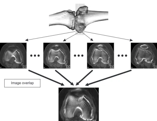
Image overlap
Figure 1. Reference points for each axis for all axial cuts obtained by computed tomography imaging were overlapped as one comprehensive image using a computer imaging program.
개, 대퇴 활차구의 최저점 1개, 후과의 최저점 2개, 내상과 고랑 부위 1개)을 표시하였다. 회전 정렬축의 기준점이 표시된 각각의 CT 축상 이미지들의 영상 투명도를 높인 후 이미지들을 중첩시 킨다. 중첩된 이미지들의 각 기준점들이 하나의 2차원적인 단일 이미지에 나타나게 된다(Fig. 1). 내/외측 후과의 최저점을 연결한 선을 PCA로, 외상과 외측 정점과 내상과 내측 정점을 연결한 선 을 해부학적 상과간 축(anatomical transepicondylar axis, aTEA)으 로, 외상과 외측 정점과 내상과 고랑 최저점을 연결하는 선을 외 과적 상과간 축(surgical transepicondylar axis, sTEA)으로 각각 나 타내었다. 그리고 대퇴 활차구의 최저점들을 연결한 연장선을 AP축으로 나타내었다(Fig. 2).
중첩된 이미지에서 도식화된 정렬축들 간의 관계에서 AP- PCA, AP-aTEA, AP-sTEA, aTEA-PCA 및 sTEA-PCA 축간 각을 측정하였다. 측정은 저자 중 한 명에 의해서 모두 시행되었으며, 측정의 관찰자 내 신뢰성 확보를 위하여 1주 이상의 간격으로 총 3회 반복 측정하여 그 평균치를 참 값으로 정의하였다. 3회 반복 된 측정의 신뢰성 확인을 위하여 intraclass correlation coefficient (ICC)를 이용하였다.16) AP-PCA, AP-aTEA, AP-sTEA, aTEA- PCA 및 sTEA-PCA 축간 각의 ICCs는 각각 0.886, 0.804, 0.759, 0.895 및 0.768이었다. 각각의 축간 각의 평균값을 OA군과 대조군 을 t-test를 이용하여 비교하였고, 나이와 H-K-A 각도에 따른 축 간 각의 상관관계를 Pearson 상관분석법을 통하여 분석하였다.
통계적 분석은 PASW Statistics ver. 17.0 (IBM Co., Armonk, NY, USA) 프로그램을 이용하였고, p값이 0.05 미만인 경우를 통계적 으로 유의한 상관관계가 있는 것으로 간주하였다.
본 연구는 제주대학교병원 생명의학연구윤리심의위원회(In- stitutional Review Board, IRB)의 승인을 받고 시행되었다(IRB No.
2013-06-008).
결 과
OA군에서 AP-PCA 축간 평균각과 AP-aTEA 축간 평균각은 각 각 92.9o와 84.5o로 대조군과 비교하여 PCA 축과 aTEA 축이 좀 더 외회전되어 있었다(p<0.01). OA군에서 aTEA-PCA 축간 평균 각은 8.4o로 대조군의 5.5o에 비하여 좀더 크게 나타났다(p<0.01).
sTEA-PCA 축간 평균각은 OA군이 대조군에 비하여 더 크게 나 타났으나 통계적으로 유의하지는 않았다(p=0.917) (Table 1).
나이가 증가함에 따라 AP-aTEA 축간 각은 보통 음성 상관성 (각각 R=-0.5447, R=-0.6288), AP-aTEA 축간 각은 뚜렷한 음성 상 관성(R=-0.7301)을 보이고, aTEA-PCA 축간 평균각 각은 보통 양 성 상관성(각 R=0.3317)을 보였다(Fig. 3). H-K-A 각도가 증가함 에 따라 AP-PCA 축간 각과 AP-aTEA 축간 각은 보통 음성 상관 성(각각 R=-0.3042, R=-0.4228)을 보이고, aTEA-PCA 축간 각은 약한 양성 상관성(R=0.2689)을 보였다(Fig. 4).
고 찰
Moreland 등13)은 TEA-PCA 축간 각이 평균 3o 정도라고 보고하였 고, 현재 많은 슬 관절 전치환술 기기에서 사용되고 있는 대퇴삽 입물 회전의 기준 역시 서양인의 측정값을 기준으로 PCA로부터 3o 외회전되어 있다. 반면에 Chang 등8)은 우리나라의 여성 중 관 절염이 없는 20명을 대상으로 MRI를 이용하여 각도를 측정한 결 과 TEA-PCA 축간 각이 평균 6.5o라고 보고하였다. 본 연구에서 도 관절염이 없는 대조군에서 TEA-PCA 축간 각이 5.5o로 일반적
Figure 2. Computed tomography images demonstrate an axial view of the distal femoral condyle. AP, anteroposterior axis; aTEA, anatomical transepicondylar axis; sTEA, surgical transepicondylar axis; PCA, posterior condylar axis.
AP
aTEA
PCA
sTEA
Table 1. Angular Measurements of the Reference Axes
Reference axis OA group (°)* Control group (°)
†p-value
AP-PCA 92.9±1.70 96.3±1.87 <0.01 AP-aTEA 84.5±2.59 90.8±1.12 <0.01 AP-sTEA 88.7±1.98 95.1±1.27 <0.01 aTEA-PCA 8.4±2.84 5.5±2.00 <0.01 sTEA-PCA 4.3±1.17 1.2±2.10 0.917 Values are presented as mean±standard deviation. *A total of 218 female patients with osteoarthritis of the knee joint, who underwent total knee arthroplasty. †A total of 50 female patients with healthy knee joint. AP-PCA, anteroposterior-posterior condylar axis; AP- aTEA, anteroposterior-anatomical transepicondylar axis; AP-sTEA, anteroposterior-surgical transepicondylar axis; aTEA-PCA, anatomical transepicondylar axis-posterior condylar axis; sTEA-PCA, surgical transepicondylar axis-posterior condylar axis.으로 알려진 것보다 더 큰 값을 보였다. AP-aTEA 축간 각은 90.8o 로 거의 직각으로 기존에 알려진 바와 유사하게 측정되었다.
OA군에서 회전 정렬축은 퇴행성 변화로 인한 해부학적 변이 로 변화가 생길 수 있으며 그 정도도 개개인에 따라 변화 정도에 차이가 있다.14,17) 이에 인공관절 전치환술 시 대퇴 삽입물의 회전 정렬 기준 축으로 TEA를 이용하여 결정하는 것이 주목받고 있
다.1,12) Chon 등15)은 골관절염 환자(OA군)와 대조군 환자의 축을
비교한 연구에서 AP축, TEA, PCA가 대조군에 비해 내회전되어 있다고 보고하였다. 본 연구에서도 OA군 환자에서 대조군에 비 해 aTEA 기준으로 APA가 6.3o 내회전되어 있었고, aTEA-PCA 축간 각이 2.9o 더 크게 나타났다. OA군의 sTEA는 aTEA에 비하 여 PCA와의 축간 각이 4.3o이고 AP축과의 축간 각이 88.7o로 기 존에 알려진 TEA와 PCA/AP축의 각도와 좀 더 유사한 값으로 나 타나나 표준편차를 고려할 때 절대적인 기준점으로 보기 어렵다.
적절한 TEA를 결정하기 위해서는 PCA에서 외회전 3o가 되는 지 점을 결정하기보다는 OA군 환자에서의 정렬축의 변화를 고려해 야 하고, 수술 전 좀더 정확한 회전 정렬축의 관계를 확인하기 위 하여 CT 촬영이 도움이 될 수 있다. 나이와 H-K-A 각이 증가할
수록 aTEA 기준으로 AP축은 내회전하고 PCA와의 축간 각의 차 이는 커지는 상관관계를 나타났으나, 나이가 많은 대상자에서 관 절염이 심한 점을 고려할때 관절염의 정도가 심할수록 정렬축의 변화가 더 큰 것으로 해석하여야 할 것이다.
관절염이 있는 환자에서 회전 정렬에서 정렬축의 정확한 기준 점을 결정하기 매우 힘들다. 수술 시에 내, 외상과를 덮고 있는 인 대 및 지방 조직으로 인해 내, 외 상과의 위치를 결정하기에는 문 제가 있으며, 특히 내상과는 외상과에 비하여 넓게 분포하기 때 문에 정확한 내상과 중앙점을 찾기 힘들다.16) 수술 전 영상 이미 지에서 정확한 회전 정렬축을 측정하기도 쉽지 않다. CT에서 얻 어낸 여러 절단면 이미지들에서 회전 정렬축들이 다르게 나타나 고, 내/외측 상과와 후과의 적절한 기준점이 하나의 절단면 이미 지에 포함된 단일 절단면 이미지를 얻을 가능성도 낮다(Fig. 5).
최근 3차원으로 재건된 CT 영상을 이용할 수도 있으나, 관절염이 심한 환자에서 과도한 골극으로 인하여 내/외측 상과 부위가 가 려지기도 하고 재건된 CT 영상에서도 정확한 AP축을 결정할 수 없다는 제한점이 있다. 하지만 본 연구에서는 컴퓨터 이미지 프 로그램을 통하여 CT로 얻은 모든 축상 절단면 상에서 각 축의 기 Figure 3. Correlation of the distribution of angles measured between the two axes (A: AP-PCA, B: AP-aTEA, C: AP-sTEA, D: aTEA-PCA, E: sTEA-PCA) and age (black dots: patients group, grey dots: control group). AP-PCA, anteroposterior-posterior condylar axis; AP-aTEA, anteroposterior-anatomical transepicondylar axis; AP-sTEA, anteroposterior-surgical transepicondylar axis; aTEA-PCA, anatomical transepicondylar axis-posterior condylar axis;
sTEA-PCA, surgical transepicondylar axis-posterior condylar axis.
A B C
100 98 96 94 92 90
AP-PCA
R= 0.5447
20 100
Age (yr) 80 60 40
D E
sTEA-PCA
8 6 4 2
0 R=0.5977
20 100
Age (yr) 80 60 40
aTEA-PCA
15 10 5
0 R=0.3317
20 100
Age (yr) 80 60 40
AP-sTEA
95 90 85 80
R= 0.7301
20 100
Age (yr) 80 60 40
AP-aTEA
95 90 85 80 75
R= 0.6288
20 100
Age (yr)
80
60
40
준점을 정하고 모든 절단면을 하나의 이미지로 만드는 작업을 통 하여 보다 정확한 회전 정렬을 얻을 수 있었으며 관절염이 있는 환자에서 대조군에 비하여 회전 정렬축의 변화를 확인할 수 있었 다. 본 연구의 회전 정렬축을 측정하는 방법으로 수술 전에 측정 함으로써 좀 더 정환한 대퇴 삽입물의 회전 정렬을 결정하는 데
도움이 될 것으로 보인다.
본 연구의 제한점으로는 CT 영상을 이용하여 연골에 따른 차 이를 고려하지 못했고 Kellgren-Lawrence grade II에 해당하는 중 등도 골관절염(moderate OA)은 고려하지 않았다는 점이다. 그리 고 대조군의 수가 환자군에 비해 4분의 1 정도로 적어서 대조군 Figure 4. Correlation of the distribution of angles measured between two axes (A: AP-PCA, B: AP-aTEA, C: AP-sTEA, D: aTEA-PCA, E: sTEA-PCA) and H-K-A angle (black dots: patients group, grey dots: control group). H-K-A angle, hip-knee-ankle angle; AP-PCA, anteroposterior-posterior condylar axis; AP-aTEA, anteroposterior-anatomical transepicondylar axis; AP-sTEA, anteroposterior-surgical transepicondylar axis; aTEA-PCA, anatomical transepicondylar axis-posterior condylar axis; sTEA-PCA, surgical transepicondylar axis-posterior condylar axis.
A B C
100 98 96 94 92 90
AP-PCA
R= 0.3042
0 30
H-K-A angle ( )
o20 10
D E
s-PCATEA
8 6 4 2 0
R=0.3506
H-K-A angle ( )
oa-PCATEA
15 10 5 0
R=0.2689
H-K-A angle ( )
oAP-sTEA
95 90 85 80
R= 0.4176
H-K-A angle ( )
oAP-aTEA
95 90 85 80 75
R= 0.4228
H-K-A angle ( )
o0 10 20 30 0 10 20 30
0 10 20 30
0 10 20 30
A B
5o 3o Figure 5. The anteroposterior-anatomical
transepicondylar axis (AP-aTEA) angle from axial computed tomography image of same patient showed subtle difference among the different axial level. (A) AP- aTEA angle: 5°, (B) AP-aTEA angle: 3°.
이 전체 정상 여성을 제대로 반영하지 못했을 가능성을 배제할 수 없다. 또한 실제로 하나의 단면을 이용하여 회전축을 정한 경 우와 중첩시킨 후 회전축을 정한 경우를 비교한 것은 아니기 때 문에 추가적으로 하나의 단면으로 회전축을 정한 경우와 중첩시 킨 단면으로 회전축을 정한 경우를 비교하는 연구를 진행해볼 필 요가 있다고 생각된다.
결 론
CT 축상 이미지들을 중첩시키는 방법으로 원위 대퇴골의 회전 정렬축 간의 관계를 정확히 측정할 수 있었으며, 이를 통해 OA 군에서 대조군보다 AP축이 aTEA에 비하여 내회전하고 aTEA- PCA 축간 각의 더 증가함을 확인할 수 있었다. 따라서 인공 슬관 절 전치환술에서 대퇴 삽입물의 회전 정렬을 결정할 때 환자에 따른 정렬축의 차이를 고려해야 한다.
CONFLICTS OF INTEREST
The authors have nothing to disclose.
ACKNOWLEDGEMENTS
This work was supported by the research grant of the Jeju National University Hospital in 2013.
REFERENCES
1. Berger RA, Rubash HE, Seel MJ, Thompson WH, Crossett LS. Determining the rotational alignment of the femoral component in total knee arthroplasty using the epicondylar axis. Clin Orthop Relat Res. 1993;286:40-7.
2. Arima J, Whiteside LA, McCarthy DS, White SE. Femoral ro- tational alignment, based on the anteroposterior axis, in total knee arthroplasty in a valgus knee. A technical note. J Bone Joint Surg Am. 1995;77:1331-4.
3. Olcott CW, Scott RD. The Ranawat Award. Femoral com- ponent rotation during total knee arthroplasty. Clin Orthop Relat Res. 1999;367:39-42.
4. Akagi M, Matsusue Y, Mata T, et al. Effect of rotational align- ment on patellar tracking in total knee arthroplasty. Clin Or- thop Relat Res. 1999;366:155-63.
5. Anouchi YS, Whiteside LA, Kaiser AD, Milliano MT. The ef- fects of axial rotational alignment of the femoral component
on knee stability and patellar tracking in total knee arthro- plasty demonstrated on autopsy specimens. Clin Orthop Relat Res. 1993;287:170-7.
6. Akagi M, Yamashita E, Nakagawa T, Asano T, Nakamura T.
Relationship between frontal knee alignment and reference axes in the distal femur. Clin Orthop Relat Res. 2001;388:147- 56.
7. Tanavalee A, Yuktanandana P, Ngarmukos C. Surgical epi- condylar axis vs anatomical epicondylar axis for rotational alignment of the femoral component in total knee arthro- plasty. J Med Assoc Thai. 2001;84 Suppl 1:S401-8.
8. Chang CB, Seong SC, Lee S, Lee MC. Anatomical assessment of the distal femur and tibia for optimal femoral rotational alignment in total knee arthroplasty. J Korean Knee Soc.
2010;22:46-55.
9. Griffin FM, Math K, Scuderi GR, Insall JN, Poilvache PL.
Anatomy of the epicondyles of the distal femur: MRI analysis of normal knees. J Arthroplasty. 2000;15:354-9.
10. Yoshino N, Takai S, Ohtsuki Y, Hirasawa Y. Computed to- mography measurement of the surgical and clinical transepi- condylar axis of the distal femur in osteoarthritic knees. J Arthroplasty. 2001;16:493-7.
11. Yoshioka Y, Siu D, Cooke TD. The anatomy and functional axes of the femur. J Bone Joint Surg Am. 1987;69:873-80.
12. Blaha JD, Mancinelli CA, Simons WH, Kish VL, Thyagarajan G. Kinematics of the human knee using an open chain ca- daver model. Clin Orthop Relat Res. 2003;410:25-34.
13. Moreland JR, Bassett LW, Hanker GJ. Radiographic analysis of the axial alignment of the lower extremity. J Bone Joint Surg Am. 1987;69:745-9.
14. Shon HC, Choi ES, Kim YM, et al. Femoral component rota- tion of total knee arthroplasty in Korean subjects: a study using grand piano sign and computed tomography. J Korean Orthop Assoc. 2012;47:432-8.
15. Chon JG, Sun DH, Jung JY, Kim TI, Jang SW. Rotational alignment of femoral component for minimal medial col- lateral ligament release in total knee arthroplasty. Knee Surg Relat Res. 2011;23:153-8.
16. Shrout PE, Fleiss JL. Intraclass correlations: uses in assessing rater reliability. Psychol Bull. 1979;86:420-8.
17. Griffin FM, Insall JN, Scuderi GR. The posterior condylar angle in osteoarthritic knees. J Arthroplasty. 1998;13:812-5.
한국의 여성 골관절염 환자에서의 대퇴골의 해부학적 회전 정렬축에 대한 컴퓨터 이미지 프로그램을 이용한 분석
김경민 • 김상림 • 배종환 • 박용근
제주대학교 의학전문대학원 제주대학교병원 정형외과학교실
목적: 대퇴골의 해부학적 정렬축들의 관계를 컴퓨터 이미지 프로그램을 이용하여 정확히 측정하는 방법을 제시하고 골관절염이 있 는 여자 환자에서 대조군과 비교하여 정렬축 간의 차이가 있는지를 비교하고자 한다.
대상 및 방법: 2013년 1월부터 2014년 12월까지 인공 슬관절 전치환술을 받은 여자 환자 218명(osteoarthritis, OA군)과 골관절염 이 없는 40세 이하의 여성 50명(대조군)을 비교하였다. 환자의 컴퓨터 단층촬영(computed tomography, CT) 영상에서 축상 영상들 을 이미지 프로그램을 이용하여 축상 영상들을 중첩하고, 중첩된 축상면에서 정렬축들을 도식하였다. 전후방 축(anteroposterior, AP 축), 후과 축(posterior condylar axis, PCA), 해부학적 대퇴 상과간 축(anatomical transepicondylar axis, aTEA) 및 외과적 대퇴 상과 간 축(surgical transepicondylar axis, sTEA)의 축간 각도의 차이를 OA군과 대조군에서 비교 분석하였다.
결과: AP-PCA 축간 평균각은 대조군에서 96.3o±1.87o이고 OA군에서 92.9o±1.70o였고 (p<0.01), AP-aTEA 축간 평균각은 각각 90.8o±1.12o, 84.5o±2.59o였으며(p<0.01), AP-sTEA 축간 평균각은 각각 95.1o±1.27o, 88.7o±1.98o였다(p<0.01). aTEA-PCA 축간 평균각은 대조군에서 5.5o±2.00o이고 OA군에서 8.4o±2.84o였으며(p<0.01), sTEA-PCA 축간 평균각은 각각 1.2o±2.10o, 4.3o±1.17o였다(p=0.917).
결론: CT 축상 이미지들을 중첩시키는 방법으로 원위 대퇴골의 회전 정렬축 간의 관계를 정확히 측정할 수 있었으며, 이를 통해 OA 군에서 대조군보다 AP축이 aTEA에 비하여 내회전하고 aTEA-PCA 축간 각이 더 증가함을 확인할 수 있었다.
색인단어: 슬관절, 관절염, 정렬, 인공 슬관절 전치환술
접수일 2016년 9월 2일 수정일 2016년 10월 24일 게재확정일 2016년 10월 26일 책임저자 박용근
63241, 제주시 아란 13길 15, 제주대학교 의학전문대학원 제주대학교병원 정형외과학교실 TEL 064-717-2710, FAX 064-717-1131, E-mail [email protected]
Copyright © 2017 by The Korean Orthopaedic Association
“This is an Open Access article distributed under the terms of the Creative Commons Attribution Non-Commercial License (http://creativecommons.org/licenses/by-nc/4.0/) which permits unrestricted non-commercial use, distribution, and reproduction in any medium, provided the original work is properly cited.”