- 57 -
KISEP Case Reports J Rhinol 4((((1)))), 1997
Primitive Neuroectodermal Tumor of Nasal Cavity: : : :Case Report
Heung Man Lee, M.D.
1, Seung Hoon Han, M.D.
1, Jeong Su Woo, M.D.
1, Seong Jin Cho, M.D.
2, Han Kyeom Kim, M.D.
2and Sang Hag Lee, M.D.
1 ABSTRACTThe primitive neuroectodermal tumor is a highly malignant small round cell tumor characterized by neuroectodermal origin and poor prognosis. The thoracopulmonary region is known to be the most common site of the disease, with peak incidence during adolescence. Primitive neuroectodermal tumors are very rarely reported in the head and neck area, and there have been only a few involving the nasal cavity. We treated a 60 year-old female patient who had nasal obstruction over a three-month period with radical surgery and postoperative radiation therapy. This paper presents our diagnostic workup and approach in treating this unique tumor, and the immunohistochemical findings. A guideline for the treatment of this aggressive neoplasm is reviewed with our experiences in treating this primitive neuroectodermal tumor of the nasal cavity by combined treatment modalities.
KEY WORDS:Primitive neuroectodermal tumor·Nasal cavity.
INTRODUCTION
The primitive neuroectodermal tumor (PNET) is rarely found in the nasal cavity. Since the initial description of PNET made in 1918 by Stout,1) who described a malignant tumor of the forearm, there have been only a few reported cases of this tumor arising in the head and neck. The most common form of PNET was first reported in 1979. The malignant small ro- und cell tumor which was found in the thoracopulmonary re- gion was neuroectodermal in origin, and is known today as Askin’s tumor.2) Since then, there have been debates and con- troversies on the terminology and classification of this very rare disease.
Since there has been no single clinical or laboratory test to aid in eliminating its often questionable diagnosis, PNET has been difficult to differentiate from other small round cell tu- mors, especially Ewing’s sarcoma.3) Although there are relat- ively few cases reported in existing literature, several studies have shown PNET to have a clinically aggressive behavior with significantly worse survival rates than other small round cell tumors.4)5)6)
CASE REPORT
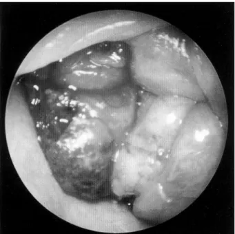
A previously healthy 60 year-old female patient was re- ferred to the Otolaryngology - Head and Neck Surgery Depa- rtment at the Korea University Hospital with a three-month history of left nasal obstruction. The patient had found the mass in the left nasal cavity two months earlier. The patient also complained of both fronto-temporal headache, however, she denied symptoms of rhinorrhea, postnasal dripping and nasal bleeding. Physical examination with a nasal endoscope revealed a smooth-surfaced, polypoid mass filled the left na- sal cavity (Fig. 1). There was subcutaneous or contralateral nasal cavity mass, nor any palpable cervical lymph nodes. No cranial nerve deficits were found either.
Radiological investigations including paranasal sinus X-rays, chest X-rays, and a high-resolution contrast-enhanced com- puted tomography (CT) of paranasal sinsuses (PNS) were performed. An axial view of PNS CT revealed a soft tissue density mass filling the left nasal cavity. There was no bony destruction and the paranasal sinus appeared unaffected (Fig. 2).
A diagnostic biopsy was performed as an out-patient proc- edure. With the initial diagnosis of malignant PNET, further studies were carried out for the purpose of a metastatic workup which would include an ultrasonogram of the abdomen, brain magnetic resonance imaging, and a bone scan. The results of these studies revealed little. Other routine laboratory test yi- elded normal results. A maxillectomy via lateral rhinotomy incision was performed for initial treatment. The mass sized 3×2 cm was attached superior to the inferior turbinate (Fig.
Department of 1Otorhinolaryngology- Head and Neck Surgery,
2Surgical Pathology, Korea University College of Medicine, Se- oul, Korea
Address correspondence and reprint requests to Heung Man Lee, M.D., Department of Otorhinolaryngology-Head and Neck Surgery, Korea University College of Medicine, 80, Gurodong, Gurogu, Seoul 152-050, Korea
Tel:82-2-818-6157, Fax:82-2-868-0475 Accepted for publication March 12, 1997
58 / J Rhinol 4(1), 1997
3). There was no invasion of the left maxillary sinus and no evidence of bone destruction.
The patient was discharged without any event on the 10th postoperative day. Postoperatively, she received a total of 61 Grey of external beam radiation for six weeks. She was con- tinually and closely followed up with nasal endoscopic exa- minations, and PNS CT for seven with no evidence of rec- urrence. The patient is alive and well.
On examing a gross section, the cut surface showed a poorly demarcated pale-gray tumor measuring 3×2 cm cross-secti- onally. Light microscopic evaluation revealed no specific ar- chitectural pattern, and the tumor was composed of small pri- mitive cells with scant cytoplasm, and round or oval nucleoli.
No definite rosettes or pseudorosettes could be identified
(Fig. 4). In immunohistochemical staining, there were positive findings for neuron-specific enolase (NSE)(Fig. 5) and syn- aptophysin (Fig. 6). However, negative findings were observed for the antibodies of neuroblastoma, MIC2, leukocyte common antigen, and HMB45.
Fig. 2. Preoperative axial CT scan of paranasal sinuses showing inhomogenous soft tissue density (*) filling the left nasal cavity.
Fig. 1. Preoperative endoscopic finding showing smooth surfa- ced, polypoid mass filling the left nasal cavity.
Fig. 4. Microscopic finding showing patterless growth of primitive cells and the high frequency of mitotic cells (H & E stain, ×400).
Fig. 3. Surgical specimen appearing irregular surfaced mass sized 3.5×3×1 cm.
Lee et al.:Primitive Neuroectodermal Tumor / 59
DISCUSSION
There have been several reports of tumors which are si- milar to PNET, but with controversiesy about their proper terminology or classification. It was not until 1973, when Hart and Earle7) applied the term‘primitive neuroectodermal tu- mor’that the controversies subsided. They described a group of small round cell tumors of the central and peripheral ne- rvous system, and identified them as being derived from fetal neuroectodermal precursor cells. PNETs have their origins ou- tside the autonomic nervous system, hence they are often called peripheral neuroectodermal tumors along with other terms such as peripheral neuroepithelioma and peripheral neurobl- astoma.5)
Incidence of these tumors is very rare, and it can occur at any age, with peak incidence during adolescence. Two cases of PNET were reported in the maxillary/ethmoid sinus of a 13 year-old patient and the nasal cavity of a 17 year-old patient.8) The predominent location of PNET is the chest
wall.2)4)5)8)9) Here we encountered a rare case of PNET arising in the nasal cavity.
Because of its aggressive nature it is important to make a correct diagnosis of PNET. However, even today, there are arguments among pathologists and oncologists in its diagn- osis. Increasingly, ultrastructural, immunohistochemical, and cytogenetic findings have been necessary to confirm the dia- gnosis of PNET.4)6)9) Differential diagnosis is imperative for PNET since even today many pathologists argue that these putative soft tissue tumors are nothing more than a metastasis from an unrecognized neuroblastoma or a carcinoma. The di- agnosis of PNET can only be made by exclusion, following an extensive search for a primary neuroblastoma.10) Differen- tial diagnosis includes taking Ewing’s sarcoma, neuroblastoma, lymphoma, and embryonic variants of rhabdo- myosarcoma into account.3)6)11)12)
Microscopically, PNETs appear as small round cell sheets or lobules with the presence of Homer-Wright rossettes.4) Our case did not show any definite rosette formations, however, small round cells were detected. Ultrastructural characteristics include sheets of primitive tumor cells with large, round, oval or irregular shaped nuclei with prominent nucleoli and scant cytoplasm. The most distinguishing findings were the pres- ence of interdigitating, long cytoplasmic processes containing glial filaments with glial differentiation, and the presence of dense core granules and microtubules with neuronal differe- ntiation.5)8)
These tumors are immunoreactive for NSE, Synaptophysin, S-100 protein, and MB2 monoclonal antibodies.6) Our case showed positive immunoreaction for NSE and synaptophysin, strongly suggesting neurogenic origin, but showed negative immunoreaction for leukocyte common antigen, UCHL-1, L26, CD68, and cytokeratin. Although previous reports have sho- wn that MIC2 expression is detected in PNET between 82%
to 97% of the time,13) our case did not.
Diagnosis of neuroblastoma could be excluded for our case even with the negative MIC2 findings due to the fact that our case did not demonstrate any neurofibrillary backgr- ound which should be presented to make a diagnosis of neu- roblastoma. Cytogenetic evaluation of the patient with PNET showed specific translocation of chromosomes 11 and 22 [+
(11:22) (q24:q12)], which can also be seen in Ewing’s sarcoma.4)5)8)11) However, our case did not show specific tra- nslocation of chromosomes 11 and 22 (data not shown). Ev- entually the confirmation of diagnosis is made by combination of all of these diagnostic modalities.
Because of its aggressiveness and poor prognosis the mu- ltimodality approach in the treatment of PNET is currently being advocated.8)11) Even with radical surgical therapy com- bined with chemotherapy and radiation therapy, the prognosis
Fig. 5. Immunohistochemical staining of NSE (×400).
Fig. 6. Immunohistochemical staining of synaptophysin (×400).
60 / J Rhinol 4(1), 1997
is still very poor.
There is a marked difference between the outcomes of patients undergoing radical and non-radical surgical proced- ures. Early initial treatment also seems to be important factor in increasing the survival rate.9) External beam radiation th- erapy using 50 to 65 Grey at the primary site and regional nodes is advocated. No studies have shown an effective si- ngle-agent regimen, therfore, multidrug therapy using drugs such as doxorubicin, vincristine, and cyclophosphamide is recommended.4) Our patient underwent resection of the tumor followed by radiation therapy using 61 Grey for six weeks.
Clinically, the tumor is a highly aggressive malignant tumor with a significantly worse survival rate than other small round cell tumors.6) Metastasis at presentation is fairly common with up to 31%.8) The most common metastatic sites are the lung, bone, and brain.4) It is important to carry out an intensive me- tastatic workup at the initial diagnosis of PNET. Our case sh- owed no definite metastatic lesion on chest X-rays, abdominal ultrasonogram, brain MRI, and whole bone scan. Unlike other studies we did not note any bone erosion due to local extension of the tumor. Some studies have emphasized routine use of bone marrow biopsy and aspiration along with chest CT and chest radiography for determination of metatstasis.8)
Complete remission following treatment is common, how- ever, long-term disease-free survival has been less encouraging.
Two year-survival rate is less than 65%8) and is dramatically reduced to 25% when the tumor size is greater than 5 cm9). Furthermore, the three-year survival rate is below 50%.5) Alt- hough our patient showed complete remission of the disease following surgery and radiation therapy, a much longer follow- up period is needed to accurately predict the prognosis of PNET in the nasal cavity.
REFERENCES
1) Stout AP. A tumor of the ulnar nerve. Proc NY Pathol Soc 1918;
12:2-12.
2) Askin FB, Rosai J, Sibley RK, Dehner LP, McAlister WH. Mali- gnant small cell tumor of the thoracopulmonary region in childhood:
a distinctive clinicopathologic entity of uncertain histogenesis.
Cancer 1979;43:2438-51.
3) Schmidt D, Harms D, Burdach S. Malignant peripheral neuroect- odermal tumors of childhood and adolescence. Virchows Arch A Pathol Anat Histopathol 1985;406:351-65.
4) Chowdhury K, Manoukian JJ, Rochon L, Begin LR. Extracranial primitive neuroectodermal tumor of the head and neck. Arch Oto- laryngol Head Neck Surg 1990;116:475-8.
5) Jurgens H, Bier V, Harma D, Beck J, Brandeis W, Etspuler G. Ma- lignant peripheral neuroectodermal tumors: a retrospective analysis of 42 patients. Cancer 1988;61:349-57.
6) Kahn HJ, Thorner PS. Monoclonal antibody MB2. A potential matter for Ewing’s sarcoma and primitive neuroectodermal tumor.
Pediatr Pathol 1989;9:153-62.
7) Hart MN, Earle KM. Primitive neuroectodermal tumors of the brain in children. Cancer 1973;32:890-7.
8) Jones JE, McGill T. Peripheral primitive neuroectodermal tumors of the head and neck. Arch Otolaryngol Head Neck Surg 1995;
121:1392-5.
9) Kushner BH, Haydu S, Gulati SC. Extracranial primitive neur- oectodermal tumors. Cancer 1991;67:1825-6.
10) Enzinger FM, Weiss SW. Soft tissue tumors. St Louis, CV Mosby Co., 1988:806-15.
11) Dehner LP. Primitive neuroectodermal tumor and Ewing’s sarcoma.
Am J Surg Pathol 1993;17 (1):1-13.
12) Shimada H, Newton WA. Pathologic features of extraosseous Ew- ing’s sarcoma: a report from the intergroup rhabdomyosarcoma study. Hum Pathol 1988;19:442-53.
13) Perlman EJ, Dickman PS, Askin FB, Grier HE, Miser JS, Link MP.
Ewing’s sarcoma-routine diagnostic utilization of MIC2 analysis:
A pediatric oncology group/children’s cancer group intergroup st- udy. Hum Pathol 1994;25:304-7.