into soft tissue via the mental foramen and is then named the mental nerve and provides lip and chin sensation. Another branch called the incisive branch goes forward in the bone to supply anterior teeth sensation6,7. In many cases, there is no distinct incisive canal because the nerves are organized in a network and are not detectable on radiographs8.
The mental foramen in the embryonic period is at the api- cal area of the canine and first deciduous molar6. During the development of the mandible until the eruption of deciduous molars, the mental foramen is displaced anteriorly but after eruption of the second deciduous molar it redirects poste- riorly. This displacement is a possible cause for develop- ment of an anterior loop of the IAN before it emerges as the mental nerve6,7. The anterior loop of the IAN is an important anatomical variation that originates from the IAN. In its first portion, it dips downwards and is then displaced upward and posteriorly to exit the mental foramen9. Failure to note this mesial loop may cause complications like sensory disorders in the lower lip. Therefore, precise evaluation of its position before surgery is essential. Pre-surgical evaluation of three- dimensional (3D) cone-beam computed tomography (CBCT)
I. Introduction
In surgical procedures, attention to anatomical structures and their precise position is paramount1. The inferior alveolar nerve (IAN) canal is an important anatomical landmark in the mandible and contains the clinically important neurovascular bundle2-5. The mental foramen is superior to the mandibular canal and is apical to the premolars. The IAN divides into two branches before exiting the foramen. One branch exits
Hamed Kermani
Department of Oral and Maxillofacial Surgery, Faculty of Dentistry, Mashhad University of Medical Sciences, Mashhad, Iran
*Current affiliation: Craniofacial and Pediatric Maxillofacial Surgery Fellow, Department of Oral and Maxillofacial Surgery, Tehran University of Medical Sciences Dental School, North Kargar Street, Tehran, Iran TEL: +00989128361094 FAX: +00982188015880
E-mail: [email protected] ORCID: http://orcid.org/0000-0003-4735-2751
This is an open-access article distributed under the terms of the Creative Commons Attribution Non-Commercial License (http://creativecommons.org/
licenses/by-nc/4.0/), which permits unrestricted non-commercial use, distribution, and reproduction in any medium, provided the original work is properly cited.
CC
Assessment of the anterior loop of the inferior alveolar nerve via cone-beam computed tomography
Baratollah Shaban1, Amin Khajavi2, Nasim Khaki3, Yones Mohiti3, Tahere Mehri3, Hamed Kermani1,*
Departments of 1Oral and Maxillofacial Surgery, 2Periodontics, and 3Oral and Maxillofacial Radiology, Faculty of Dentistry, Mashhad University of Medical Sciences, Mashhad, Iran
Abstract(J Korean Assoc Oral Maxillofac Surg 2017;43:395-400)
Objectives: The aim of this study was to evaluate different anatomical variants of the anterior loop of the inferior alveolar nerve (IAN) via cone-beam computed tomography (CBCT).
Materials and Methods: CBCT images of 71 patients (36 males and 35 females) were evaluated. We used the classification described by Solar for IAN evaluation. In this classification, three different types of IAN loops were introduced prior to emerging from the mental foramen. We classified pa- tients according to this system and introduced a new, fourth type.
Results: Type I was seen in 15 sites (10.6%), type II in 39 sites (27.5%), and type III in 50 sites (35.2%). We found a new type in 38 sites (26.8%) that constituted a fourth type.
Conclusion: We found that type III was the most common variant. In the fourth type, the IAN was not detectable because the main nerve was adjacent to the cortical plate and the incisive branch was thinner than the main branch and alongside it. In this type, more care is needed for surgeries including inferior alveolar and mental nerve transposition.
Key words: Anterior, Mental loop, Cone-beam computed tomography, Iran
[paper submitted 2017. 1. 21 / revised 2017. 4. 7 / accepted 2017. 5. 7]
Copyright © 2017 The Korean Association of Oral and Maxillofacial Surgeons. All rights reserved.
and incisive branch thickness is similar to the main branch.
In type II, the anterior loop is absent but the anatomy is T- shaped. The incisive branch is perpendicular to the main branch and the mental branch enters the mental foramen in a perpendicular direction. In type III, the anterior loop is de- tectable and the anatomy is Y-shaped. The incisive branch is thicker than the main branch and the mental branch diverges images plays an important role in prevention of probable
damage. Several cadaver studies using radiological images have classified different shapes of the anterior loops of the IAN. We used the classification described by Solar et al.10. In this classification, three different types have been introduced for the anterior loop. According to this classification, the anterior loop is not seen in type I. The anatomy is Y-shaped
Mental for amen Mental for amen
Fig. 1. Both left and right mental fo- ramina on the axial section and pan- oramic curve drawn on the axial sec- tion.
Baratollah Shaban et al: Assessment of the anterior loop of the inferior alveolar nerve via cone-beam computed tomography. J Korean Assoc Oral Maxillofac Surg 2017
1
122334455667788
A
B
1
5
2 3 4
6 7 8
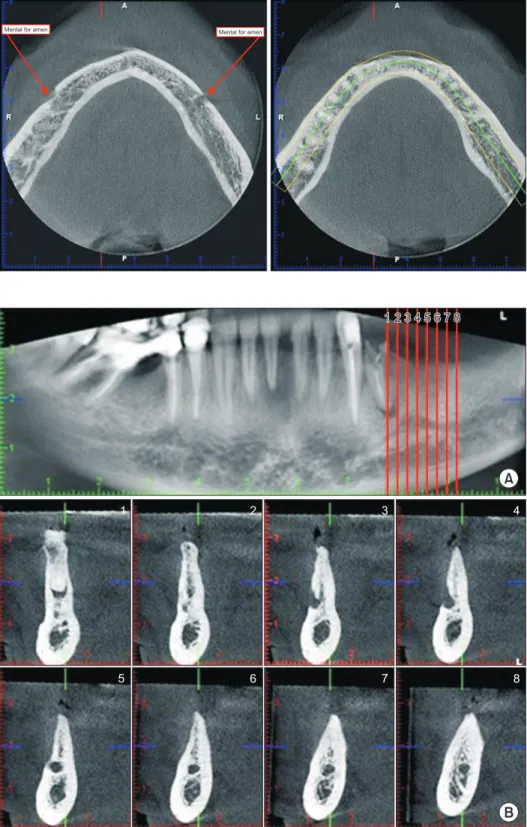
Fig. 2. A. Panoramic section showing type I on both sides; the guide plans for cross sections are also seen. B.
Cross-sectional images showing type I.
Baratollah Shaban et al: Assessment of the anterior loop of the inferior alveolar nerve via cone-beam computed tomography. J Korean Assoc Oral Maxillofac Surg 2017
graduate students who were trained to evaluate the IAN loop and its different types. All data were checked by an associate professor of oral and maxillofacial radiology. Both sides of all images were scanned with 0.2 mm3 voxel size and 2 mm interslice distances. Axial, sagittal, and coronal sections were obtained. All images were similarly evaluated to standard- ize the procedure. First, the axial section was generated in a way that both left and right mental foramina could be seen.
Panoramic curves were then drawn from the right to the left mental foramen.(Fig. 1) Panoramic sections were used to generate the cross-sections.(Fig. 2, 3)
III. Results
In our study, CBCT images of the mandible obtained from 71 patients, 36 male patients (50.7%) and 35 female patients from the IAN anterior to the mental foramen. The aim of this
study was to evaluate different IAN loop anatomical variants in an Iranian population via CBCT.
II. Materials and Methods
CBCT images of 71 patients (36 males and 35 females) from the radiology department archive of Mashhad Dental School were evaluated. The mean age of the patients was 43.54±9.72 years (range, 20-68 years). All images were taken by the same radiographic device (Planmeca Oy, Helsinki, Finland) and under the same exposure settings of 90 kVp, 10 mA, 12 seconds, and 18×18 cm field of view. All pa- tients with history of trauma to the mandible, developmental anomalies, and pathological lesions were excluded. To ensure precise evaluation, all images were assessed by three post-
A
B
1 12233445566
1 2 3
5
4 6
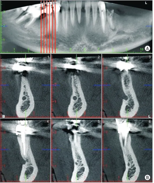
Fig. 3. A. Panoramic view of type IV. B.
Cross-sectional images of type IV.
Baratollah Shaban et al: Assessment of the anterior loop of the inferior alveolar nerve via cone-beam computed tomography. J Korean Assoc Oral Maxillofac Surg 2017
dose compared to computed tomography (CT)18. However, decreased resolution and contrast in comparison to CT are disadvantages. The American Academy of Oral and Maxil- lofacial Radiology (AAOMR) suggested CBCT for evalu- ation of periodontal and orthodontic treatment and implant surgeries14,19-22. According to the results of our study, type I was seen in 10.6%, type II in 27.5%, and type III in 35.2%
of patients. Most patients had anterior loops. The results of our study were similar to a study on a Turkish patient popula- tion23. They showed that type III was the most prevalent type (59.5%), and type I (8.6%) was the least prevalent type. Sev- eral studies (anatomical, radiographic, and combination of both) have evaluated the prevalence of an anterior loop and its morphological characteristics. The findings of these stud- ies are summarized in Table 12,8,9,11,23-26.
In our study, this anatomical structure was visible in 50 sites (35.2%). But the prevalence of the anterior loop cannot provide enough information for surgeons because determina- tion of the safety margin for interforaminal surgeries is a ma- jor issue. Despite many studies, a distinct distance from the mesial aspect of the mental foramen is still not recommended as a guideline. Different studies suggested a safety margin of 1-9 mm from the anterior margin of the mental fora- men10,24,27-34. In a study by Wismeijer et al.13, a protocol with a 3 mm safety margin was considered for all patients, and sen- sory disorders due to damage to the anterior loop was seen in 7% of cases. A standard distance from the mental foramen as a safe margin still cannot be recommended. Due to fear from this complication, many clinicians place implants anterior to their ideal position to avert damage to the mental nerve and sensory disorders in the lower lip35. In our study, we found a new variant of the anterior loop in 26.8% of sites that could not be classified into any defined type. In this new variation, the nerve was not detectable because the main branch was ad- jacent to the cortical plate and the incisive branch was along (49.3%), were evaluated bilaterally. Type I was seen at 15
sites (10.6%), type II at 39 sites (27.5%), and type III at 50 sites (35.2%). We identified a new type of the anterior loop at 38 sites (26.8%) that could not be classified into any of the defined types. We found that in some radiographs the shape of the mental nerve was different from type III. In this new type, the inferior alveolar canal is adjacent to the buc- cal cortical plate of the mandible. The IAN divided into two branches but the mental nerve was not detectable because the main branch was adjacent to the cortical plate in the mental foramen area. The anatomy of this type was neither Y nor T- shaped and the incisive branch was along the main branch and this branch was thinner than the main branch. Panoramic and cross-sectional images of type IV are shown in Fig. 3.
IV. Discussion
Many studies have shown the unreliability of plain radio- graphs for detection of anatomical structures11. Panoramic radiography is an appropriate imaging modality for observa- tion of the mandibular canal. Vazquez et al.12 evaluated the efficiency of panoramic radiographs for treatment planning of implant surgery. These images effectively evaluated the bone height available for posterior mandibular implants and 3D imaging was not necessary12. However, panoramic radi- ography is not an efficient imaging modality for evaluation of nerve loop morphology because processing errors and incor- rect patient position strongly affect image quality. In addition, the anterior loop is an intermedullary structure that is covered with thick cortical bone13. The absence of the anterior loop in panoramic radiographs does not mean that it does not exist in those cases14,15. 3D imaging is necessary for evaluation of the anterior IAN loop16,17. CBCT is an imaging method that has advantages including easy technique, precise images, de- creased artifacts, lower costs, and decreased patient radiation
Table 1. Results of anatomical and radiographic studies on the prevalence of the anterior mental loop and its morphological characteristics Author Year Country Evaluation tools Sample size (n) Prevalence of anterior mental loop Rosenquist24
Arzouman et al.9 Jacobs et al.2 Kaya et al.25 Ngeow et al.8 Benninger et al.26 Kajan and Salari11 Demir et al.23 Our study
1996 1993 2002 2008 2009 2011 2012 2015
Belgium Turkey Malaya USA Iran Turkey Iran
Cadaver
Cadaver (anatomic and radiographic evaluation) Panoramic
Panoramic, CBCT Panoramic Cadaver CBCT CBCT CBCT
58 25 545 73 33 15 84 279 91
15%
Anatomic (96%), radiographic (12%) 11%
Anatomic (28%), CBCT (34%) 40.2%
26%
36.9%
59.5%
48%
(CBCT: cone-beam computed tomography)
Baratollah Shaban et al: Assessment of the anterior loop of the inferior alveolar nerve via cone-beam computed tomography. J Korean Assoc Oral Maxillofac Surg 2017
cial Orthop 2009;136:764.e1-11; discussion 764-5.
5. White SC. Cone-beam imaging in dentistry. Health Phys 2008;95:
628-37.
6. Chávez-Lomeli ME, Mansilla Lory J, Pompa JA, Kjaer I. The hu- man mandibular canal arises from three separate canals innervating different tooth groups. J Dent Res 1996;75:1540-4.
7. de Villiers H. The skull of the South African Negro: a biometrical and morphological study. Johannesburg: Wits University Press;
1970.
8. Ngeow WC, Dionysius DD, Ishak H, Nambiar P. A radiographic study on the visualization of the anterior loop in dentate subjects of different age groups. J Oral Sci 2009;51:231-7.
9. Arzouman MJ, Otis L, Kipnis V, Levine D. Observations of the anterior loop of the inferior alveolar canal. Int J Oral Maxillofac Implants 1993;8:295-300.
10. Solar P, Ulm C, Frey G, Matejka M. A classification of the intraos- seous paths of the mental nerve. Int J Oral Maxillofac Implants 1994;9:339-44.
11. Kajan ZD, Salari A. Presence and course of the mandibular inci- sive canal and presence of the anterior loop in cone beam com- puted tomography images of an Iranian population. Oral Radiol 2012;28:55-61.
12. Vazquez L, Saulacic N, Belser U, Bernard JP. Efficacy of panoram- ic radiographs in the preoperative planning of posterior mandibular implants: a prospective clinical study of 1527 consecutively treated patients. Clin Oral Implants Res 2008;19:81-5.
13. Wismeijer D, van Waas MA, Vermeeren JI, Kalk W. Patients' perception of sensory disturbances of the mental nerve before and after implant surgery: a prospective study of 110 patients. Br J Oral Maxillofac Surg 1997;35:254-9.
14. Kalender A, Orhan K, Aksoy U. Evaluation of the mental foramen and accessory mental foramen in Turkish patients using cone-beam computed tomography images reconstructed from a volumetric rendering program. Clin Anat 2012;25:584-92.
15. Kuzmanovic DV, Payne AG, Kieser JA, Dias GJ. Anterior loop of the mental nerve: a morphological and radiographic study. Clin Oral Implants Res 2003;14:464-71.
16. Chen JC, Lin LM, Geist JR, Chen JY, Chen CH, Chen YK. A ret- rospective comparison of the location and diameter of the inferior alveolar canal at the mental foramen and length of the anterior loop between American and Taiwanese cohorts using CBCT. Surg Ra- diol Anat 2013;35:11-8.
17. Parnia F, Moslehifard E, Hafezeqoran A, Mahboub F, Mojaver- Kahnamoui H. Characteristics of anatomical landmarks in the man- dibular interforaminal region: a cone-beam computed tomography study. Med Oral Patol Oral Cir Bucal 2012;17:e420-5.
18. Danforth RA, Dus I, Mah J. 3-D volume imaging for dentistry: a new dimension. J Calif Dent Assoc 2003;31:817-23.
19. Haghanifar S, Rokouei M. Radiographic evaluation of the men- tal foramen in a selected Iranian population. Indian J Dent Res 2009;20:150-2.
20. Bushong SC. Radiologic science for technologists: physics, biol- ogy, and protection. 8th ed. New York: Elsevier; 2004:29-30.
21. Katakami K, Mishima A, Shiozaki K, Shimoda S, Hamada Y, Ko- bayashi K. Characteristics of accessory mental foramina observed on limited cone-beam computed tomography images. J Endod 2008;34:1441-5.
22. Koh KJ, Kim KA. Observation of the anterior loop and mental fo- ramen of the mandibular canal using cone beam computed tomog- raphy. Korean J Oral Maxillofac Radiol 2009;39:81-7.
23. Demir A, Izgi E, Pekiner FN. Anterior loop of the mental foramen in a Turkish subpopulation with dentate patients: a cone beam computed tomography study. J Marmara Univ Inst Health Sci 2015;5:231-8.
24. Rosenquist B. Is there an anterior loop of the inferior alveolar nerve? Int J Periodontics Restorative Dent 1996;16:40-5.
25. Kaya Y, Sencimen M, Sahin S, Okcu KM, Dogan N, Bahcecitapar
the main branch and this branch was thinner than the main branch. The presence of this type is very important in nerve transpositioning. Superficial IAN positioning in this variant can more likely render damage to the nerve.
In our study, all radiographs available in the archive that matched inclusion criteria were evaluated. Future multicenter studies with a larger sample size can provide more accurate information regarding the prevalence of different mental nerve variants.
V. Conclusion
We found type III to be the most common variant. In the fourth type, the IAN was not detectable because the main nerve was adjacent to the cortical plate and the incisive branch was thinner than the main branch and alongside it. For this type, more care is needed in surgeries including inferior alveolar and mental nerve transposition.
Conflict of Interest
No potential conflict of interest relevant to this article was reported.
ORCID
Baratollah Shaban, http://orcid.org/0000-0002-1857-1990 Amin Khajavi, http://orcid.org/0000-0003-3916-3088 Nasim Khaki, http://orcid.org/0000-0003-2017-7703 Yones Mohiti, http://orcid.org/0000-0002-8143-3814 Tahere Mehri, http://orcid.org/0000-0001-8755-1056 Hamed Kermani, http://orcid.org/0000-0003-4735-2751
References
1. Mraiwa N, Jacobs R, van Steenberghe D, Quirynen M. Clinical assessment and surgical implications of anatomic challenges in the anterior mandible. Clin Implant Dent Relat Res 2003;5:219-25.
2. Jacobs R, Mraiwa N, vanSteenberghe D, Gijbels F, Quirynen M.
Appearance, location, course, and morphology of the mandibular incisive canal: an assessment on spiral CT scan. Dentomaxillofac Radiol 2002;31:322-7.
3. Agbaje JO, Sun Y, De Munter S, Schepers S, Vrielinck L, Lam- brichts I, et al. CBCT-based predictability of attachment of the neu- rovascular bundle to the proximal segment of the mandible during sagittal split osteotomy. Int J Oral Maxillofac Surg 2013;42:308- 4. Alqerban A, Jacobs R, Souza PC, Willems G. In-vitro comparison 15.
of 2 cone-beam computed tomography systems and panoramic imaging for detecting simulated canine impaction-induced external root resorption in maxillary lateral incisors. Am J Orthod Dentofa-
M. Retrospective radiographic evaluation of the anterior loop of the mental nerve: comparison between panoramic radiography and spiral computerized tomography. Int J Oral Maxillofac Implants 2008;23:919-25.
26. Benninger B, Miller D, Maharathi A, Carter W. Dental implant placement investigation: is the anterior loop of the mental nerve clinically relevant? J Oral Maxillofac Surg 2011;69:182-5.
27. Mraiwa N, Jacobs R, Moerman P, Lambrichts I, van Steenberghe D, Quirynen M. Presence and course of the incisive canal in the hu- man mandibular interforaminal region: two-dimensional imaging versus anatomical observations. Surg Radiol Anat 2003;25:416-23.
28. Bavitz JB, Harn SD, Hansen CA, Lang M. An anatomical study of mental neurovascular bundle-implant relationships. Int J Oral Max- illofac Implants 1993;8:563-7.
29. Mardinger O, Chaushu G, Arensburg B, Taicher S, Kaffe I. Ante- rior loop of the mental canal: an anatomical-radiologic study. Im- plant Dent 2000;9:120-5.
30. Yu SK, Kim S, Kang SG, Kim JH, Lim KO, Hwang SI, et al. Mor- phological assessment of the anterior loop of the mandibular canal in Koreans. Anat Cell Biol 2015;48:75-80.
31. Uchida Y, Noguchi N, Goto M, Yamashita Y, Hanihara T, Takamori H, et al. Measurement of anterior loop length for the mandibular canal and diameter of the mandibular incisive canal to avoid nerve damage when installing endosseous implants in the interforaminal region: a second attempt introducing cone beam computed tomog- raphy. J Oral Maxillofac Surg 2009;67:744-50.
32. Oguz O, Bozkir MG. Evaluation of location of mandibular and mental foramina in dry, young, adult human male, dentulous man- dibles. West Indian Med J 2002;51:14-6.
33. Rosa MB, Sotto-Maior BS, Machado Vde C, Francischone CE.
Retrospective study of the anterior loop of the inferior alveolar nerve and the incisive canal using cone beam computed tomogra- phy. Int J Oral Maxillofac Implants 2013;28:388-92.
34. Apostolakis D, Brown JE. The dimensions of the mandibular incisive canal and its spatial relationship to various anatomical landmarks of the mandible: a study using cone beam computed tomography. Int J Oral Maxillofac Implants 2013;28:117-24.
35. Lee JK, Kim YG. An anatomical study on the mandibular medial surface by CBCT analysis for safer implant placement. J Korean Assoc Oral Maxillofac Surg 2011;37:43-8.