Cone-beam computed tomography (CBCT) has been widely used in dental cilincs for over ten years. While the benefits of CBCT examination have been reported widely, the radiation dose to the patient is also becoming a major concern. In 2010, an article entitled “Radiation worries for children in dentist’s chair” was published in The New York Times newspaper. It was the first time a major news- paper brought the radiation dosage of CBCT to the atten- tion of the public.
Then, one may ask: What radiation dose is received by a patient who undergoes a CBCT examination? How high is the radiation dose compared with those obtained with conventional dental radiography and a helical CT examina- tion? Are there methods for reducing the radiation dose without affecting the image quality? Answering these ques- tions requires information on how a radiation dose is mea- sured. Thus, this report includes the following three com- ponents: 1) measurement of radiation dosage; 2) compari-
son of patient radiation dose among CBCT, helical CT, and conventional dental radiography; and 3) patient protection from CBCT radiation.
Measurement of radiation dose
There are three basic concepts associated with the radia- tion dose: the absorbed dose, equivalent dose, and effective dose. The absorbed dose is used to describe the amount of X-ray energy absorbed by a unit mass (total weight) of tissue. The SI unit is the Gray (Gy). The equivalent dose is used to compare the biologic effect of different types of radiation on tissue or an organ. The SI unit is Sievert (Sv).
For a diagnostic X-ray examination, the abosorbed dose is equal to the equivalent dose, that is, 1 Gray equals 1 Sievert. For the estimation of radiation risk, which is the possibility of biological consequences after radiation expo- sure to human beings, the concept of effective dose is used. The effective dose is a measurement of the degree of harmful effects on the human body of one kind of radi- ation. The SI unit for the effective dose is the Sievert, but in practice, milli- or micro-Sievert is often used.
To determine the effective dose, a direct method is the use of an anthropomorphic phantom (Fig. 1). The phantom
Patient radiation dose and protection from cone-beam computed tomography
Gang Li
Department of Oral and Maxillofacial Radiology, Peking University School and Hospital of Stomatology, Beijing, China ABSTRACT
After over one decade development, cone beam computed tomography (CBCT) has been widely accepted for clini- cal application in almost every field of dentistry. Meanwhile, the radiation dose of CBCT to patient has also caused broad concern. According to the literature, the effective radiation doses of CBCTs in nowadays market fall into a considerably wide range that is from 19 μSv to 1073 μSv and closely related to the imaging detector, field of view, and voxel sizes used for scanning. To deeply understand the potential risk from CBCT, this report also reviewed the effective doses from literatures on intra-oral radiograph, panoramic radiograph, lateral and posteroanterior cephalo- metric radiograph, multi-slice CT, and so on. The protection effect of thyroid collar and leaded glasses were also reviewed.
KEY WORDS: Radiation Dosage; Cone-Beam Computed Tomography; Tomography, Spiral Computed; Radiation Protection
Received January 28, 2013; Revised March 4, 2013; Accepted March 11, 2013 Correspondence to : Prof. Gang Li
Department of Oral and Maxillofacial Radiology, Peking University School and Hospital of Stomatology, #22 Zhongguancun Nandajie, Haidian District 100081, Beijing, China
Tel) 86-10-8219-5328, Fax) 86-10-8219-5328, E-mail) [email protected]
Copyright ⓒ 2013 by Korean Academy of Oral and Maxillofacial Radiology
This is an Open Access article distributed under the terms of the Creative Commons Attribution Non-Commercial License (http://creativecommons.org/licenses/by-nc/3.0) which permits unrestricted non-commercial use, distribution, and reproduction in any medium, provided the original work is properly cited.
Imaging Science in Dentistry∙pISSN 2233-7822 eISSN 2233-7830
can be made with a real human skull covered with soft tissue-equivalent materials or only made with bone- and soft tissue-equivalent materials. The phantom usually com- prises nine sections and in each section there are holes in the places where the tissues are being measured. Thermolu- minescent dosimeters (TLDs), optically stimulated lumi- nescent dosimeters (OSLs), or radiophotoluminescence
glass dosimeters can be positioned in the holes for the measurement of the absorbed dose of the corresponding tissue. The tissues measured can include the bone mar- row, thyroid gland, esophagus, salivary glands, skin, bone surface, brain, pituitary, and eyes, and in most studies,1-14 TLDs are used for the mesasurement of the radiation dose (Fig. 2).
It is worth noticing that at this stage only the absorbed dose is measured. In order to determine the effective dose, which is used to estimate the risk in human beings, the absorbed dose for individual organs must be translated to an equivelant dose and then multiplied with a weighting factor defined by the International Commission on Radio- logical Protection (ICRP) for the specific organ. Later, the effective doses of these organs are summed up to obtain a total effective dose. The total dose is used to represent the potential risk of the whole body exposed to radiation. The effective dose is a calculated value rather than a directly measured value.
Comparison of patient radiation dose among CBCT, helical CT, and conventional dental
radiograpahy
We have thus shown that the effective dose is a repre- sentation of the potential risk of radiation dose to the pati- ent, and in most studies, the effective dose is drived from absorbed dose, which is measured with TLDs by the use of an anthropomorphic phantom. Thus, in the following discussion, only the reported effective dose derived from a phantom is addressed.
Effective doses obtained with different CBCT units Many studies have been performed to estimate the effec- tive doses of different CBCT units. However, a simple comparison cannot be made since, as one researcher has noted, “significant differences in dose for the same exami- nation have been reported for different CBCT units, and significant differences in dose have been reported for dif- ferent examinations or techniques with the same unit”13and another has observed that “the results are often difficult to compare when a number of different phantoms and dosime- ters have been used together with different assumptions.”15
To avoid these research limitations, Ludlow et al5and Pauwels et al14investigated the effective dose of 8 and 14 CBCT units, respectively, by using the same phantom and TLD dosimeters. The results from the two studies are sum- marized in Table 1.
Fig. 1. The image of one anthropomorphic phantom.
Fig. 2. The example image of thermoluminescent dosimeters (TLDs).
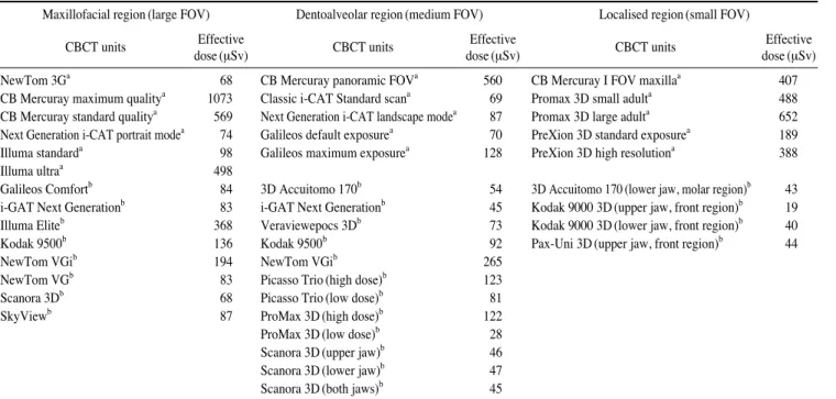
Although the phantoms and dosimeters employed in the two studies were different, from Table 1 we can still see that the effective dose is quite different from one CBCT unit to another, irrespective of the size of the field of view (FOV) used. The highest effective dose is 1073 μSv for CB MercuRay with a large FOV scanning for maxillofacial region, while the lowest effective dose is only 19 μSv for the Kodak 9000 3D with a scanning area of the front region of the upper jaw. This is a difference of almost 500 times between the highest and lowest effective doses.
When we look futher into the data in Table 1, we can see that the effective dose is closely related to the protocol used for scanning. Since a protocol is a combination of kVp, mAs, and voxel sizes and other factors, the effective dose is in reality closely related to the chosen exposure parameters. In the study performed by Ludlow et al, the effective dose for maximum quality CB MercuRay (1073 μSv) is almost twice that of standard quality CB MercuRay (569 μSv), the effective dose for CBCT unit Galileos ob- tained at the default exposure (70 μSv) is almost half of that obtained at maximum exposure (128 μSv), and the effective dose for standard and high resolution images from the PreXion 3D were 189 μSv and 388 μSv, respec- tively. The later data also indicate that with an increase in the spatial resolution, the effective dose is increased as well. This is also confirmed by the study conducted by Davies et al.16
Field of view (FOV) is another factor that plays an impor- tant role in the assessment of the effective dose of one CBCT examination. When the exposure parameters such as the kVp and mAs are maintained at the same level, the larger the FOV used, the higher the effective dose obtain- ed. This is substantiated by the effective doses for CB Mercuay in Table 1, where the effective dose is 1073 μSv for a large FOV with maximum quality, 560 μSv for a medium FOV, and 407 μSv for a small FOV. The expo- sure parameters for all of the three FOV examinations were kept at 120 kVp and 150 mAs. A study by Qu et al13 further discloses the positive relationship between the FOV and effective dose. In this study, 12 protocols that combined different patient size, FOV, kVp, mA, and expo- sure times were employed for the estimation of effective doses of the ProMax 3D CBCT unit. While holding all of the other exposure parameters constant, the researchers found that for a scanning area of full volume height with a full volume diameter (8 cm×8 cm), the effective dose (298 μSv) is much higher than the effective doses obtain- ed from used other scanning FOVs, specifically, half the volume height (upper jaw) with a full volume diameter (4 cm×8 cm, 131 μSv), half the volume height (lower jaw) with a full volume diameter (4 cm×8 cm, 171 μSv), a full volume height with half the volume diameter (anterior region, 8 cm×4 cm, 127 μSv), and a full volume height with half the volume diameter (posterior region, 8 cm×4
Table 1.Effective doses from different CBCT units
Maxillofacial region (large FOV) Dentoalveolar region (medium FOV) Localised region (small FOV)
CBCT units Effective CBCT units Effective CBCT units Effective
dose (μSv) dose (μSv) dose (μSv)
NewTom 3Ga 68 CB Mercuray panoramic FOVa 560 CB Mercuray I FOV maxillaa 407
CB Mercuray maximum qualitya 1073 Classic i-CAT Standard scana 69 Promax 3D small adulta 488 CB Mercuray standard qualitya 569 Next Generation i-CAT landscape modea 87 Promax 3D large adulta 652 Next Generation i-CAT portrait modea 74 Galileos default exposurea 70 PreXion 3D standard exposurea 189
Illuma standarda 98 Galileos maximum exposurea 128 PreXion 3D high resolutiona 388
Illuma ultraa 498
Galileos Comfortb 84 3D Accuitomo 170b 54 3D Accuitomo 170 (lower jaw, molar region)b 43 i-GAT Next Generationb 83 i-GAT Next Generationb 45 Kodak 9000 3D (upper jaw, front region)b 19
Illuma Eliteb 368 Veraviewepocs 3Db 73 Kodak 9000 3D (lower jaw, front region)b 40
Kodak 9500b 136 Kodak 9500b 92 Pax-Uni 3D (upper jaw, front region)b 44
NewTom VGib 194 NewTom VGib 265
NewTom VGb 83 Picasso Trio (high dose)b 123
Scanora 3Db 68 Picasso Trio (low dose)b 81
SkyViewb 87 ProMax 3D (high dose)b 122
ProMax 3D (low dose)b 28
Scanora 3D (upper jaw)b 46
Scanora 3D (lower jaw)b 47
Scanora 3D (both jaws)b 45
aData from the study by Ludlow et al (2008), bData from the study by Pauwels et al (2012)
cm, 197 μSv).
The above demonstrates that the effective dose is differ- ent from one CBCT unit to another and closely related to the exposure parameters used for scanning; for a given model of a CBCT unit, the larger the FOV used for scann- ning, the higher the effective dose derived when all the other exposure parameters are kept at the same level. Sim- ilarly, the higher the spatial resolution chosen for scanning, the higher the effective dose is.
Effective dose of CBCT and conventional dental radiography
There are few studies focusing on the direct comparison
of the effective doses obtained from CBCT and conven- tional dental radiography. The results from the direct com- parison studies were summarized in Table 2, where the effective dose for panoramic radiography is about 22.0 μSv, for lateral cephalometric examination about 4.5 μSv and for CBCT examnation the effective dose is 61-134 μSv.
No study has performed a direct comparison of the effec- tive dose from intraoral and CBCT examinations. In the guidelines19provided by the European Academy of Dento- Maxillofacial Radiology, the suggested effective dose of one intraoral radiograph is 1.5 μSv. Other studies20-26that exclusively estimated the effective dose of conventional dental radiography have demonstrated that the range of the effective dose for a panoramic radiograph is 3.85-38.0
Table 3. Effective doses from panoramic radiography
Authors Panoramic machine Exposure parameters Effective dose (μSv)
Danforth et al20 Planmeca PM 2002 60 kVp, 4 mA, 18 s 3.85a
Gijbels et al21 Cranex tone, SPP 70 kVp, 4 mA, 15 s 8.1a
Cranex Excel, CCD 65 kVp, 6 mA, 19 s 12.3a
Veraviewepocs 5D, CCD 70 kVp, 4 mA, 8.2 s 5.5a
EC Proline, CCD 64 kVp, 7 mA, 18.3 s 14.9a
Orthoralix 9200 DDE, CCD 74 kVp, 4 mA, 12 s 4.7a
Ludlow et al1 Sirona Orthophos Plus DS, CCD 66 kVp, 16 mA, 14.1 s 22a
Gavala et al22 Planmeca Promax, film 66 kVp, 6 mA, 16 s 26a
Planmeca PM 2002, CCD 66 kVp, 8 mA, 18 s 38a
Planmeca PM 2002, CCD 60 kVp, 4 mA, 18 s 12a
Ludlow et al23 Orthophos XG, CCD 64 kV, 8 mA, 14.1 s 14.2b
ProMax, CCD 68 kV, 13 mA, 16 s 24.3b
a: ICRP60 1990, b: ICRP103 2007, CCD: charge-coupled device, SPP: storage phosphor plate
Table 4.Effective doses from lateral cephalometric radiography
Authors Instrument Exposure parameters Effective doses (μSv)
Visser et al24 Siemens Orthophos C, film 77 kV, 14 mA, 0.5 s 2.3a
Siemens Orthophos DS Ceph, CCD 73 kV, 15 mA, 15.8 s 1.1a
Gijbels et al25 Cranex Tome, SPP 70 kV, 4 mAs 2.2a
Proline Ceph CM, CCD 70 kV, 10 mA, 23 s 3.4a
Ludlow et al23 unknown 77 kVp, 6.5 mAs 5.6b
a: ICRP60 1990, b: ICRP103 2007, CCD: charge-coupled device, SPP: storage phosphor plate
Table 2.Comparison of effective dose (μSv) of CBCT, panoramic and later cephalometric (ceph.) radiography
Authors
Panoramic Lateral ceph. Panoramic CBCT
radiography ++lateral ceph.
OP-100 Orthophos OC-100 Orthophos i-CAT 0.3 voxel i-CAT 0.2 voxel NewTom i-CAT
Plus DS DS landscape landscape 9000
Grünheid et al17 21.5 4.5 65 134.2
Ludlow et al13 22 77.9
Silva et al18 10.4 56.2 61.1
μSv (Table 3), for a lateral cephalometric examination is 1.1-5.6 μSv (Table 4), for posteroanterior cephalometric radiograph, 5.1 μSv, and for one introal examination, 0.65- 9.5 μSv (Table 5).
These data indicate that the effective dose of CBCT is several to hundreds of times higher than the effective dose from a conventional dental radiographic examination.
Effective dose of CBCT and helical CT
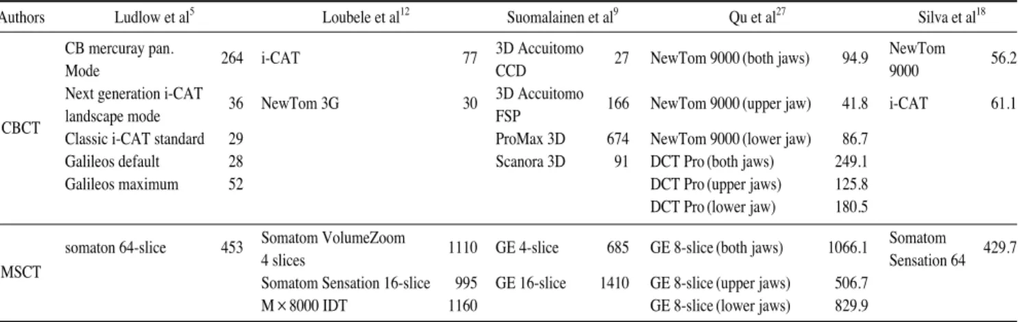
More attention is paid to the effective dose of CBCT and multislice CT (MSCT) since both techniques provide three dimensional images. The effective doses from the litera- ture on CBCT and MSCT are shown in Table 6. Generally, the effective dose of MSCT is much higher than that of CBCT. However, in some of the studies, the scanning area, i.e. the FOV was not well defined. To avoid the effect of the FOV on the assessment of effective dose, Qu et al13 strictly defined the scanning area for both MSCT and CBCT examinations in their study. The results showed that the effective doses of MSCTs are about several to ten
times higher than those of CBCTs. For example, when scaning both the maxilla and mandible, the effective dose is about 94.9 μSv for CBCT NewTom 9000, 249.1 μSv for CBCT DCT-Pro, and 1066.1 for GE 8-slice MSCT.
Similar results were also observerd in other studies, as shown in Table 6.
However, it should be borne in mind that although the effective dose of MSCT is much higher than that of CBCT, the image qualities for the two techniques are quite differ- ent. For hard tissue, such as bone and tooth, the image quality of CBCT is equal to or better than the image qua- lity of MSCT, but for soft tissues, the image from CBCT is not satisfactory due to the inherent drawbacks of the technique.
Patient radiation protection from CBCT To perform one medical X-ray examination, three main factors must be taken into account: the X-ray unit, patient for examination, and receptor used for capturing the image
Table 5.Effective doses from intra-oral examinations
Authors Exposure parameters Total (average) effective dose (μSv)
Gibbs26 70 kVp, short cone bisecting angle, round collimation, 18 E-speed films 100 (5.6)a 70 kVp, long cone parallel, round collimation, 21 E-speed films 74 (3.5)a 70 kVp, long cone parallel, rectangular collimation, 21 E-speed films 14 (0.67)a 70 kVp, short cone bisecting angle, round collimation, 4 E-speed bitewings 14 (3.5)a 70 kVp, long cone parallel, round collimation, 4 E-speed bitewings 12 (3)a 70 kVp, long cone parallel, rectangular collimation, 4 E-speed bitewings 2.6 (0.65)a Ludlow et al23 70 kVp, 8 mA, round collimation, 18 D-speed films 388 (21.6)b 70 kVp, 8 mA, round collimation, 18 SPP or F-speed films 170.7 (9.5)b 70 kVp, 8 mA, rectangular collimation, 18 SPP or F-speed films 34.9 (1.9)b 70 kVp, 8 mA, rectangular collimation, 4 SPP or F-speed bitewings 5.0 (1.25)b
a: ICRP60 1990, b: ICRP103 2007, SPP: storage phosphor plate
Table 6.Effective dose (μSv) of CBCT and MSCT from literatures
Authors Ludlow et al5 Loubele et al12 Suomalainen et al9 Qu et al27 Silva et al18
CB mercuray pan. 264 i-CAT 77 3D Accuitomo 27 NewTom 9000 (both jaws) 94.9 NewTom 56.2
Mode CCD 9000
Next generation i-CAT 36 NewTom 3G 30 3D Accuitomo 166 NewTom 9000 (upper jaw) 41.8 i-CAT 61.1
CBCT landscape mode FSP
Classic i-CAT standard 29 ProMax 3D 674 NewTom 9000 (lower jaw) 86.7
Galileos default 28 Scanora 3D 91 DCT Pro (both jaws) 249.1
Galileos maximum 52 DCT Pro (upper jaws) 125.8
DCT Pro (lower jaw) 180.5
somaton 64-slice 453 Somatom VolumeZoom 1110 GE 4-slice 685 GE 8-slice (both jaws) 1066.1 Somatom 429.7
MSCT 4 slices Sensation 64
Somatom Sensation 16-slice 995 GE 16-slice 1410 GE 8-slice (upper jaws) 506.7
M×8000 IDT 1160 GE 8-slice (lower jaws) 829.9
MSCT: multi-slice CT
of the patient. Therefore, when an X-ray examination is indicated for a patient, the patient dose can be reduced by the reduction of the X-ray intensity emitted from the employed x-ray unit, increasing of the imaging receptor capturing speed and collimation, or shielding of the x-ray beam to the patient. This section will only focus on the shielding devices for the reduction of the radiation dose.
The shielding devices include a leaded thyroid collar for the protection of the thyroid gland, leaded glasses for the protection of the eye lens, a leaded hat for the protection of the brain, and a leaded apron for the protection of the body trunk. It is well known that a thyroid collar is effec- tive for the protection of the thyroid gland in an intraoral examination. However, for a CBCT examination, is it still effective when the X-ray unit rotates around the patient?
With this question in mind, two studies were conducted.
One study was mainly aimed to identify the effectiveness of a thyroid collar on the dose redution of the thyroid gland.28 In this study, five conditions were tested as follows: 1) without a collar around the neck; 2) with one collar loosely on the front of the neck; 3) with two collars loosely on the front and back of the neck; 4) with one collar tightly on the front of the neck; and 5) with two collars tightly on the front and back of the neck. The results showed that when the thyroid collars were used loosely around the neck, no effective organ dose reduction was observed. When one thyroid collar was used tightly on the frontof the neck, the effective organ dose to the thyroid gland and esophagus were reduced to 15.9 μSv (48.7% reduction) and 1.4 μSv (41.7% reduction), respectively. A similar organ dose reduction (46.5% and 41.7%) was achieved when CBCT scanning was performed with two collars tightly affixed to the front and back of the neck. The study supported the use of a thyroid collar during a CBCT scan. In a subsequent study, different oral and maxillofacial regions were scann- ed with the phantom tightly wearing one or two thyroid collars.29The results also supported the use of thyroid col- lars (61% thyroid dose reduction for a large view examina- tion, 72% thyroid dose reduction for a medium FOV, and 70% thyroid dose reduction for a small FOV) and further disclosed that the total effective dose for medium and small FOV examinations were also significantly reduced by the use of a thyroid collar.
The use of leaded glasses during a CBCT examinaiton was also investigated.30In the study peformed by Prins et al, three phantoms representing an adult male, an adult female, and a child were employed. The results showed that the radiation dose to the eye lens could be reduced by over 60% without having a deleterious effect on the image qual-
ity in the area of clinical significance for dental imaging.
Considering the above, one conclusion that could be drawn was that a thyroid collar and leaded glasses should be used during a CBCT examination, given that diagnostic information and image quality are not reduced.
Summary
The effective dose of CBCT, conventional dental radio- graphy, and multislice CT and the effect of a thyroid collar and leaded glasses on the dose reduction was presented in this paper. Based on the above analysis, we can conclude the following:
1. The patient radiation dose is much lower for CBCT than for helical CT;
2. The patient radiation dose is closely related to the FOV and exposure parameters used for a CBCT examination.
Without alteration of any other exposure parameters, the larger the FOV used for scanning, the higher the radiation dose is;
3. Compared with conventional dental radiography, the effective dose of CBCT is several to hundreds of times higher;
4. To reduce the patient dose to the greatest possible extent, the chosen CBCT scanning protocol should be in accor- dance with the dignostic task at hand;
5. A thyroid collar should be used for CBCT scanning;
wearing leaded glasses is recommended when it does not detract from imaging quality.
References
1. Ludlow JB, Davies-Ludlow LE, Brooks SL. Dosimetry of two extraoral direct digital imaging devices: NewTom cone beam CT and Orthophos Plus DS panoramic unit. Dentomaxillofac Radiol 2003; 32: 229-34.
2. Schulze D, Heiland M, Thurmann H, Adam G. Radiation expo- sure during midfacial imaging using 4- and 16-slice computed tomography, cone beam computed tomography systems and conventional radiography. Dentomaxillofac Radiol 2004; 33:
83-6.
3. Tsiklakis K, Donta C, Gavala S, Karayianni K, Kamenopoulou V, Hourdakis CJ. Dose reduction in maxillofacial imaging using low dose Cone Beam CT. Eur J Radiol 2005; 56: 413-7.
4. Ludlow JB, Davies-Ludlow LE, Brooks SL, Howerton WB.
Dosimetry of 3 CBCT devices for oral and maxillofacial radio- logy: CB Mercuray, NewTom 3G and i-CAT. Dentomaxillofac Radiol 2006; 35: 219-26.
5. Ludlow JB, Ivanovic M. Comparative dosimetry of dental CBCT devices and 64-slice CT for oral and maxillofacial radiology. Oral Surg Oral Med Oral Pathol Oral Radiol Endod 2008; 106: 106-14.
6. Palomo JM, Rao PS, Hans MG. Influence of CBCT exposure conditions on radiation dose. Oral Surg Oral Med Oral Pathol Oral Radiol Endod 2008; 105: 773-82.
7. Lofthag-Hansen S, Thilander-Klang A, Ekestubbe A, Helmrot E, Grondahl K. Calculating effective dose on a cone beam computed tomography device: 3D Accuitomo and 3D Accui- tomo FPD. Dentomaxillofac Radiol 2008; 37: 72-9.
8. Hirsch E, Wolf U, Heinicke F, Silva MA. Dosimetry of the cone beam computed tomography Veraviewepocs 3D compared with the 3D Accuitomo in different fields of view. Dentomax- illofac Radiol 2008; 37: 268-73.
9. Suomalainen A, Kiljunen T, Kaser Y, Peltola J, Kortesniemi M. Dosimetry and image quality of four dental cone beam com- puted tomography scanners compared with multislice computed tomography scanners. Dentomaxillofac Radiol 2009; 38: 367- 78.
10. Chau AC, Fung K. Comparison of radiation dose for implant imaging using conventional spiral tomography, computed tomography, and cone-beam computed tomography. Oral Surg Oral Med Oral Pathol Oral Radiol Endod 2009; 107: 559-65.
11. Roberts JA, Drage NA, Davies J, Thomas DW. Effective dose from cone beam CT examinations in dentistry. Br J Radiol 2009; 82: 35-40.
12. Loubele M, Bogaerts R, Van Dijck E, Pauwels R, Vanheusden S, Suetens P, et al. Comparison between effective radiation dose of CBCT and MSCT scanners for dentomaxillofacial applications. Eur J Radiol 2009; 71: 461-8.
13. Qu XM, Li G, Ludlow JB, Zhang ZY, Ma XC. Effective radia- tion dose of ProMax 3D cone-beam computerized tomography scanner with different dental protocols. Oral Surg Oral Med Oral Pathol Oral Radiol Endod 2010; 110: 770-6.
14. Pauwels R, Beinsberger J, Collaert B, Theodorakou C, Rogers J, Walker A, et al. Effective dose range for dental cone beam computed tomography scanners. Eur J Radiol 2012; 81: 267- 71.
15. Thilander-Klang A, Helmrot E. Methods of determining the effective in dental radiology. Radiat Prot Dosimetry 2010;
139: 306-9.
16. Davies J, Johnson B, Drage NA. Effective doses from cone beam CT investigation of the jaws. Dentomaxillofac Radiol 2012; 41: 30-6.
17. Grünheid T, Kolbeck Schieck JR, Pliska BT, Ahmad M, Lar- son BE. Dosimetry of a cone-beam computed tomography machine compared with a digital x-ray machine in orthodontic imaging. Am J Orthod Dentofacial Orthop 2012; 141: 436-43.
18. Silva MA, Wolf U, Heinicke F, Bumann A, Visser H, Hirsch E. Cone-beam computed tomography for routine orthodontic treatment planning: a radiation dose evaluation. Am J Orthod
Dentofacial Orthop 2008; 133: 640.e1-5.
19. SEDENTEXCT Guideline Development Panel. Radiation pro- tection No 172. Cone beam CT for dental and maxillofacial radiology. Evidence based guidelines. Luxembourg: European Comminssion Directorate-General for Energy; 2012.
20. Danforth RA, Clark DE. Effective dose from radiation absorb- ed during a panoramic examination with a new generation machine. Oral Surg Oral Med Oral Pathol Oral Radiol Endod 2000; 89: 236-43.
21. Gijbels F, Jacobs R, Bogaerts R, Debaveye D, Verlinden S, Sanderink G. Dosimetry of digital panoramic imaging. Part I:
Patient exposure. Dentomaxillofac Radiol 2005; 34: 145-9.
22. Gavala S, Donta C, Tsiklakis K, Boziari A, Kamenopoulou V, Stamatakis HC. Radiation dose reduction in direct digital pan- oramic radiography. Eur J Radiol 2009; 71: 42-8.
23. Ludlow JB, Davies-Ludlow LE, White SC. Patient risk related to common dental radiographic examinations: the impact of 2007 International Commission on Radiological Protection recommendations regarding dose calculation. J Am Dent Assoc 2008; 139: 1237-43.
24. Visser H, Rödig T, Hermann KP. Dose reduction by direct- digital cephalometric radiography. Angle Orthod 2001; 71:
159-63.
25. Gijbels F, Sanderink G, Wyatt J, Van Dam J, Nowak B, Jacobs R. Radiation doses of indirect and direct digital cephalometric radiography. Br Dent J 2004; 197: 149-52.
26. Gibbs SJ. Effective dose equivalent and effective dose: com- parison for common projections in oral and maxillofacial radio- logy. Oral Surg Oral Med Oral Pathol Oral Radiol Endod 2000;
90: 538-45.
27. Qu XM, Li G, Zhang ZY, Ma XC. Comparative dosimetry of dental cone-beam computed tomography and multi-slice com- puted tomography for oral and maxillofacial radiology. Zhong- hua Kou Qiang Yi Xue Za Zhi 2011; 46: 595-9.
28. Qu XM, Li G, Sanderink GC, Zhang ZY, Ma XC. Dose reduc- tion of cone beam CT scanning for the entire oral and maxil- lofacial regions with thyroid collars. Dentomaxillofac Radiol 2012: 41: 373-8.
29. Qu X, Li G, Zhang Z, Ma X. Thyroid shields for radiation dose reduction during cone beam computed tomography scan- ning for different oral and maxillofacial regions. Eur J Radiol 2012; 81: e376-80.
30. Prins R, Dauer LT, Colosi DC, Quinn B, Kleiman NJ, Bohle GC, et al. Significant reduction in dental cone beam computed tomography (CBCT) eye dose through the use of leaded glasses.
Oral Surg Oral Med Oral Pathol Oral Radiol Endod 2011; 112:
502-7.