ABSTRACT
Purpose: The laparoscopic transhiatal approach (LA) for adenocarcinoma of the
esophagogastric junction (AEJ) is advantageous since it allows better visualization of the surgical field than the open approach (OA). We compared the surgical outcomes of the 2 approaches.
Materials and Methods: We analyzed 108 patients with AEJ who underwent transhiatal distal esophagectomy and gastrectomy with curative intent between 2003 and 2015. Surgical outcomes were reviewed using electronic medical records.
Results: The LA and OA were performed in 37 and 71 patients, respectively. Compared to the OA, the LA was associated with significantly shorter duration of postoperative hospital stay (9 vs. 11 days, P=0.001), shorter proximal resection margins (3 vs. 7 mm, P=0.004), and extended operative times (240 vs. 191 min, P=0.001). No significant difference was observed between the LA and OA for intraoperative blood loss (100 vs. 100 mL, P=0.392) or surgical morbidity rate (grade≥II) for complications (8.1% vs. 23.9%, P=0.080). Two cases of anastomotic leakage occurred in the OA group. The number of harvested lymph nodes was not significantly different between the LA and OA groups (54 vs. 51, P=0.889). The 5-year overall and 3-year relapse-free survival rates were 81.8% and 50.7% (P=0.024) and 77.3% and 46.4% (P=0.009) for the LA and OA groups, respectively. Multivariable analyses revealed no independent factors associated with overall survival.
Conclusions: The LA is feasible and safe with short- and long-term oncologic outcomes similar to those of the OA.
Keywords: Stomach neoplasms; Esophagogastric junction; Laparoscopy; Gastrectomy
INTRODUCTION
The incidence of adenocarcinoma of the esophagogastric junction (AEJ) has been increasing in Western countries [1]. In the East, AEJ has historically been relatively rare; however, an increasing incidence has recently been reported because of increasingly westernized dietary intake and decreased Helicobacter pylori infection [2-4]. A retrospective analysis showed that AEJ in Western countries was diagnosed at a more advanced stage and had a poorer prognosis compared with that in the East [2]. Furthermore, most cases of AEJ in Eastern countries are Siewert types II and III [5,6].
Original Article
Received: Oct 23, 2018 Revised: Jan 8, 2019 Accepted: Jan 10, 2019 Correspondence to Hyung-Ho Kim
Department of Surgery, Seoul National University Bundang Hospital, 166 Gumi-ro, Bundang-gu, Seongnam 13620, Korea.
E-mail: [email protected]
*Present address: Department of Surgery, Korea University Ansan Hospital, Ansan, Korea.
Copyright © 2019. Korean Gastric Cancer Association
This is an Open Access article distributed under the terms of the Creative Commons Attribution Non-Commercial License (https://
creativecommons.org/licenses/by-nc/4.0) which permits unrestricted noncommercial use, distribution, and reproduction in any medium, provided the original work is properly cited.
ORCID iDs Yoontaek Lee
https://orcid.org/0000-0003-2643-2007 Ki Bum Park
https://orcid.org/0000-0001-5404-5667 Young Suk Park
https://orcid.org/0000-0002-6352-9759 Sang-Hoon Ahn
https://orcid.org/0000-0001-8827-3625 Do Joong Park
https://orcid.org/0000-0001-9644-6127 Hyung-Ho Kim
https://orcid.org/0000-0002-8916-0048
Yoontaek Lee *, Sa-Hong Min, Ki Bum Park , Young Suk Park , Sang-Hoon Ahn , Do Joong Park , Hyung-Ho Kim
Department of Surgery, Seoul National University Bundang Hospital, Seongnam, Korea
Long-term Outcomes of Laparoscopic
Versus Open Transhiatal Approach
for the Treatment of Esophagogastric
Junction Cancer
Author Contributions
Conceptualization: K.H.H.; Data curation: L.Y.;
Formal analysis: L.Y.; Investigation: M.S.H.;
Methodology: P.Y.S.; Project administration:
A.S.H.; Resources: P.D.J.; Software: P.K.B.;
Supervision: K.H.H.; Writing - original draft:
L.Y.; Writing - review & editing: K.H.H.
Conflict of Interest
No potential conflict of interest relevant to this article was reported.
The thoracoabdominal approach is generally accepted in cases of Siewert type I AEJ, and the transhiatal approach is proposed as the method of choice for patients with Siewert type III tumors. However, there is controversy regarding whether the thoracoabdominal approach is superior to the transhiatal approach for the treatment of patients with Siewert type II cancer. The transhiatal approach, popularized by Orringer [7], has advantages in terms of pain and pulmonary complications compared with the thoracoabdominal approach. In contrast, the thoracoabdominal approach has the advantage of direct exposure of the lower mediastinal lymph nodes. In Japan, the JCOG 9502 trial, which compared the left-sided thoracoabdominal and transhiatal approaches for patients with gastric cancer or AEJ with esophageal invasion of 3 cm or less, showed that the left-sided thoracoabdominal approach did not increase survival and had higher surgical morbidity despite the better view of the lower mediastinal area obtained using this approach [8].
Despite the increasing popularity of laparoscopic distal gastrectomy, laparoscopic procedures are not yet widely accepted in total gastrectomy or proximal gastrectomy owing to their technical difficulty. However, laparoscopic surgery has been performed for AEJ with the accumulation of experience in certain specialized institutions. The theoretical advantage of the laparoscopic transhiatal approach (LA) for AEJ is that it provides a better surgical view compared to the open approach (OA). However, there are currently no randomized controlled trials comparing the laparoscopic approach with the open transhiatal approach, and few reports have focused on outcomes of the LA for AEJ. In the present study, we focused on the short- and long-term outcomes of the LA compared with the OA.
MATERIALS AND METHODS
Patients
The review of the electronic medical records from the Seoul National University Bundang Hospital for the period 2003–2015 allowed identification of 121 patients with AEJ who underwent transhiatal distal esophagectomy and gastrectomy with curative intent. We excluded 13 patients who had other malignancies or could not undergo R0 resection. The final analysis thus included 108 patients.
Surgical procedure
All patients underwent total gastrectomy with lymph node dissection, with the exception of 1 patient who underwent proximal gastrectomy. The patient with early gastric cancer underwent D1+ lymph node dissection, and those with advanced gastric cancer underwent D2 lymph node dissection based on the Japanese gastric cancer treatment guidelines [9].
In the LA, a 3- or 2-dimensional, flexible, high-definition laparoscope and an ultrasonic- activated device were used. Roux-en-Y esophagojejunostomy and jejunojejunostomy
reconstruction were performed. The lower esophagus was divided using a laparoscopic purse- string clamp (Lap-jack; Eterne, Seongnam, Korea). The trocar incision in the left lower area was extended 4 to 5 cm transversely to retrieve the specimen and perform reestablishment of bowel continuity, extracorporeal jejunojejunostomy, and intracorporeal end-to-side esophagojejunostomy with a single-stapling technique using a circular stapler. In 4 cases, a transorally inserted anvil (OrVil™; Covidien Ltd., Mansfield, MA, USA) was used to achieve an adequate proximal resection margin. The lower esophagus was divided using a linear stapler, and a small hole was made in the corner of the staple line for passage of an OrVil™ connecting
tube. Intracorporeal end-to-side esophagojejunostomy was accomplished using the hemi- double-stapling technique to prevent anastomotic stricture by the OrVil™ system.
In the OA, an upper midline incision was made from the xiphoid process to the umbilicus, and the open procedure was performed in the usual manner.
Postoperative evaluation
Based on pathologic reports, the tumors were classified according to the Siewert system, as follows: type I, 1 to 5 cm above the esophagogastric junction (EGJ); type II, 1 cm above to 2 cm below the EGJ; and type III, 2 to 5 cm below the EGJ. TNM staging was performed according to the guidelines of the American Joint Committee on Cancer classification, 7th edition. Postoperative complications, including wound infection, leakage, and intestinal obstruction, occurring within 30 days of surgery were evaluated based on the Clavien-Dindo classification. For analysis based on the World Health Organization classification, papillary, well-differentiated, and moderately differentiated types were classified as the differentiated group, and poorly differentiated, mucinous, and poorly cohesive types were classified as the undifferentiated group. Overall survival (OS) was calculated from surgery to death, and relapse-free survival (RFS) was defined as the time from surgery to the first recurrence or death from the same cancer and all treatment-related deaths.
Statistical analysis
The baseline characteristics of each group were compared using the χ2 or Fisher's exact test. The Kaplan-Meier method was used to estimate the survival rate, and differences between survival curves were analyzed using the log-rank test. To assess the confounding factors for survival, a multivariable Cox proportional hazards model was applied by incorporating the prognostic factors identified in the univariable log-rank test. All variables with P<0.2 in the univariable analysis were included in the multivariable analysis. A P-value threshold of 0.05 was considered statistically significant. All statistical analyses were performed with R software (R Foundation for Statistical Computing, Vienna, Austria; http://cran.r-project.org/).
Ethics statement
The Institutional Review Board of the Seoul National University Bundang Hospital (B- 1808/484-117) approved the present study. All procedures followed were in accordance with the ethical standards of the responsible committees on human experimentation (institutional and national) and with the Helsinki Declaration of 1964 and later versions. This retrospective study was waived patient's informed consent.
RESULTS
Patient characteristics
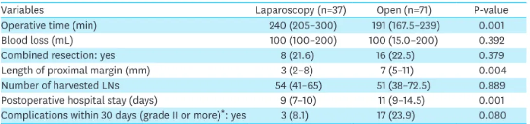
We included 108 patients with AEJ who underwent transhiatal distal esophagectomy and gastrectomy with curative intent. The clinicopathological characteristics of the patients are shown in Table 1, and the surgical outcomes are shown in Table 2. The LA and OA were utilized in 37 and 71 patients, respectively. Compared with the OA, the LA was associated with significantly shorter postoperative hospital stay (9 vs. 11 days, P=0.001), shorter proximal resection margins (3 vs. 7 mm, P=0.004), and extended operative times (240 vs. 191 min, P=0.001). No significant difference was found between the LA and OA in intraoperative blood loss (100 vs. 100 mL, P=0.392) or total number of harvested lymph nodes (54 vs. 51,
P=0.889). Although there was a tendency for pathologic stage progression in the OA group, no significant difference was found between the 2 groups in stage or Siewert type (P=0.100 and 0.991, respectively).
Postoperative complications
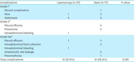
The rate of surgical morbidity (grade≥II) for complications was not significantly different (8.1 vs. 23.9%, P=0.080) between the 2 groups. Table 3 shows the details of postoperative complications within 30 days in each group based on the Clavien-Dindo classification. There were 2 cases of anastomotic leakage in the OA group, but none in the LA group. Both patients recovered after percutaneous catheter drainage under radiologic intervention.
Table 1. Clinicopathogical patient characteristics
Variables Laparoscopy (n=37) Open (n=71) P-value
Age (yr) 0.474
<70 25 (67.6) 54 (76.4)
≥70 12 (32.4) 17 (23.9)
Sex 0.720
Female 11 (29.7) 25 (35.2)
Male 26 (70.3) 46 (64.8)
Body mass index (kg/m2) 22.0 (2.8) 22.0 (2.8) 0.953
ASA performance status 0.163
1 18 (48.6) 22 (31.0)
2 18 (48.6) 44 (62.0)
3 1 (2.7) 5 (7.0)
Tumor size 0.057
pT stage 0.059
T1 8 (21.6) 4 (5.6)
T2 3 (8.1) 4 (5.6)
T3 14 (37.8) 28 (39.4)
T4 12 (32.4) 35 (49.3)
pN stage 0.085
N0 9 (24.3) 7 (9.9)
N+ 28 (75.7) 64 (90.1)
pTNM stage 0.100
I 5 (13.5) 4 (5.6)
II 16 (43.2) 22 (31.0)
III 16 (43.2) 45 (63.4)
Siewert classification 0.991
Type II 18 (48.6) 33 (46.5)
Type III 19 (51.4) 38 (53.5)
Histologic type 1.000
Differentiated 16 (43.2) 31 (43.7)
Undifferentiated 21 (56.8) 40 (56.3)
Data shown are number (%), mean (SD), or median (IQR).
ASA = American Society of Anesthesiologists; SD = standard deviation; IQR = interquartile range.
Table 2. Surgical outcomes
Variables Laparoscopy (n=37) Open (n=71) P-value
Operative time (min) 240 (205–300) 191 (167.5–239) 0.001
Blood loss (mL) 100 (100–200) 100 (15.0–200) 0.392
Combined resection: yes 8 (21.6) 16 (22.5) 0.379
Length of proximal margin (mm) 3 (2–8) 7 (5–11) 0.004
Number of harvested LNs 54 (41–65) 51 (38–72.5) 0.889
Postoperative hospital stay (days) 9 (7–10) 11 (9–14.5) 0.001
Complications within 30 days (grade II or more)*: yes 3 (8.1) 17 (23.9) 0.080 Data shown are number (%), mean (SD), or median (IQR).
LNs = lymph nodes; SD = standard deviation; IQR = interquartile range.
*According to the Clavien-Dindo grading system.
Survival outcomes
The median follow-up duration for all 108 patients was 34.5 months (range, 3–162 months). The 5-year OS rates were 81.8% and 50.7% for the LA and OA, respectively (P=0.024) (Fig. 1). The 3-year RFS rates were 77.3% and 46.4% for the LA and OA, respectively (P=0.009) (Fig. 2). In subgroup analysis of patients with stage III tumors, there were no significant differences in OS and RFS between the LA and OA groups (P=0.490 and 0.366, respectively) (Fig. 3). Regarding patients with stage I or II tumors, we could not analyze survival rates because there were no death or recurrence events in the LA group.
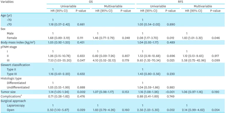
The factors associated with OS and RFS are listed in Table 4. In a univariable analysis for OS, patients with pathologic stage III and larger tumors, and those in the OA group, showed worse survival rates (hazard ratio [HR], 7.53; 95% confidence interval [CI], 1.03–55.20;
P=0.047; HR, 1.14; 95% CI, 1.05–1.24; P=0.002; and HR, 2.50; 95% CI, 1.10–5.67; P=0.029;
respectively). Multivariable analyses identified no independent prognostic factors for OS.
Table 3. Postoperative complications within 30 days
Complications Laparoscopy (n=37) Open (n=71) P-value
Grade I*
Wound complication 1
Ileus 1 3
Atelectasis 4 2
Grade II*
Pleural effusion 4
Pneumonia 2
Intraabdominal bleeding 1
Grade IIIa*
Pleural effusion 1 3
Intraabdominal fluid collection 3
Intraabdominal bleeding 1
Anastomotic site leakage 2
Pneumothorax 1
Total complications 21 (21.6%) 21 (29.6%) 0.199
*According to the Clavien-Dindo grading system.
No. at risk
Laparoscopy 37 34 28 20 15 8 5 2 2 0 0 0 0 0
Open 71 67 48 31 24 18 13 10 5 5 4 2 2 1
Time (mo) 0.75
0 24
Survival probability
1.00
0.50
0.25
12 36 48 60 72 84 96 108 120 132 144 156
Laparoscopy P=0.024
Open
Fig. 1. OS rates according to the surgical approach. The 5-year OS rates were 81.8% for the laparoscopic transhiatal approach and 50.7% for the open approach (P=0.024).
OS = overall survival.
In a univariable analysis for RFS, female patients and patients with pathologic stage III and larger tumors showed worse survival rates (HR, 2.08; 95% CI, 1.17–3.70; P=0.012; HR, 9.65; 95% CI, 1.32–70.34; P=0.025; HR, 1.16; 95% CI, 1.08–1.26; P<0.001, respectively).
Multivariable analyses revealed that female sex was the only independent prognostic factor for RFS (HR, 1.83; 95% CI, 1.01–3.32; P=0.046).
The number of patients with recurrence was 8 (21.6%) in the LA group and 29 (40.8%) in the OA group. The most common recurrence site was the peritoneum in both groups (Table 5).
The pattern of recurrence was similar in the 2 groups.
No. at risk
Laparoscopy 37 33 26 19 15 7 4 2 2 0 0 0 0 0
Open 71 55 36 26 21 17 13 10 5 4 4 2 2 1
Time (mo) 0.75
0 24
Survival probability
1.00
0.50
0.25
12 36 48 60 72 84 96 108 120 132 144 156
Laparoscopy P=0.009
Open
Fig. 2. RFS rate according to the surgical approach. The 3-year RFS rates were 77.3% for the laparoscopic transhiatal approach and 56.1% for the open approach (P=0.009).
RFS = relapse-free survival.
No. at risk
Laparoscopy 16 14 10 5 3 2 1 0 0 0 0
Open 45 41 25 13 11 10 6 4 2 2 1
Time (mo) A
0 24
Survival probability
12 36 48 60 72 84 96 108 120
Laparoscopy Open 0.75
1.00
0.50
0.25 P=0.490
No. at risk
Laparoscopy 16 13 8 4 3 1 0 0 0 0 0
Open 45 30 17 11 9 9 6 4 2 1 1
Time (mo) B
0 24
Survival probability
12 36 48 60 72 84 96 108 120
Laparoscopy Open 0.75
1.00
0.50
0.25 P=0.366
Fig. 3. OS and RFS rates for stage III patients. OS and RFS rates for stage III patients were not significantly different between the laparoscopic transhiatal approach and open approach groups (P=0.49 and 0.366, respectively). (A) Overall survival, (B) relapse-free survival.
OS = overall survival; RFS = relapse-free survival.
DISCUSSION
The present study was conducted to evaluate the feasibility and safety of the LA for type II and III AEJ. Since the Japan Clinical Oncology Group (JCOG9502) revealed the advantage of the transhiatal approach compared to the thoracoabdominal approach for the treatment of type II and III AEJ, the transhiatal approach has been considered the optimal procedure. Considering that lower mediastinal lymph nodes might be difficult to visualize in the transhiatal approach, a laparoscopic view can overcome this drawback of the transhiatal approach.
In our institution, the indication for the laparoscopic procedure was initially confined to distal gastrectomy for clinical T1-T2 stage cancer without suspected lymph node metastasis.
Based on experience, however, the indications were gradually extended to total gastrectomy for clinical T4 stage cancer. Consequently, the first LA for AEJ was performed in 2007.
Table 4. Univariable and multivariable analysis of prognostic factors for OS and RFS
Variables OS RFS
Univariable Multivariable Univariable Multivariable
HR (95% CI) P-value HR (95% CI) P-value HR (95% CI) P-value HR (95% CI) P-value Age (yr)
<70 1 1
≥70 1.18 (0.57–2.42) 0.661 1.05 (0.54–2.02) 0.890
Sex
Male 1 1 1 1
Female 1.68 (0.89–3.19) 0.111 1.46 (0.77–2.79) 0.248 2.08 (1.17–3.70) 0.012 1.83 (1.01–3.32) 0.046
Body mass index (kg/m2) 1.05 (0.92–1.20) 0.451 1.04 (0.93–1.17) 0.489
pTNM stage
I 1 1 1 1
II 1.26 (0.15–10.78) 0.833 0.82 (0.09–7.26) 0.857 1.53 (0.18–12.68) 0.696 1.12 (0.13–9.65) 0.917
III 7.53 (1.03–55.20) 0.047 4.10 (0.52–32.13) 0.179 9.65 (1.32–70.34) 0.025 5.58 (0.72–42.96) 0.099 Siewert classification
Type II 1 1
Type III 1.16 (0.61–2.20) 0.652 1.43 (0.80–2.56) 0.230
Histologic type
Differentiated 1 1
Undifferentiated 1.05 (0.55–1.99) 0.888 1.04 (0.59–1.86) 0.883
Tumor size 1.14 (1.05–1.24) 0.002 1.07 (0.98–1.17) 0.153 1.16 (1.08–1.26) <0.001 1.06 (0.97–1.16) 0.190
Complications* 0.71 (0.28–1.82) 0.476 0.88 (0.41–1.89) 0.749
Surgical approach
Laparoscopy 1 1 1 1
Open 2.50 (1.10–5.67) 0.029 1.83 (0.79–4.26) 0.160 2.56 (1.23–5.30) 0.052 2.14 (0.99–4.62) 0.054
OS = overall survival; RFS = relapse-free survival; HR = hazard ratio; CI = confidence interval.
*Complications within 30 days (grade II or more) according to the Clavien-Dindo grading system.
Table 5. Recurrence pattern
Recurrence site Laparoscopy (n=8) Open (n=29) P-value
Peritoneum 6 (75.0) 15 (51.7) 0.555
Liver 0 (0.0) 2 (6.9)
Bone 1 (12.5) 1 (3.4)
Colon 0 (0.0) 1 (3.4)
Pancreas 0 (0.0) 1 (3.4)
Adrenal 0 (0.0) 1 (3.4)
Ovary 1 (12.5) 1 (3.4)
Ureter 0 (0.0) 1 (3.4)
Lymph nodes 0 (0.0) 3 (10.3)
Locoregional 0 (0.0) 3 (10.3)
Data shown are number (%).
Laparoscopic transhiatal mediastinal anastomosis after lower mediastinal lymph node dissection was first reported by Kinoshita et al. [10]. Previously, all reports on the LA had only described laparoscopic transhiatal esophagectomy and lymph node dissection with cervical anastomosis [11-14]. Compared to the thoracic approach, esophagojejunal anastomosis with the transhiatal approach is technically difficult. However, the development of stapling devices has allowed surgeons to safely perform esophagojejunal anastomosis in the mediastinum. Anastomotic leakage in the mediastinum is the most important and potentially life-threatening complication after esophagojejunal anastomosis. From a technical viewpoint, laparoscopic-enhanced visualization of the mediastinal space through the hiatus not only reduces the risk of hemorrhage or other complications, but also enables proper lymph node dissection in the lower mediastinum. These benefits of laparoscopy might reduce the postoperative complications and offer potential advantages for survival.
Recently, a Japanese group reported the safety and feasibility of the LA for Siewert type II AEJ [15]. The authors reported that the LA was associated with significantly reduced blood loss, but had longer operative times compared with the OA. The anastomotic leakage rate was almost the same in the 2 groups (approximately 4.5%). In China, Huang et al. [16]
demonstrated that the LA was associated with better short-term outcomes, including less blood loss and shorter hospitalization periods for Siewert types II and III AEJ compared with the OA. However, no significant differences were detected in the rate and severity of postoperative complications in Huang's study. In the present study, no significant differences were observed between the postoperative complication rates, similar to the findings of Huang. However, anastomotic leakage only occurred in the OA group.
It is not known whether the appropriate number of lymph nodes can be retrieved in total gastrectomy using the LA [17,18]. With regard to the lower mediastinal area, all lymph nodes can be visualized during dissection during the LA. Huang et al. demonstrated a significantly increased number of retrieved lymph nodes and superior survival rates for Siewert type II AEJ [16] with the LA compared to the OA. However, the number of harvested lymph nodes did not differ between the LA and OA in the present study. Furthermore, the LA did not independently affect the survival rate, a finding that might result from the relatively small sample size and earlier TNM stage of the LA group compared to the OA group.
Complete resection with negative margins is the most successful curative method in gastric cancer surgery. The safe length of resection margins has been reported in several studies [19,20], and the Japanese gastric cancer treatment guidelines recommend a proximal margin of at least 2 cm for early gastric cancer and 3 cm (expansive growth type) or 5 cm (infiltrative growth type) for advanced gastric cancer [9]. However, other studies have reported that the length of free resection margins does not affect prognosis [21-23]. Lee et al. [23] analyzed the correlation between the proximal margin and survival in 1,788 patients who had undergone curative surgery for gastric cancer. The authors reported that when a negative resection margin is pathologically confirmed, additional resections are not necessary, even in cases with proximal margins less than 0.5 cm. In the present study, the median length of proximal margins was 0.3 cm in the LA group. Regarding anvil-side esophageal tissue, the actual proximal margin was more than 1 cm. Since intraoperative frozen-section examinations were always performed at the authors' institution, the length of proximal margins in the present study was acceptable.
Although the current Japanese gastric cancer treatment guidelines recommend lower mediastinal lymph node dissection for patients with AEJ [9], the therapeutic effect of
complete lower mediastinal lymph node dissection remains unclear. Hosoda et al. [24]
compared patients with esophageal invasion of less than or equal to 3 cm and more than 3 cm to evaluate the therapeutic value of mediastinal lymphadenectomy. Lower mediastinal lymph node dissection showed benefits only in patients with esophageal invasion depth of more than 3 cm. Suh et al. [25] reported the role of mediastinal dissection using the validation index of recurrence. The authors demonstrated that routine complete mediastinal lymph node dissection was not essential in terms of recurrence in mediastinal lymph nodes. In the present study, we routinely performed lower mediastinal lymph node dissection for AEJ in the later study period. However, lower mediastinal lymph node dissection was not routinely performed in the early study period. Therefore, we could not evaluate the role of lower mediastinal lymph node dissection.
The current study has some limitations. First of all, its retrospective and single-institution design may have led to patient selection bias. Second, the sample sizes were very small and not well distributed between the groups. Lastly, this was a case control study; therefore, the surgical extent of the entire case series, particularly the extent of lymphadenectomy, was not standardized.
In conclusion, for patients with Siewert type II/III AEJ, the LA seems feasible and safe in comparison to the OA, not only with respect to the short-term but also with respect to the long-term oncologic outcomes. With respect to anastomotic leakage, the LA might have an advantage over the OA.
REFERENCES
1. Buas MF, Vaughan TL. Epidemiology and risk factors for gastroesophageal junction tumors:
understanding the rising incidence of this disease. Semin Radiat Oncol 2013;23:3-9.
PUBMED | CROSSREF
2. Hasegawa S, Yoshikawa T, Cho H, Tsuburaya A, Kobayashi O. Is adenocarcinoma of the esophagogastric junction different between Japan and western countries? The incidence and clinicopathological features at a Japanese high-volume cancer center. World J Surg 2009;33:95-103.
PUBMED | CROSSREF
3. Chung JW, Lee GH, Choi KS, Kim DH, Jung KW, Song HJ, et al. Unchanging trend of esophagogastric junction adenocarcinoma in Korea: experience at a single institution based on Siewert's classification.
Dis Esophagus 2009;22:676-681.
PUBMED | CROSSREF
4. Hatta W, Tong D, Lee YY, Ichihara S, Uedo N, Gotoda T. Different time trend and management of esophagogastric junction adenocarcinoma in three Asian countries. Dig Endosc 2017;29 Suppl 2:18-25.
PUBMED | CROSSREF
5. Kodera Y, Yamamura Y, Shimizu Y, Torii A, Hirai T, Yasui K, et al. Adenocarcinoma of the
gastroesophageal junction in Japan: relevance of Siewert's classification applied to 177 cases resected at a single institution. J Am Coll Surg 1999;189:594-601.
PUBMED | CROSSREF
6. Suh YS, Han DS, Kong SH, Lee HJ, Kim YT, Kim WH, et al. Should adenocarcinoma of the
esophagogastric junction be classified as esophageal cancer? A comparative analysis according to the seventh AJCC TNM classification. Ann Surg 2012;255:908-915.
PUBMED | CROSSREF
7. Orringer MB. Transhiatal esophagectomy without thoracotomy for carcinoma of the thoracic esophagus.
Ann Surg 1984;200:282-288.
PUBMED | CROSSREF
8. Kurokawa Y, Sasako M, Sano T, Yoshikawa T, Iwasaki Y, Nashimoto A, et al. Ten-year follow-up results of a randomized clinical trial comparing left thoracoabdominal and abdominal transhiatal approaches to total gastrectomy for adenocarcinoma of the oesophagogastric junction or gastric cardia. Br J Surg 2015;102:341-348.
PUBMED | CROSSREF
9. Japanese Gastric Cancer Association. Japanese gastric cancer treatment guidelines 2014 (ver. 4). Gastric Cancer 2017;20:1-19.
PUBMED | CROSSREF
10. Kinoshita T, Gotohda N, Kato Y, Takahashi S, Konishi M, Okazumi S, et al. Laparoscopic transhiatal resection for Siewert type II adenocarcinoma of the esophagogastric junction: operative technique and initial results. Surg Laparosc Endosc Percutan Tech 2012;22:e199-e203.
PUBMED | CROSSREF
11. DePaula AL, Hashiba K, Ferreira EA, de Paula RA, Grecco E. Laparoscopic transhiatal esophagectomy with esophagogastroplasty. Surg Laparosc Endosc 1995;5:1-5.
PUBMED
12. Swanstrom LL, Hansen P. Laparoscopic total esophagectomy. Arch Surg 1997;132:943-947.
PUBMED | CROSSREF
13. Perry KA, Enestvedt CK, Pham T, Welker M, Jobe BA, Hunter JG, et al. Comparison of laparoscopic inversion esophagectomy and open transhiatal esophagectomy for high-grade dysplasia and stage I esophageal adenocarcinoma. Arch Surg 2009;144:679-684.
PUBMED | CROSSREF
14. Montenovo MI, Chambers K, Pellegrini CA, Oelschlager BK. Outcomes of laparoscopic-assisted transhiatal esophagectomy for adenocarcinoma of the esophagus and esophago-gastric junction. Dis Esophagus 2011;24:430-436.
PUBMED | CROSSREF
15. Sugita S, Kinoshita T, Kaito A, Watanabe M, Sunagawa H. Short-term outcomes after laparoscopic versus open transhiatal resection of Siewert type II adenocarcinoma of the esophagogastric junction. Surg Endosc 2018;32:383-390.
PUBMED | CROSSREF
16. Huang CM, Lv CB, Lin JX, Chen QY, Zheng CH, Li P, et al. Laparoscopic-assisted versus open total gastrectomy for Siewert type II and III esophagogastric junction carcinoma: a propensity score-matched case-control study. Surg Endosc 2017;31:3495-3503.
PUBMED | CROSSREF
17. Lee MS, Lee JH, Park DJ, Lee HJ, Kim HH, Yang HK. Comparison of short- and long-term outcomes of laparoscopic-assisted total gastrectomy and open total gastrectomy in gastric cancer patients. Surg Endosc 2013;27:2598-2605.
PUBMED | CROSSREF
18. Kim HS, Kim BS, Lee IS, Lee S, Yook JH, Kim BS. Comparison of totally laparoscopic total gastrectomy and open total gastrectomy for gastric cancer. J Laparoendosc Adv Surg Tech A 2013;23:323-331.
PUBMED | CROSSREF
19. Kim JH, Park SS, Kim J, Boo YJ, Kim SJ, Mok YJ, et al. Surgical outcomes for gastric cancer in the upper third of the stomach. World J Surg 2006;30:1870-1876.
PUBMED | CROSSREF
20. Bissolati M, Desio M, Rosa F, Rausei S, Marrelli D, Baiocchi GL, et al. Risk factor analysis for involvement of resection margins in gastric and esophagogastric junction cancer: an Italian multicenter study. Gastric Cancer 2017;20:70-82.
PUBMED | CROSSREF
21. Jang YJ, Park MS, Kim JH, Park SS, Park SH, Kim SJ, et al. Advanced gastric cancer in the middle one-third of the stomach: Should surgeons perform total gastrectomy? J Surg Oncol 2010;101:451-456.
PUBMED
22. Lee JH, Kim YI. Which is the optimal extent of resection in middle third gastric cancer between total gastrectomy and subtotal gastrectomy? J Gastric Cancer 2010;10:226-233.
PUBMED | CROSSREF
23. Lee CM, Jee YS, Lee JH, Son SY, Ahn SH, Park DJ, et al. Length of negative resection margin does not affect local recurrence and survival in the patients with gastric cancer. World J Gastroenterol 2014;20:10518-10524.
PUBMED | CROSSREF
24. Hosoda K, Yamashita K, Katada N, Moriya H, Mieno H, Sakuramoto S, et al. Impact of lower mediastinal lymphadenectomy for the treatment of esophagogastric junction carcinoma. Anticancer Res 2015;35:445-456.
PUBMED
25. Suh YS, Lee KG, Oh SY, Kong SH, Lee HJ, Kim WH, et al. Recurrence pattern and lymph node metastasis of adenocarcinoma at the esophagogastric junction. Ann Surg Oncol 2017;24:3631-3639.
PUBMED | CROSSREF