* Corresponding author:
Bae Dong JungCollege of Veterinary Medicine, Kangwon National University, 1 Gangwondaehak-gil, Chuncheon 24341, Korea
E-mail:
[email protected]* ORCID:
https://orcid.org/0000-0002-6653-9928ORIGINAL ARTICLE
Berberine Induces p53-Dependent Apoptosis through Inhibition of DNA Methyltransferase3b in Hep3B Cells
Dae-Yeon Kim 1 , Seon-Hyoung Kim 1 , Hee-Tae Cheong 1 , Chang-Six Ra 2 , Ki-Jong Rhee 3 , Bae Dong Jung 1
1
College of Veterinary Medicine & Institute of Veterinary Science, Kangwon National University, Chuncheon, Korea
2
College of Animal Life Sciences, Kangwon National University, Chuncheon, Korea
3
Department of Biomedical Laboratory Science, College of Health Sciences, Yonsei University at Wonju, Wonju, Korea
Hep3B 세포에서 베르베린은 DNA methyltransferase3b 억제를 통해 p53을 발현시켜 세포사멸을 유도
김대연 1 , 김선형 1 , 정희태 1 , 라창식 2 , 이기종 3 , 정배동 1
1
강원대학교 춘천캠퍼스 수의과대학 동물의학종합연구,
2강원대학교 춘천캠퍼스 동물생명과학대학,
3연세대학교 원주캠퍼스 보건과학대학 생명의학연구소
ARTICLE INFO ABSTRACT
Received
January 9, 2020Revised
January 30, 2020Accepted
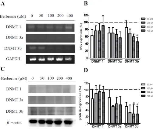
January 31, 2020The tumor suppressor gene, p53, is inactivated in the human hepatocellular carcinoma cells line, Hep3B. Berberine has been reported to inhibit the proliferation of cancer cells. This study examined whether apoptosis was induced in berberine-treated Hep3B cells and observed the association between apoptosis and the expression of p53 and DNA methyltransferase (DNMT). The cell viability was measured using an MTT assay. Apoptosis of Hep3B was measured using annexin V flow cytometry. Berberine-treated cells were examined for their DNMT enzymatic activity, mRNA expression, and protein synthesis. The p53 levels were examined by Western blot analysis. The berberine treatment resulted in increased Hep3B cell death and apoptosis in a time- and dose-dependent manner. The DNMT3b activity, mRNA expression, and protein levels all decreased after the berberine treatment. In contrast, the p53 protein levels increased with a concomitant decrease in DNMT3b. No change in the expression of ERK was observed, but the P-ERK levels decreased in a dose dependent manner. These results indicate that a treatment of Hep3B cells with berberine can reduce the expression of DNMT3b, leading to an increase in the tumor suppressant gene p53 and an increase in cell apoptosis. This shows that berberine can effectively suppress the proliferation of liver cancer cells.
Copyright © 2020 The Korean Society for Clinical Laboratory Science. All rights reserved.
Key words Apoptosis Berberine
DNA methyltransferase Hepatocellular carcinoma p53
INTRODUCTION
Liver cancer is one of the most common cancers worldwide [1]. Genetic alteration is one of the key contributing factors for liver carcinogenesis and
genetic alterations is heavily influenced by DNA methylation [2]. Specific DNA methylations are regulated by expression of DNA methyltransferase (DNMT) and as such, DNMT expression are associated with liver carcinogenesis [3, 4]. Three forms of DNMT exist. DNMT1 acts on nascent DNA during cellular differentiation resulting in formation of hemimethylated CpG dinucleotides (maintenance DNMT) [5, 6]. DNMT3a and DNMT3b methylate CpG dinucleotides during early
Korean Society for Clinical Laboratory Science