HepG2 간암세포에서 미토콘드리아 경로를 통한 개똥쑥 추출물의 Apoptosis 유도 효과
김보민1․김근태1․김은지1․임은경1․김상용2․김영민1
1한남대학교 생명나노대학 생명시스템과학과
2신안산대학교 식품생명과학과
Extract from Artemisia annua Linné Induces Apoptosis through the Mitochondrial Signaling Pathway in HepG2 Cells
Bo Min Kim1, Guen Tae Kim1, Eun Ji Kim1, Eun Gyeong Lim1, Sang-Yong Kim2, and Young Min Kim1
1Department of Biological Science and Biotechnology, College of Life Science and Nano Technology, Hannam University
2Department of Food Science & Bio Technology, Shinansan University
ABSTRACT The Akt/mammalian target of the rapamycin (mTOR) pathway is activated in the majority of human cancers. Activation of the Akt/mTOR pathway confers resistance to many types of cancer therapy. In this study, we evaluated the apoptotic effect of ethanol extract of Artemisia annua L. through down-regulation of Akt signal pathways and the mitochondrial pathway in hepato-carcinoma cells (HepG2). A. annua extract is known as a medicinal herb that is effective against cancer. We evaluated anti-proliferative activity by MTT-based viability assay and apoptotic effect by Annexin-V/PI staining, mitochondrial membrane potential (MMP), and caspase-3/7 activity as determined by flow cytometry. A. annua treatment led to loss of MMP, resulting in cytochrome c-inducible activation of caspase-3/7.
Treatment with A. annua extract reduced activities of Akt/mTOR/anti-apoptotic proteins (such as Bcl-2 and Bcl-XL), leading to increased activation of tumor suppressor p53 and pro-apoptotic proteins (such as Bax and Bak). We applied LY294002 (inhibitor of Akt) and rapamycin (inhibitor of mTOR) to determine the relationship between signal trans- duction of proteins associated with apoptosis. LY294002 and rapamycin significantly reduced cell viability and increased apoptosis. These results indicate that Bcl-2 and caspase-3 are key regulators in A. annua extract-induced apoptosis in HepG2 cells and are controlled through the Akt/mTOR signaling pathway.
Key words: HepG2, Akt, mTOR, Artemisia annua L., apoptosis
Received 4 August 2016; Accepted 13 September 2016 Corresponding author: Young Min Kim, Department of Biological Science and Biotechnology, College of Life Science and Nano Technology, Hannam University, Daejeon 34054, Korea
E-mail: [email protected], Phone: +82-42-629-8753
서 론
암 치료의 가장 중요한 전략 중 하나는 apoptosis 경로를 통해 암세포만 특이적으로 사멸시키는 것이다. Apoptosis 의 경로는 크게 외인성 경로(extrinsic pathway)와 내인성 경로(intrinsic pathway)로 구분된다(1). 외인성 경로는 세 포막에 존재하는 death receptor(DR)에 death ligand가 결 합하여 initiator caspase의 활성을 유도하여 이루어진다.
반면에 내인성 경로는 Bcl-2 family 단백질에 의해 주로 조절된다. Anti-apoptotic 단백질에는 Bcl-2/Bcl-XL, pro- apoptotic 단백질에는 Bax와 Bak이 있는데 이들은 서로 결
합하여 동형이량체(homodimer)와 이형이량체(heterodimer) 를 이룬다. 이 단백질들의 상대적 비율은 apoptosis의 억제 와 촉진 효과에 영향을 준다(2,3).
암 치료법에는 화학요법 및 외과적인 수술요법 등이 있지 만, 정상세포에 대한 독성 및 부작용을 초래하고 지속적인 항암제 사용에 따른 약제 내성을 일으키는 문제점이 제기되 고 있어 최근에는 여러 가지 천연 추출물을 이용한 암 예방 및 치료에 관한 연구가 활발하게 이루어지고 있다(4,5). 항 암 및 항균 효과를 지니고 있는 것으로 알려져 있는 개똥쑥 (Artemisia annua L.)은 국화과의 쑥속에 속한다. 개똥쑥은 항산화력이 높은 약용식물 중의 하나로 시료 중에 포함되어 있는 페놀 화합물에 의한 것으로 보고되어 있다. 개똥쑥 추 출물의 페놀 화합물을 phenolic acid, flavonol 및 catechin 류로 분류하여 분석한 결과 개똥쑥 추출물의 항산화 활성은 catechin류와 관련성이 높은 것으로 밝혀졌다(6,7).
Protein kinase B(PKB)라 불리는 Akt는 serin/threonine
kinase로 다른 단백질을 인산화시킴으로써 세포 증식이나 분화, 성장에 관여한다. 이러한 Akt 신호경로의 조절은 비정 상적인 암세포의 증식을 억제하는 데 큰 역할을 한다(8).
Akt는 Tuberous Sclerosis Complex 2(TSC2)를 인산화시 켜 Mammalian target of rapamycin(mTOR)의 활성을 증 가시킨다(9). 상위조절 단백질인 Akt에 의하여 활성화되는 mTOR은 세포의 성장, 증식에 관여하는 중요 조절자로 종양 형성에 관여하는 단백질이다(10). 이러한 mTOR의 저해는 COX-2의 발현을 감소시키고 p53의 활성을 조절하는 것으 로 보고되었다(11,12).
따라서 본 연구에서는 HepG2 간암세포에 개똥쑥 추출물 을 처리했을 때 apoptosis 효과를 확인하고자 하였고, 이에 관여하는 Akt/mTOR 신호경로의 저해와 Bcl-2 family 단 백질 간의 상호관계를 규명하고자 하였다.
재료 및 방법
실험재료
본 실험에서 사용된 개똥쑥은 대전 한약재시장에서 구입 한 개똥쑥 분쇄가루 100 g에 95% 에탄올 800 mL를 가하여 72시간 동안 상온에서 환류 추출하였다. 이러한 방법으로 추출된 개똥쑥 추출물을 감압농축기(N-1200A, Tokyo Rikakikai Co., Tokyo, Japan)를 이용하여 감압 농축과정을 거친 후, DMSO(dimethyl sulfoxide, Samchun, Pyeong- taek, Korea)로 100 mg/mL stock으로 만들어 -20°C에 보관하여 사용하였다. 3-(4,5-Dimethylthiazol-2-yl)-2,5 -diphenyltetrazolium bromide(MTT)는 Sigma-Aldrich Co.(St. Louis, MO, USA)에서 구입하여 5 mg/mL stock으 로 만들어서 사용하였다. 또한, pifithrin-α와 celecoxib는 Sigma-Aldrich Co.에서 구입하여 DMSO에 녹인 후 각각 20 mM과 100 mM로 만들어 사용하였다.
세포배양
본 실험에 사용된 HepG2 세포는 American Type Culture Collection(ATCC, Rockville, MD, USA)에서 분양받았으 며, wild-type p53을 가지는 간암세포이다(13). 10% FBS (HyClone Laboratories Inc., Logan, UT, USA)와 1% an- tibiotics(HyClone Laboratories Inc.)가 포함된 RPMI 1640 배지(HyClone Laboratories Inc.)를 사용하였고 fi- broblast 세포는 ATCC에서 분양받았으며 10% FBS와 1%
antibiotics가 포함된 DMEM(HyClone Laboratories Inc.) 배지를 사용하여 5% CO2, 37°C 조건하에 배양(MCO-15AC, SANYO Electric Biomedical Co., Osaka, Japan)하였다.
매 48시간마다 Trypsin-EDTA(HyClone Laboratories Inc.)를 이용하여 세포를 부유 상태로 만든 다음, 세포를 1×10⁶ cells/mL로 분주하고 계대 배양하였다.
MTT assay에 의한 세포독성 및 생존율 측정
세포배양용 12 well plate에 HepG2 세포를 1×10⁴ cells/
mL로 분주하고 24, 48시간 배양하였으며, Fibroblast 세포 를 1×10⁴ cells/mL로 분주하고 24시간 동안 배양시킨 후 개똥쑥 추출물을 처리하였다. LY294002(Sigma-Aldrich Co.)와 rapamycin(Sigma-Aldrich Co.) 처리 시에는 LY 294002, rapamycin을 30분 먼저 처리한 후 개똥쑥 추출물 을 농도별로 처리하여 24시간 동안 배양하였다. MTT 용액 을 30 μL씩 첨가하여 30분 동안 CO2 incubator에서 배양한 후, MTT 시약이 들어있는 배지를 제거하고 DMSO 150 μL 를 넣어 well에 생성된 formazan을 모두 녹여 96 well plate 에 100 μL씩 옮겨서 Microplate Reader(Bio-Rad Labo- ratories Inc., Tokyo, Japan)로 595 nm에서 흡광도를 측정 하였다.
Lactate dehydrogenase(LDH) assay를 의한 세포독성 측정
Apoptosis 시 세포막의 손상으로 인해 발생하는 LDH의 양을 측정하기 위해 LDH cytotoxicity assay kit(Thermo Scientific, Hudson, NH, USA)을 사용하여 세포독성을 확 인하였다. 배양된 HepG2 세포를 24-well plate에 1×10⁴ cells/well의 농도로 분주하여 24시간 배양하여 부착 및 안 정화한 후, 개똥쑥 추출물을 처리하였다. LY294002와 ra- pamycin 처리 시에는 LY294002, rapamycin을 30분 먼저 처리한 후 개똥쑥 추출물을 농도별로 처리하여 24시간 동안 배양하였다. 상층액 50 μL와 LDH reagent 50 μL를 혼합하 여 30분간 암 조건에서 반응시킨 후 stop solution 25 μL를 가한 다음 490 nm, 655 nm에서 흡광도를 측정하여 LDH 방출량을 측정하였다.
HepG2 세포의 형태 관찰
6 well plate에 HepG2 세포를 well당 1×10⁶ cells/mL로 분주하여 24시간 동안 배양한 후 개똥쑥 추출물을 농도별로 처리하였다. 물질을 최종 처리하여 24시간 동안 CO2 in- cubator에서 배양한 후 HepG2 세포에서 일어나는 형태 변 화를 inverted microscope(Zeiss M, Carl Zeiss, Oberko- chen, Germany)으로 관찰하였다.
Annexin V-Fluorescein Isothiocyanate(FITC) staining 에 의한 apoptosis 측정
Apoptosis를 정량적으로 분석하기 위해 MuseTM Annexin V & Dead Cell Kit(MCH100105, Merck Millipore Co., Billerica, MA, USA)을 사용하였다. HepG2 세포는 DMEM media 100 μL를 이용하여 1×10⁵ cells/mL로 부유시키고 MuseTM Annexin V & Dead Cell Reagent 100 μL를 혼합 하여 37°C, CO2 incubator에서 20분 동안 반응시켰다. 반응 후 Cell Analyzer(PB4455ENEU, MuseTM Cell Analyzer, Merck Millipore Co.)를 이용하여 분석하였다.
Hoechst 33342 staining에 의한 핵 형태 관찰
Cover glass가 놓여진 12 well plate에 HepG2 세포를 1×10⁴ cells/mL로 분주하여 24시간 동안 배양 후 개똥쑥 추출물을 농도별로 처리하여 24시간 동안 CO2 incubator에 서 배양하였다. Hoechst 33342 시약을 10 μM의 농도로 처리하여 30분 동안 반응시켰다. 3.5% formaldehyde로 15 분간 고정시키고 phosphate buffered saline(PBS)으로 두 번 세척 후 slide glass에 aqueous mounting medium (Dako Faramount, Dako North America Inc., Santa Clara, CA, USA)을 이용해 부착시키고 형광현미경(Axio- skop 50, Carl Zeiss)을 이용하여 관찰하였다.
Mitochondrial membrane potential(MMP, Δψm)의 분석 미토콘드리아의 막 1전위의 변화를 조사하기 위해 MuseTM Mitopotential Kit(MCH100110, Millipore Co.)을 사용하였다. HepG2 세포는 1× assay buffer를 이용하여 1×10⁵ cells/mL로 부유시키고 mitopotential working solution 95 μL를 혼합하여 37°C, CO2 incubator에서 20분 동안 반응시켰다. 반응 후 Muse Mitopotential 7-AAD re- agent 5 μL를 혼합하여 5분 동안 실온에서 염색하고 Cell Analyzer (PB4455ENEU, MuseTM Cell Analyzer, Merck Millipore Co.)를 이용하여 분석하였다.
Caspase-3/7 activity 분석
Caspase-3/7의 활성을 조사하기 위해 MuseTM Mitopo- tential Kit(MCH100108, Merck Millipore Co.)을 사용하 였다. HepG2 세포는 1× assay buffer BA 50 μL를 이용하 여 1×10⁵ cells/mL로 부유시키고 MuseTM Caspase-3/7 Reagent working solution 5 μL를 혼합하여 37°C, CO2 incubator에서 30분 동안 반응시켰다. MuseTM Caspase 7-AAD working solution 150 μL를 혼합하여 상온에서 5 분간 반응시킨 후 Cell Analyzer(PB4455ENEU, MuseTM Cell Analyzer, Merck Millipore Co.)를 이용하여 분석하였다.
Western blotting에 의한 단백질 발현의 분석
6 well plate에 HepG2 세포를 well당 1×10⁶ cells/mL로 분주하여 24시간 동안 배양한 후 개똥쑥 추출물을 농도별로 처리하였다. LY294002, rapamycin과 병행 처리 시에는 inhibitor를 30분 먼저 처리하였으며, 모든 물질을 최종 처 리한 후 24시간 동안 CO2 incubator에서 배양하였다. 배양 이 끝난 세포에 RIPA lysis buffer[25 mM Tris-Cl(pH 7.4), 1% NP40, 0.5% sodium deoxycholate, 150 mM NaCl, 1 mM PMSF]를 각 well에 150 μL씩 첨가하여 단백 질을 분리한 후, 14,000 rpm, 4°C에서 20분 동안 원심분리 하여 상등액을 취하였다. 추출한 단백질은 ELISA-reader를 이용하여 595 nm에서 흡광도를 측정하여 정량하였다. 그다 음에 8%, 12% acrylamide gel을 이용하여 전기영동을 실 시한 후 nitrocellulose membrane에 transfer 하였다. 2%
bovine serum albumin(BSA)을 이용해 blocking 하고, 1차 항체를 4°C에서 overnight 처리한 후 2차 항체를 결합시켜 1시간 반 동안 반응시킨 다음 Blue X-ray film에 감광하여 실험 결과를 측정하였다.
통계처리
실험 설계에 대한 분산분석은 t-test(SPSS 20.0 soft- ware, SPSS Inc., Chicago, IL, USA)를 실시하여 유의성을 검정하였다. 각 자료는 3번 이상의 반복된 실험을 통하여 얻어진 결과로 검정하였고 P<0.05인 경우 통계적으로 유의 한 것으로 판정하였다.
결과 및 고찰
개똥쑥 추출물이 HepG2 간암 세포의 증식에 미치는 효과 확인
생명체의 생명유지와 발생단계에 관여하는 apoptosis는 정상적인 발달과 분화뿐만 아니라 체내 비정상적인 세포들 을 제거하는 기전으로 세포 내외 인자들이 관여하여 일어나 는 능동적인 세포 자멸 현상이다(14,15). 개똥쑥 추출물의 단독 처리가 HepG2 간암세포의 생존에 미치는 영향을 알아 보기 위해 MTT assay와 LDH release assay를 실시하였 고, 이를 통해 세포독성과 세포증식 억제 효과를 각각 확인 하였다. 정상세포인 Fibroblast 세포에 대한 개똥쑥 추출물 의 세포독성을 측정한 결과는 Fig. 1A에 나타내었다. 개똥 쑥 추출물을 농도별(10, 20, 40, 60, 80, 100 μg/mL)로 처 리한 후 24시간 동안 반응시켰을 때 정상세포의 생존율이 90% 이상으로 유지되는 것으로 보아 개똥쑥 추출물의 독성 이 없음을 확인하였다. Fig. 1B에 나타난 바와 같이 개똥쑥 추출물을 농도별(10, 20, 40, 60, 80, 100 μg/mL)로 처리한 후 24, 48시간 동안 반응시켰을 때 개똥쑥 추출물의 농도와 시간 의존적으로 HepG2 간암 세포의 생존율이 감소하는 것을 확인하였다. 다음으로 LDH release assay를 실시하 였다. 세포질에 존재하는 효소인 LDH는 세포막이 손상을 받 으면 세포 내에 존재하는 LDH 방출이 증가한다(16). 따라서 세포의 손상 정도를 확인하기 위하여 배양액 중에 들어있는 LDH 방출량을 측정한 결과는 Fig. 1C와 같다. 개똥쑥 추출 물을 농도별(20, 40, 60, 80, 100 μg/mL)로 처리한 후 24시 간 동안 반응시켰을 때 LDH 방출량이 개똥쑥 추출물의 농도 에 의존적으로 증가하는 것을 확인하였다. 그리고 개똥쑥 추출물을 농도별로 처리했을 때 세포 증식이 억제되는 것을 현미경으로 관찰한 결과는 Fig. 1D에 나타낸 바와 같다.
개똥쑥 추출물에 의한 HepG2 간암 세포의 apoptosis 유도 효과 확인
HepG2의 증식 억제 효과가 apoptosis 유도에 의한 것인 지를 알아보기 위해 Annexin-V/PI 염색 후 flow cytometry 를 이용하여 실시하였다. Fig. 2A에 나타난 바와 같이 개똥
A B
C
D
Fig. 1. Artemisia annua extract (AAE) inhibits cell proliferation and induces apoptosis in HepG2 liver cancer cells. (A) Cell pro- liferation rate was measured by MTT assay. The statistical analysis of the data was carried out by use of a t-test. N.S.: not significant (each experiment’s, n=3). (B) HepG2 cells were treated with A. annua extract for 24∼48 h. The statistical analysis of the data was carried out by use of a t-test. **,##P<0.01, ***,###P<0.001 compared to control. N.S.: not significant (each experiment’s, n=3).
(C) A. annua extract increased the LDH release in HepG2 liver cancer cells. The statistical analysis of the data was carried out by use of a t-test. *P<0.05, **P<0.01, ***P<0.001 compared to control. N.S.: not significant (each experiment’s, n=3). (D) Phase Contrast Image-Based Monitoring of Apoptosis Induction. 40× phase contrast images HepG2 cells after 24 h incubation.
A
B
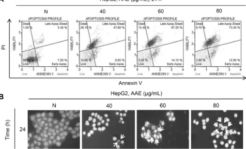
Fig. 2. A. annua extract induces apoptosis in HepG2 liver cancer cells. Cells were treated with the indicated concentrations of A.
annua extract for 24 h. (A) Cells stained with MuseTM Annexin V and Dead Cell Assay kit and analyzed by MuseTM Cell Analyzer.
Data shows four cell populations-Live, Dead, Late Apop./Dead, Early Apop. (B) Cell apoptosis observed using Hoechst 33342 staining. Cells were treated with the indicated concentrations of A. annua extract for 24 h. Fluorescence was detected using a fluorescence microscope. Arrows indicate apoptotic bodies, which were DNA fragments produced when apoptosis occurred.
A
B
C
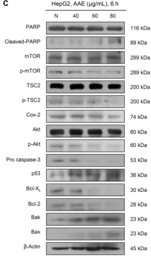
Fig. 3. Evaluation of the effect of A. annua extract on the apoptosis through the mitochondrial signaling pathway. Cells were treated with the indicated concentrations of A. annua extract for 24 h. (A) Cells stained with MuseTM Mitopotential Kit and analyzed by MuseTM Cell Analyzer. Data shows four cell populations-Live, Depolarized/Live, Depolarized/Dead, and Dead cells. (B) Cells stained with MuseTM Caspase-3/7 kit and analyzed by MuseTM Cell Analyzer. Data shows four cell populations-Live, Dead, Apoptotic, Apoptotic/Dead. (C) Protein level was measured by western blotting. The β-actin probe served as protein-loading control.
쑥 추출물을 농도별(40, 60, 80 μg/mL)로 처리한 후 24시간 동안 반응시켰을 때 정상 배지의 apoptosis 유발 빈도는 약 12.63%로 매우 낮았으나 개똥쑥 추출물 처리 농도가 증가 할수록 빈도가 증가하여 80 μg/mL를 처리한 군에서는 약 86.23%에 해당하는 세포에서 apoptosis가 유도된 것으로 관찰되었다. 그리고 개똥쑥 추출물 처리에 따른 염색체 응축 현상을 관찰하기 위해 Hoechst 33342 staining을 실시하였 다. Fig. 2B에 나타난 바와 같이 개똥쑥 추출물의 처리 농도 가 증가할수록 염색체 응축 현상인 apoptotic body가 더 많이 발생한 것을 확인할 수 있었다. 이를 통해 개똥쑥 추출 물 처리에 의한 HepG2 간암 세포의 생존율 및 증식 억제는 apoptosis에 의한 현상임을 알 수 있었다.
개똥쑥 추출물에 의한 미토콘드리아 막 전위 변화와 관련 단백질 발현 확인
Apoptosis가 미토콘드리아 경로를 통해 유도되면 세포질 에 존재하는 칼슘 이온 및 apoptosis 관련 조절 단백질들이 미토콘드리아로 이동하여 미토콘드리아 단편화(fragmen- tation)를 일으킨다. 이로 인해 미토콘드리아 막의 탈분극화 가 일어나면 투과성 조절 구멍(permeability transition pore)이 열려 apoptosis에 작용하는 단백질들이 밖으로 유 출되어 핵의 응집(condensation)과 DNA 단편화를 일으켜 apoptosis가 일어나게 된다(17,18). 개똥쑥 추출물 처리에 따른 apoptosis 유도에 있어서 미토콘드리아의 역할을 확인 하기 위하여 미토콘드리아 막 전위의 변화를 나타낸 결과는 Fig. 3A와 같다. 막 전위 변화는 MuseTM Mitopotential Kit 을 사용하여 Cell Analyzer로 분석하였다. 개똥쑥 추출물을
농도별(40, 60, 80 μg/mL)로 처리한 후 24시간 동안 반응시 켰을 때 정상 배지의 apoptosis 유발 빈도는 약 15.36%로 매우 낮았으나 개똥쑥 추출물의 처리 농도가 증가할수록 빈 도가 증가하여 80 μg/mL 처리군에서는 약 95%에 해당하는 세포에서 apoptosis가 유도된 것으로 관찰되었다.
미토콘드리아의 막 전위를 유지하는 데 중요한 역할을 하 는 Bcl-2 family 단백질은 기능적으로 구분된다. Anti- apoptotic 단백질인 Bcl-2, Bcl-XL과 pro-apoptotic 단백 질인 Bax, Bak가 이형이량체를 형성하게 되면 apoptosis를 억제하고 동량이량체를 이루게 되면 막 투과성을 증가시켜 cytochrome c의 유리와 caspase 활성을 유도하게 된다 (19). Apoptosis 과정에서 중요한 조절인자로 알려진 cas- pase는 apoptosis 신호를 세포 밖에서 안으로 매개하거나 세포 내 단백질을 직접 분해하는 역할을 하는데 이 중 cas- pase-3/7은 수행 caspase(effector caspase)로 PARP를 분절시켜 apoptosis를 일으킨다(20,21). 따라서 본 연구에 서는 caspase activity assay와 western blotting을 통해 개똥쑥 추출물 처리에 따른 caspase의 활성 정도와 Akt, mTOR, TSC2, COX-2, p53뿐만 아니라 미토콘드리아 경 로를 구성하는 단백질들의 발현 정도를 확인하였다. Fig.
3B에 나타난 바와 같이 개똥쑥 추출물을 농도별(40, 60, 80 μg/mL)로 처리한 후 24시간 동안 반응시켰을 때 정상 배지 에서는 약 0.82%로 매우 낮았으나 개똥쑥 추출물 80 μg/mL 처리군에서는 약 69.57%로 caspase-3/7의 활성이 증가하 여 apoptosis가 일어남을 확인할 수 있었다. 다음으로 Fig.
3C에 나타난 바와 같이 개똥쑥 추출물을 농도별(40, 60, 80 μg/mL)로 처리하였을 때 그 결과 세포증식에 관여하는 p-Akt, p-mTOR, COX-2와 암 억제자이지만 불활성화 상 태인 p-TSC2의 감소, 그리고 anti-apoptotic 단백질인 Bcl-2, Bcl-XL, pro caspase-3의 감소를 확인할 수 있었 다. 또한, 암 억제유전자인 p53과 pro-apoptotic 단백질인 Bax, Bak, 그리고 PARP의 불활성형인 cleaved-PARP의 증가를 확인하였다. 이상의 결과를 살펴볼 때 개똥쑥 추출물 이 HepG2 간암세포에서 내인성 경로에 관여함으로써 apop- tosis를 일으키는 단백질들의 활성을 유도한다는 것을 알 수 있었다.
Akt 및 mTOR 저해제가 HepG2 간암 세포의 증식 억제 및 apoptosis 유도 효과 확인
개똥쑥 추출물 처리에 따른 Akt와 mTOR의 저해가 HepG2 간암 세포의 증식에 미치는 영향을 확인하기 위해 Akt와 mTOR의 저해제인 LY294002와 rapamycin, 그리고 개똥쑥 추출물 단독 혹은 병행 처리하였다. LY294002는 Akt(Ser473)의 인산화를 억제시키고 rapamycin은 cyto- solic protein FK-binding protein 12(FKBP12)와 결합하 여 mTOR을 억제시켜 세포의 증식 억제를 유도한다. 이러한 mTOR의 억제는 S6K1과 4E-binding protein 1(4EBP1)의 활성과 인산화를 감소시켜 RNA에서 단백질로 번역되는 과
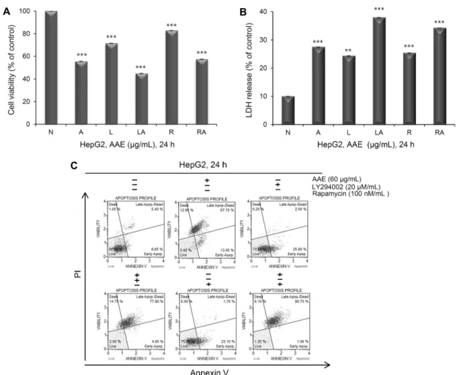
정을 억제시킴으로써 apoptosis를 유도한다(22,23). MTT assay를 통해 세포의 생존율과 세포증식 억제를 확인한 결 과는 Fig. 4A와 같다. LY294002 20 nM과 rapamycin 100 μM, 개똥쑥 추출물 60 μg/mL를 24시간 동안 단독 혹은 병 행 처리했을 때 암세포 증식을 확인하였다. 개똥쑥 추출물을 단독으로 처리했을 때에는 약 56%, LY294002를 단독 처리 했을 때에는 약 71%, LY294002와 개똥쑥 추출물을 병행 처리했을 때에는 약 45%, rapamycin 단독으로 처리했을 때에는 약 83%, rapamycin과 개똥쑥 추출물 병행 처리했을 때에는 약 58%의 생존율을 보였다. 이를 통해 LY294002/
rapamycin을 단독 처리했을 때보다 개똥쑥 추출물과 병행 처리했을 때 생존율의 감소 효과가 더 크게 나타남을 확인하 였다.
다음으로 LDH release assay를 실시하였다. Fig. 4B에 나타난 바와 같이 개똥쑥 추출물을 단독으로 처리했을 때에 는 약 27%, LY294002를 단독 처리했을 때에는 약 24%, LY294002와 개똥쑥 추출물을 병행 처리했을 때에는 약 38%, rapamycin 단독으로 처리했을 때에는 약 25%, rapa- mycin과 개똥쑥 추출물 병행 처리했을 때에는 약 34%의 LDH 방출량을 나타냈다. Akt 및 mTOR의 저해가 apopto- sis를 유도하는 것인지를 알아보기 위해 Annexin-V/PI 염 색 후 flow cytometry를 이용하여 실시하였다. Fig. 4C에 나타난 바와 같이 LY294002와 rapamycin을 단독 혹은 개 똥쑥 추출물을 24시간 동안 병행 처리했을 때의 apoptosis 정도를 확인하였다. 개똥쑥 추출물을 단독으로 처리했을 때 에는 약 82%, LY294002를 단독 처리했을 때에는 약 28%, LY294002와 개똥쑥 추출물을 병행 처리했을 때에는 약 83%, rapamycin 단독으로 처리했을 때에는 약 25%, rapa- mycin과 개똥쑥 추출물 병행 처리했을 때에는 약 93%의 생존율을 보였다.
Akt 및 rapamycin 저해제에 의한 미토콘드리아 막 전위 변화와 관련 단백질 발현 확인
Akt와 mTOR 저해제와 개똥쑥 추출물을 처리했을 때 미 토콘드리아 막 전위의 변화를 알아보기 위해 MuseTM Mito- potential Kit을 사용하여 Cell Analyzer로 분석하였다.
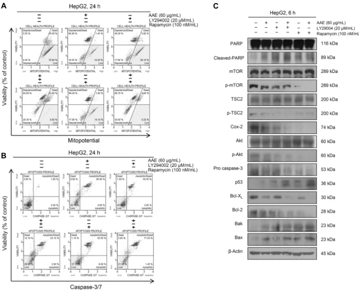
Fig. 5A에 나타난 바와 같이 개똥쑥 추출물을 단독으로 처리 했을 때에는 약 84%, LY294002를 단독 처리했을 때에는 약 67%, LY294002와 개똥쑥 추출물을 병행 처리했을 때에 는 약 84%, rapamycin 단독으로 처리했을 때에는 약 85%, rapamycin과 개똥쑥 추출물 병행 처리했을 때에는 약 90%
에 해당하는 세포에서 apoptosis가 유도된 것으로 관찰되었 다. 그리고 Fig. 5B에 나타난 바와 같이 개똥쑥 추출물을 단독으로 처리했을 때에는 약 61%, LY294002를 단독 처리 했을 때에는 약 53%, LY294002와 개똥쑥 추출물을 병행 처리했을 때에는 약 58%, rapamycin 단독으로 처리했을 때에는 약 62%, rapamycin과 개똥쑥 추출물 병행 처리했을 때에는 약 73%로 caspase-3/7의 활성이 증가하여 Akt,
A B
C
Fig. 4. Co-treatment of LY294002 (L), rapamycin (R), and A. annua extract (A) inhibits cell proliferation and induces apoptosis.
Cells were pre-treated with 20 μM LY294002 or 100 nM rapamycin for 30 min and co-treated with 60 μg/mL A. annua extract 24 h. (A) The statistical analysis of the data was carried out by use of a t-test. ***P<0.001 compared to control (each experiment’s, n=3). (B) A. annua extract increased the LDH release in HepG2 liver cancer cells. The statistical analysis of the data was carried out by use of a t-test. **P<0.01, ***P<0.001 compared to control (each experiment’s, n=3). (C) Cells stained with MuseTM Annexin V and Dead Cell Assay kit and analyzed by MuseTM Cell Analyzer.
mTOR의 저해와 개똥쑥 추출물에 의해 apoptosis가 일어남 을 확인하였다.
끝으로 Akt, mTOR 저해제와 개똥쑥 추출물의 처리에 따 른 apoptosis 관련 신호 단백질들 간의 관계를 알아보기 위 해 western blotting을 실시하였다. Fig. 5C에 나타난 바와 같이 LY294002와 rapamycin을 개똥쑥 추출물과 단독 혹 은 병행 처리했을 때 p-Akt, p-mTOR, COX-2, pro cas- pase-3, anti-apoptotic 단백질인 Bcl-2, Bcl-XL 그리고 암 억제자임에도 불구하고 불활성화 상태인 p-TSC2의 감 소와 pro-apoptotic 단백질인 Bax, Bak 그리고 cleaved- PARP, p53의 증가를 확인할 수 있었다.
다수의 선행연구에 의하면 LY294002와 rapamycin은 HepG2 간암세포 혹은 다른 암세포에 처리했을 때 p53의 발현이 증가한다고 보고되었다(8,24). 결과적으로 이러한 선행연구와 비교해 보았을 때 개똥쑥 추출물에 의한 Akt, mTOR의 저해는 p53의 발현을 촉진하여 미토콘드리아 경
로를 통한 apoptosis가 유발됨을 확인하였다. 이 연구를 바 탕으로 실제로 간암 치료제로서의 사용 가능성을 확인하기 위해서는 앞으로 활성 성분 분석 및 다양한 추가 실험 등이 수행되어야 할 것이다.
요 약
Akt 및 mTOR는 세포 생존에 필수적인 경로로 세포 성장과 증식 등에서 중요한 역할을 하는 것으로 알려져 있다. 본 연구에서는 항암 및 항균 효과가 있는 것으로 알려진 개똥쑥 (Artemisia annua L.)에 의한 HepG2 간암세포의 apopto- sis 유도 효과를 확인하였다. 본 연구 결과에 의하면 개똥쑥 추출물의 처리 농도가 증가함에 따라 HepG2 세포의 생존율 은 억제되었으며, 이는 apoptosis 유도 효과에 의한 것임을 세포의 형태적 변화와 flow cytometry를 통해 확인하였다.
그리고 mitopotential assay와 caspase-3/7 activity as-
A
B
C
Fig. 5. Co-treatment of LY294002, rapamycin and A. annua extract induces apoptosis. Cells were pre-treated with 20 μM LY294002 or 100 nM rapamycin for 30 min and co-treated with 60 μg/mL A. annua extract 24 h. (A) Cells stained with MuseTM Mitopotential Kit and analyzed by MuseTM Cell Analyzer. (B) Cells stained with MuseTM Caspase-3/7 kit and analyzed by MuseTM Cell Analyzer.
(C) Protein level was measured by western blotting. Cells were treated with 40∼80 μg/mL of A. annua extract for 24 h. The β- actin probe served as protein-loading control.
say, western blotting으로 Bcl-2 family 단백질을 확인함 으로써 apoptosis 경로 중 내인성 경로(intrinsic pathway) 에 의해 apoptosis가 일어남을 알 수 있었다. 이러한 효과는 Akt/mTOR의 활성 저해와 연관이 있었으며 Akt/mTOR의 저해제인 LY294002/rapamycin을 개똥쑥 추출물과 병행 처리하였을 경우 개똥쑥 추출물에 의한 apoptosis 효과를 더욱 증대시켰다. 따라서 Akt/mTOR의 저해는 개똥쑥 추출 물의 apoptosis 효과를 상승시켰으며 이에 따라 미토콘드리 아의 기능 손상과 caspase 활성의 증가를 통해 이루어짐을 확인하였다.
REFERENCES
1. Jin Z, El-Deiry WS. 2005. Overview of cell death signaling pathways. Cancer Biol Ther 4: 50-74.
2. Hwang JY, Choi YH. 2015. Sanguinarine induces apoptosis
in human hepatocellular carcinoma HepG2 cells through the generation of ROS and modulation of Akt/ERK signaling pathways. J Life Sci 25: 984-992.
3. Soung YH, Lee JW, Kim SY, Nam SW, Park WS, Lee JY, Yoo NJ, Lee SH. 2005. Mutational analysis of proapoptotic bcl-2 family genes in colon carcinomas. Korean J Pathol 39: 168-171.
4. Kwon HN, Bang WS, Kim JY, Park JR, Jeon JR. 2011.
Effect of Acer tegmentosum M. extracts on hepatocarcinoma cell. Korean J Food Sci Technol 43: 787-790.
5. Lee HN, Jang HY, Kim HJ, Shin SA, Choo GS, Park BK, Kim BS, Jung JY. 2015. Induction of apoptosis by picea- tannol in YD-15 human oral cancer cells. J Korean Soc Food Sci Nutr 44: 975-982.
6. Ryul JH, Lee SJ, Kim MJ, Shin JH, Kang SK, Cho KM, Sung NJ. 2011. Antioxidant and anticancer activities of Artemisia annua L. and determination of functional com- pounds. J Korean Soc Food Sci Nutr 40: 509-516.
7. Brisibe EA, Umoren UE, Brisibe F, Magalhaes PM, Ferreira JFS, Luthria D, Wu X, Prior RL. 2009. Nutritional charac-
terisation and antioxidant capacity of different tissues of Artemisia annua L.. Food Chem 115: 1240-1246.
8. Park SY, Lee SH, Park OJ, Kim YM. 2011. Apoptotic ef- fects of curcumin and EGCG via Akt-p53 signaling pathway in HCT116 colon cancer cells. J Life Sci 21: 89-95.
9. Choi JY, Jo MW, Lee EY, Choi DS. 2014. AKT is involved in granulosa cell autophagy regulation via mTOR signaling during rat follicular development and atresia. Reproduction 147: 73-80.
10. Lee SH, Kim IS, Park SY, Park OJ, Kim YM. 2011. Querce- tin induces apoptosis via regulation of mTOR-VASP signal- ing pathway in HT-29 colon cancer cells. Cancer Prev Res 16: 340-347.
11. Lee SH, Lee H, Park SY, Park OJ, Kim YM. 2011. Apoptotic effects of resveratrol via mTOR and COX-2 signal pathways in MCF-7 breast cancer cells. J Life Sci 9: 1288-1294.
12. Choi EM, Heo JI, Oh JY, Kim YM, Ha KS, Kim JI, Han JA. 2005. COX-2 regulates p53 activity and inhibits DNA damage-induced apoptosis. Biochem Biophys Res Commun 328: 1107-1112.
13. Roudier E, Mistafa O, Stenius U. 2006. Statins induce mam- malian target of rapamycin (mTOR)-mediated inhibition of Akt signaling and sensitize p53-deficient cells to cytostatic drugs. Mol Cancer Ther 5: 2706-2715.
14. Lee HH, Jeon JW, Choi YH. 2016. Inhibition of PI3K/AKT signaling pathway enhances cordycepin-induced apoptosis in human gastric cancer cells. J Korean Soc Food Sci Nutr 45: 835-842.
15. Hwang WD, Im YG, Son BY, Park C, Park DI, Choi YH.
2013. Induction of apoptosis by ethanol extract of Scutellaria baicalensis in renal cell carcinoma caki-1 cells. J Life Sci 23: 518-528.
16. Kim I, Om AS, Kim JH, Kim HR. 2012. Screening of etha-
nol extracts of Korean fruits and vegetables for cell viability and antioxidant enzyme activity on alloxan-induced oxida- tive stress in pancreatic beta cell. J Korean Soc People Plant Environ 15: 85-92.
17. Kang DY, Lee JY, Kim MS, Kim CH, Kim HK, Lee SM, Kwon YM, Lee JW, Baik HS, Yu BP, Chung HY. 2008.
4-Hydroxynonenal induces endothelial apoptosis through mitochondrial depolarization. J Life Sci 18: 1513-1520.
18. Gottlieb E, Armour SM, Harris MH, Thompson CB. 2003.
Mitochondrial membrane potential regulates matrix config- uration and cytochrome c release during apoptosis. Cell Death Differ 10: 709-717.
19. Park C, Yoon KH, Lee YJ, Kim YK, Choi YC, Shin JH, Cho JH, Park R. 2002. 5-FU induces apoptosis of Fas (+), HepG2 cells via activation of Fas-mediated caspase and mi- tochondria dysfunction. Cancer Res Treat 34: 128-138.
20. Boatright KM, Salvesen GS. 2003. Mechanisms of caspase activation. Curr Opin Cell Biol 15: 725-731.
21. Kim TH, Kim PH, Jeon BK. 2011. Effect of Anemarrhenae rhizoma ethanol extract on apoptosis induction of HT-29 human colon cancer cells. J Korean Orient Med Ophthalmol Otolaryngol Dermato 24: 16-24.
22. Jiang H, Fan D, Zhou G, Li X, Deng H. 2010. Phosphatidy- linositol 3-kinase inhibitor (LY294002) induces apoptosis of human nasopharyngeal carcinoma in vitro and in vivo.
J Exp Clin Cancer Res 22: 29-34.
23. Ballou LM, Lin RZ. 2008. Rapamycin and mTOR kinase inhibitors. J Chem Biol 1: 27-36.
24. Nakano M, Nakashima A, Nagano T, Ishikawa S, Kikkawa U, Kamada S. 2013. Branched-chain amino acids enhance premature senescence through mammalian target of rapamy- cin complex Ⅰ-mediated upregulation of p21 protein. PLoS One: e80411.