레스베라트롤에 의한 인간 암세포주, A549와 SKOV3의 p53 의존적 Apoptosis 유발
이슬기, 남주옥*
경북대학교농업생명과학대학식품공학부식품공학전공
Received: December 30, 2015 / Revised: March 10, 2016 / Accepted: March 10, 2016
서 론
암은전세계적으로인류의건강과생명을위협하는사망 률이 높은질병중하나이며, 암관련 사망자의수는 향후 50년간 2배증가할것으로예상된다[3]. 따라서암치료를위 한연구가지속적으로이루어지고있고, 최근프로그램된세 포죽음을표현하는용어인세포자멸사(apoptosis)에대한분 자기전연구가활발히진행되고있다[14]. Apoptosis는세포 스스로조절이 가능한능동적과정이지만, 암세포의경우
apoptosis에대한저항성을갖기위해세포내다양한분자
기전교란을일으킴으로써항암제에대한저항성을가질수 있다[1, 6]. Anoikis는 apoptosis의아류형이며부적절한세
포부착혹은부착의손실에의해일어난다[17]. 일반세포는
부착되지않은상태에서생존하지못하고 Anoikis라는세포
사멸과정을거치게되는데이에반해, 암세포의경우부착되 지않은상태에서도 spheroid 형태를형성하여성장할수있 으며, 생체내에서혈류를통해다른기관으로이동할수있
으므로, 암세포의 Anoikis 저항성은세포의전이능력과연
관성이있다[10].
Resveratrol은자연계에널리존재하는폴리페놀류의한
종류이며와인, 땅콩, 오디등의식품에함유되어있다[9]. 또 한백혈병과전립선암, 유방암과같은다양한상피암종의세 포증식억제효과를가진다고보고되었다[13]. 이러한효과 는세포주기의억제와 apoptosis 유도를통해나타나며, 이 과정에서 p53 유전자가관여한다고보고된바있다[5]. 종양 억제유전자로널리알려진 p53의돌연변이는인간암세포에 서흔히관찰되고정상적인 p53은 FAS, Bax, Bcl-2 등다양 한 apoptosis 관련유전자들의발현을조절함으로써 apoptosis
를유발한다[12, 18]. 최근항암효과를가지는다양한천연소
재에서 apoptosis를조절하는중요한메커니즘을규명하는
연구가 꾸준히 이루어지고 있으며[8, 14], 이전의 연구는 Induction of p53-dependent Apoptosis by Resveratrol in Human Cancer Cells, A549 and SKOV3
Seul Gi Lee and Ju-Ock Nam*
Department of Food Engineering, Kyungpook National University, Daegu 41566, Republic of Korea
Resveratrol, a polyphenolic compound present in many fruits and vegetables such as grapes, mulberries, and peanuts, has been reported to have various biological effects. However, the molecular mechanisms underlying resveratrol-induced apoptosis in A549 ovarian cancer cells are not well understood. In this study, we investigated the effect of resveratrol on A549 lung cancer cells (expressing wild-type p53) and compared it with that observed for SKOV3 ovarian cancer cells (expressing null-type p53). Resveratrol sig- nificantly inhibited the viability and proliferation of A549 cells in a concentration- and time-dependent manner, compared with its effects on SKOV3 cells. It also induced A549 cell apoptosis, but did not affect anoikis resistance. Furthermore, the viability and proliferation of p53-knockdown A549 cells were unaf- fected by the presence of resveratrol. Therefore, we demonstrate that the anticancer effect of resveratrol against A549 lung cancer cells is dependent on the presence of functional p53.
Keywords: Resveratrol, apoptosis, anoikis, p53, A549, SKOV3
*Corresponding author
Tel: +82-53-950-7760, Fax: +82-53-950-7762 E-mail: [email protected]
© 2016, The Korean Society for Microbiology and Biotechnology
resveratrol이 A549 세포의성장을억제하고 apoptosis를유도 하는등의항암효과를가진다는것을보고한바있다[16]. 그 러나, resveratrol이정상적인 p53 유전자를가지는 A549 세
포의 anoikis 저항성에어떠한영향을미치는지에대한연구
와 p53 유전자가결실된 SKOV3 세포와의차이점에대해서
는보고된바없다.
그러므로본연구에서는 resveratrol이 A549 폐암세포와
SKOV3 자궁암세포의생존및사멸에어떠한영향을미치
는지알아보고또한이러한과정에서 p53이관여하는조사 하였다.
재료 및 방법
시약 및 재료
본실험에서사용한 resveratrol은 Sigma Aldrich (Sigma Aldrich, MO, USA)에서 구입하였고 dimethyl sulfoxide (DMSO, 5 mg/ml) 에용해한뒤 -20oC에보관하였다. p53 small interfering RNA (siRNA), 대조군 siRNA, beta- actin antibody는 Santa cruz biotechnology사(CA, USA)에 서 구입하였다. Pro-caspase3, p53 antibodies는 Cell signaling technology사(MA, USA)에서구입하였다.
세포배양
A549 세포는 wild-type의 p53 유전자를가지고있는폐암 세포주이며 SKOV3는 p53 유전자가결실된자궁암세포주
이다[2, 11]. A549, SKOV3 세포는 한국 세포주 은행
(KCLB)으로부터분양받았고, 10% fetal bovine serum (FBS), 100 U/ml penicillin 그리고 100 µg/ml streptomycin을포 함하는 RPIM-1640 배지에 5% CO2가 공급되는 37oC incubator에서배양하였다. 배양중인세포의배지는 2−3일 에한번새로운배지로교체해주었다.
세포 생존율 측정(MTT assay)
A549, SKVO3 세포를 96-well plates에 각 well 당 4 × 10³으로 100 µl의 medium과함께분주하고 5% CO2가 공급되는 incubator에서 24시간동안배양한후, 각 well 마 다 resveratrol을 20, 40, 60, 80, 100 (µM) 농도로처리하고 24, 48시간배양하였다. Control group에는 resveratrol을넣 지않고동일한양의 DMSO를넣었다. 배양후 medium을 제거하고 MTT (0.4 mg/ml) 를 20 µl/well 넣고 incubator에 서 3시간 배양한뒤 iso-propyl alcohol을 100 µl/well 넣어 생성된 formazan을용해시킨뒤, infinite reader를이용하 여 595 nm에서흡광도를측정했다. 측정값은 3번은반복하 여평균값으로얻었다.
세포사멸 검출
A549, SKOV3 세포를 chamber slide (Lab-Tak II chamber slider system, USA)에 각 well 당 4 × 10⁴로 분주한 뒤 24시간동안부착시키고, resveratrol을각농도별로처리 후 48시간 동안 배양하였다. 배지를 제거하고 4% para- formaldehyde로 상온에서 10분간 세포를 고정시킨 후, terminal deoxynucleotidyl transferase-mediated nick-end labeling (TUNEL) assays는 Red in situ apoptosis Detection kit (Millipore Corporation, MA, USA)를사용하여제조사 의매뉴얼에 따라수행하였다. DAPI로 DNA를대조염색 한후, 형광현미경을이용하여각농도에따른핵의형태를 관찰하였고, 무작위적으로 5개의필드를선택하여평균치 를구하였다.
부착성 상실 세포사멸 실험(anoikis assay)
Anoikis assay는 CytoSelect 96-well anoikis assay kit (Biolab Diagnostics, South Africa)를사용하여제조사의매 뉴얼에따라수행하였다. Resveratrol을농도별로포함하는 medium에 A549, SKOV3 세포를부유시킨뒤, 각 well 당 0.5 × 10⁵으로 hydrogel이코팅된 96-well plate에분주했다. Resveratrol 처리 48시간후, 각 well에 MTT reagent를첨 가하고 3시간반응시킨뒤, detergent solution를첨가하였 다. 3시간배양후, infinite reader를이용하여 570 nm에서 흡광도를측정하였다.
siRNA를 이용한 p53 유전자 일시적 침묵
A549 세포에서 siRNA transfection은 p53에 특이적인 siRNA (p53 siRNA)와 양성대조군인 scrambled control siRNA (control siRNA)를 이용하여, 각각의 siRNA를 lipofectamine 2000 (Invitrogen, CA, USA)과섞어제조사 의매뉴얼에따라수행하였다. Transfection시킨뒤, resveratrol 을농도별로 48시간처리하였고, 단백질발현분석을통해
siRNA의효율성을검증하였다.
Western blot 분석
각실험조건에따라 resveratrol을농도별로처리한 A549, SKOV3 세포를 lysis buffer에용해한뒤, 15,000 rpm에서
10분간원심분리하여상층액에있는전체단백질을얻었다.
100oC에서 10분간가열하여 sample을얻었고동일한양의 단백질을 sodium dodecyl sulphate polyacrylamide gel에 의해분리시켰다. 분리된단백질을 Nitrocellulose membranes 에전달시킨뒤, TBST에용해시킨 5% non-fat skim milk를 이용하여 blocking하였다. 그후, 1차항체를 4oC에서 overnight 처리하였고 2차항체는상온에서 1시간 반응시켰다. 반응
후, enhanced chemiluminescence reagent (GEHealthcare, UK)를 membrane에도포하고 X-ray film에노출시켜시각 화하였다.
결과 및 고찰
Resveratrol이 A549, SKOV3 세포의 생존과 성장에 미치는 영향
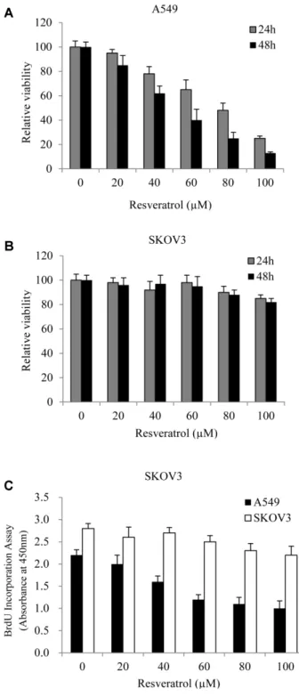
Resveratrol이 A549, SKVO3 세포의생존에미치는영향 을알아보기위하여 MTT assay를시행하였다. Resveratrol 을농도 별(0, 20, 40, 60, 80, 100 µM)로각각 24, 48시간 처리한후세포생존율을측정한결과, resveratrol은 A549 세포의 생존을농도, 시간의존적으로 억제하였다. 반면에 SKOV3 세포의생존에는영향을미치지않았다(Fig. 1A, B).
Resveratrol이세포의성장에도영향을미치는지알아보기
위해, BrdU incorporation assay를 시행하였다. 그 결과 A549 세포의증식비율은 resveratrol 처리농도에따라점 차적으로감소하였다. 반면에 SKOV3 세포의증식에는영향 을미치지않는것으로나타났다(Fig. 1C). 이러한결과를통 해 resveratrol이농도의존적을 A549 세포의생존과증식
을억제하고, SKOV3 세포에는영향을미치지않는다는것
을확인하였다.
Resveratrol이 A549 세포의 apoptosis에 미치는 영향 다음으로, resveratrol이 A549, SKOV3 세포의 apoptosis 를 유도하는지 조사하였다. Resveratrol을농도 별(0, 40, 100 µM)로 48시간처리후, 세포의핵에염색되어형태적변
화를관찰할수있는 DAPI 염색과 DNA의분절여부를파악
할수있는 TUNEL 염색을시행하였다. 그결과, A549 세포
에서는 DAPI에의해염색된세포의수가확실하게감소하
였다는것을관찰할수있었고, TUNEL에의해염색된세
포는 resveratrol 처리농도에따라증가하는것을확인하였 다. 반면에, SKOV3 세포에서는 TUNEL에의해염색된세 포가거의없는것으로나타났고염색된세포의형광강도와 면적또한현저히적었다(Fig. 2A-B). Resveratrol에의해유 도된세포 apoptosis 비율을확인하기위해, TUNEL에염색
된 세포/DAPI에 염색된 세포의 비율을 분석한 결과
resveratrol을 100 µM의농도로 처리하였을 때, 확실하게 apoptosis가일어난 A549 세포의비율이증가했다는것을확 인하였다(Fig. 2C). 이러한 결과들을통해 A549 세포에서 resveratrol의생존억제효과는 apoptosis 유도와연관성이
있다고보여지며, SKOV3 세포에서는크게영향을미치지
않는것으로보여진다.
세포와세포외기질과의결합이끊어질때, 정상세포는
anoikis에빠지게되어대부분의세포가죽는반면암세포는
Fig. 1. Effect of resveratrol on the viability and proliferation of A549 and SKOV3 cells. Human lung carcinoma A549 cells (A) and human ovarian carcinoma SKOV3 cells (B) treated with res- veratrol at various concentrations for 24 and 48 h. The effect of resveratrol on cell viability was determined by MTT assay.
(C) Cells were exposed to various concentrations of resvera- trol for 48 h. The effect of resveratrol on cell proliferation was determined by BrdU assay. Values are mean ± S.D. of three exper- iments, with triplicate of each experiment.
Fig. 2. Apoptotic cell death of A549 and SKOV3 cells by resveratrol. A549 cells (A) and SKOV3 cells (B) were treated with resveratrol (0, 40, 100 μM) for 48 h. A representative images of DAPI (blue)-, TUNEL (red)-stained cells and merged image are shown on the left, middle, and light, respectively. While DAPI stains the entire nuclei, TUNEL stains the apoptotic and necrotic cells. (C) Quantification of number of TUNEL-stained cells/number of DAPI stained cells. (D) suspension-cultured A549 and SKOV3 cells were treated with resver- atrol for 48 h in hydrogel coated-96 well plate. Cell survival was assessed by the anoikis assay. Values are mean ± S.D. of three experi- ments, with triplicate of each experiment.
고착 비의존성성장(anchorage-independent growth)을함 으로써부착할수있는기층(substratum) 없이생존이가능 하다[4]. 그러므로우리는 resveratrol이 A549, SKOV3 세포
의 anoikis 저항성에 영향을 미치는지 조사하기 위해
resveratrol을농도별로 48시간처리한후 anoikis assay를 시행하였다. 그결과두가지세포모두에서 resveratrol에 의한 anoikis 유도효과가없었다(Fig. 2D). 이러한결과는 resveratrol이 A549 세포가부착상태일때는 apoptosis를유 도하지만부유상태일때는세포생존에영향을미치지않는 다는것을보여준다. 또한이러한결과는 resveratrol이일 차종양세포의증식은억제하지만, 전이과정에서다른기관 으로이동하는세포의증식을억제하는효과는없다는것을 의미한다.
Resveratrol이 apoptosis 관련 단백질의 발현에 미치는 영향 Caspases는세포내에서불활성화된 proenzyme의형태로
존재하여세포사멸신호에의해분절되어활성화된다[15].
그 중 caspase3는 apoptosis 유발인자들에 의해 caspase cascade가 일어나면 다른 caspase에 의해 활성화되며,
apoptosis 유도의 최종 단계에서 중 중요한 역할을 하는
effecter caspase이다[7]. 그러므로, resveratrol을 A549 세포 에처리했을때농도의존적으로 apoptosis가유도되는현상 의분자적기전분석을위하여 pro-caspase3 단백질의발현 변화를 western blot을통해확인하였다. 그결과, A549 세 포에서 resveratrol에농도가증가함에따라 pro-caspase3의 발현이억제되었고, SKOV3 세포에서는변화가없었다(Fig.
3A-B). pro-caspase3의 발현량의 감소로 A549 세포의 apoptosis 유도효과에 caspase3가관여한다는것을확인하 였다.
P53 knock-down(유전자 기능의 불완전 소실)에 의한 resveratrol의 A549 세포 증식억제 및 caspase 발현 조절효과 위실험들을통해 resveratrol이 A549 세포의 apoptosis를 유도한다는것과반면에 SKOV3 세포에는어떠한영향도미 치지않는다는것을알아냈다. P53은 apoptosis를유발하는
것으로잘알려져있으므로, 이러한결과가 p53 유전자발
현에의존한것인지알아보기위해정상적인 p53 유전자를
가지는 A549 세포에 siRNA를이용하여일시적으로 p53 유 전자의발현을억제시켰다. P53 유전자가확실하게억제되
었음을 확인한 후(Fig. 4A), p53 유전자가 억제된 세포에
Fig. 2. Continued.
Fig. 3. Effect of resveratrol on the expression of apoptosis-related protein. A549 cells (A) and SKOV3 cells (B) were treated with resveratrol at various concentrations for 48 h. After treatment with resveratrol, the cells were lysed and the expression of pro-spase3 was confirmed by western blot analysis. Equal amount of loading was confirmed by the quantification of β-actin.
resveratrol을 농도별(0, 40, 100µM)로 48시간 처리하여 pro-caspase3의발현량을확인하였다. 그결과 resveratrol 처리에의한 pro-caspase3의발현량에변화가없었다(Fig.
4B). 또한 p53 유전자가억제된 A549 세포의생존율과성장 률모두변화가없었다(Fig. 4C, D). 반면 control siRNA를 처리한세포는상기의결과들과동일하게세포생존과증식 모두 resveratrol 처리농도에따라감소하였다. 이러한결과 는 resveratrol에의한 A549 세포의생존및증식억제효과
는 p53 유전자에의존한다는것을보여준다.
결론적으로 본 연구에서는 resveratrol에 의해 유도된 A549 세포의증식억제및 apoptosis 유도효과는 p53 유전 자에의존적임을 SKOV3 세포와의 비교를통해알아냈고, 또한세포가부유상태일때는억제효과가나타나지않는다
는 것을 보여준다. 본 연구 결과는 A549 폐암세포에서
resveratrol의항암효과에대한기전을밝히는데기여할수
있다고사료된다.
요 약
Resveratrol은포도, 오디, 땅콩과같은많은과일과채소 에존재하는폴리페놀화합물로써다양한생물학적효과를 가진다고보고되어있다. 그러나, resveratrol이 A549 폐암세
포에서 유도하는 apoptosis에 관여하는 분자적 기전 및
anoikis에관해서는명백하게밝혀지지않았다. 본연구에서, 우리는정상적인 p53 유전자를갖는 A549 세포에서 resveratrol
의효과를조사하고, p53 유전자가결실된 SKOV3 난소암
세포와그효과를비교했다. Resveratrol은확실하게 SKOV3 세포에비해농도, 시간의존적으로 A549 세포의생존과증 식을억제했다. 또한 resveratrol은 A549 세포의 apoptosis 를유도했지만세포의 anoikis 저항성에는영향을미치지않 았다. 더불어, p53 유전자 기능이 불완전 소실된(knock- down) A549 세포의생존과증식은 resveratrol에의해변하 지않았다. 그러므로, 본연구의결과는 resveratrol의항암 Fig. 4. Effect of resveratrol in A549 cells transfected with p53 siRNA. (A) A549 cells transfected with control siRNA or p53 siRNA for 16 h and p53 siRNA efficiency was confirmed by western blot analysis. (B) Transfected cells were treated with resveratrol for 48 h and then cell lysates were prepared for western bolt analysis. Transfected cells were seeded in 96-well plate and treated group was treated with resveratrol for 48 h. Cell viability (C) and proliferation (D) were determined by MTT assay and BrdU assay, respectively. Values are mean ± S.D. of three experiments, with triplicate of each experiment.
효과가기능을하는 p53 유전자의존재에의존한다는것을 보여준다. 결론적으로, 우리는 resveratrol이 p53 유전자에 의존적으로 A549 세포에대해항암효과를가진다는것을입 증했다.
Acknowledgments
This research was supported by Basic Science Research Program through the National Research foundation of Korea (NRF) funded by Ministry of Education (NRF-2013R1A1A2064784); by Kyungpook National University Research Fund, 2012.
References
1. Chen J, Peng H, Ou-Yang X, He X. 2008. Research on the antitu- mor effect of ginsenoside Rg3 in B16 melanoma cells. Mela- noma Res. 18: 322−329.
2. Corney DC, Flesken-Nikitin A, Godwin AK, Wang W, Nikitin AY.
2007. MicroRNA-34b and MicroRNA-34c are targets of p53 and cooperate in control of cell proliferation and adhesion-inde- pendent growth. Cancer Res. 67: 8433−8438.
3. Ferraz da Costa DC, Casanova FA, Quarti J, Malheiros MS, Sanches D, Dos Santos PS, et al. 2012. Transient transfection of a wild-type p53 gene triggers resveratrol-induced apoptosis in cancer cells. PLoS One 7: e48746.
4. Fiucci G, Ravid D, Reich R, Liscovitch M. 2002. Caveolin-1 inhib- its anchorage-independent growth, anoikis and invasiveness in MCF-7 human breast cancer cells. Oncogene 21: 2365−2375.
5. Hsieh TC, Juan G, Darzynkiewicz Z, Wu JM. 1999. Resveratrol increases nitric oxide synthase, induces accumulation of p53 and p21(WAF1/CIP1), and suppresses cultured bovine pulmo- nary artery endothelial cell proliferation by perturbing pro- gression through S and G2. Cancer Res. 59: 2596−2601.
6. Kang SW. 2013. Role of reactive oxygen species in cell death pathways. Hanyang Med. Rev. 33: 77−82.
7. Kim BW, Kwon HJ, Jin K-S, Park H-J, Lee J-Y, Oh YN, et al. 2013.
Induction of G2/M arrest and apoptosis by the methanol extract of typha orientalis in human colon adenocarcinoma
HT29 cells. Korean J. Microbiol. Biotechnol. 41: 425−432.
8. Kim HH, Min G. 2015. Inhibitory effects of S-allylcysteine on cell proliferation of human cervical cancer cell line, HeLa. J. Life Sci.
25: 397−405.
9. Lee SH, Park SY, Kim I-S, Park OJ, Kim YM. 2012. Effects of res- veratrol on migration and proliferation in HT-29 colon cancer cells. KSBB J. 27: 289−294.
10. Min SO, Lee SW, Bak SY, Kim KS. 2015. Sphere-forming cultiva- tion of liver cancer cell line in culture media without growth factors.
Korean J. Clinical Oncol. 11: 20−23.
11. Mukhopadhyay T, Roth JA. 1997. Induction of apoptosis in human lung cancer cells after wild-type p53 activation by methoxyestradiol. Oncogene 14: 379−384.
12. NaveenKumar SK, Thushara RM, Sundaram MS, Hemshekhar M, Paul M, Thirunavukkarasu C, et al. 2015. Unconjugated bilirubin exerts pro-apoptotic effect on platelets via p38-MAPK activa- tion. Sci. Rep. 5: 15045.
13. Opipari AW Jr, Tan L, Boitano AE, Sorenson DR, Aurora A, Liu JR.
2004. Resveratrol-induced autophagocytosis in ovarian cancer cells. Cancer Res. 64: 696−703.
14. Park H-J, Jin S, Oh YN, Kim BW, Kwon HJ. 2015. Induction of apoptosis by methanol extract of endlicheria anomala in human lung and liver cancer cells. J. Life Sci. 25: 441−449.
15. Park S-Y, Kim E-J, Lim D-Y, Kim J-S, Lim S-S, Shin H-K, et al. 2008.
Inhibitory effect of the hexane extract of Saussurea lappa on the growth of LNCaP human prostate cancer cells. J. Korean Soc.
Food Sci. Nutr. 37: 8−15.
16. Wang H, Zhang H, Tang L, Chen H, Wu C, Zhao M, et al. 2013. Res- veratrol inhibits TGF-beta1-induced epithelial-to-mesenchymal transition and suppresses lung cancer invasion and metastasis.
Toxicology 303: 139−146.
17. Wang X, Lin G, Martins-Taylor K, Zeng H, Xu RH. 2009. Inhibition of caspase-mediated anoikis is critical for basic fibroblast growth factor-sustained culture of human pluripotent stem cells. J. Biol. Chem. 284: 34054−34064.
18. Yoo J, Park S, Kang CS, Kang SJ, Kim BK. 2002. Expression of E- cadherin and p53 proteins in human soft tissue sarcomas. Arch Pathol. Lab. Med. 126: 33−38.