MCF-7 유방암 세포에서 AMPK 활성에 의한 conjugated linoleic acid의 apoptosis 유도에 관한 연구
인선교1․김현숙2․박옥진3․김영민*
한남대학교 생명나노과학대학 생명과학과, 1iBIO 마케팅부, 2경희대학교 의과대학 병리학교실, 3한남대학교 생명나노과학대학 식품영양학과
Received August 7, 2008 /Accepted November 20, 2008
Conjugated Linoleic Acid Induces Apoptosis by Activating AMPK in MCF-7 Breast Cancer Cells.
Sun Kyo Lin
1, Hyun Sook Kim
2, Ock Jin Park
3and Young Min Kim*. Department of Biological Sciences, College of Life Science and Nano Technology, Hannam University of Daejeon, 306-791, Korea,
1Department of Marketing iBIO, Daejeon, Korea,
2Department of Pathology College of Medicine, Kyung Hee University, Seoul, Korea,
3Department of Food and Nutrition, College of Life Science and Nano Technology, Hannam University of Daejeon, 306-791, Korea - Conjugated linoleic acid (CLA) is a naturally occurring compound found in dairy and beef products. It has been shown to suppress cancer cells and to induce apoptosis.
Practically, there is emerging evidence that CLA can inhibit chemically induced carcinogenesis in vari- ous tissues. However, the molecular mechanisms of CLA on human MCF-7 breast cancer cells have not been clearly explained yet. In this report, we investigated the anti-cancer activity of CLA in MCF-7 cells. It was found that CLA could inhibit the growth of the MCF-7 cells and induce apoptosis, through modulating AMP-activated protein kinase (AMPK) and cyclooxygenase-2 (COX-2). AMPK acts as a cellular fuel gauge and responds to decreased cellular energy status by inhibiting ATP-con- suming pathways and increasing ATP-synthesis. CLA treatment with variable concentrations and dif- ferent time of same-dose CLA on MCF-7 cells resulted in a strong activation of AMPK and an in- hibition of COX-2 expression. It supports that CLA induces apoptosis in CLA-treated MCF 7 cells.
Therefore, the effects of CLA induced COX-2 expression via activating AMPK can provide new possi- bility into the understanding the molecular mechanisms of anti-cancer component.
Key words : Conjugated linoleic acid, MCF-7 cells, apoptosis, AMP-activated protein kinase, cyclooxygenase-2
*Corresponding author
*Tel:+82-42-629-8753, Fax:+82-42-629-8751
*E-mail : [email protected]
서 론
우리나라 여성의 유방암 발병률은 서구 국가에 비해서 아 직은 낮은 편이지만, 생활양식의 서구화로 인한 비만의 증가, 출산율 및 수유의 감소, 만혼, 조기초경 및 폐경기 지연과 같 은 이유로 점차 증가하고 있는 추세이다. 유방암을 포함한 암의 발생은 다양한 환경적 요인에 기인하며, 이러한 요인 중에서 식품 내 존재하는 발암 및 항암 성분에 대한 연구가 활발하게 진행되고 있다[6]. 이 가운데 하나의 항암제로 연구 되고 있는 것이 conjugated linoleic acids (CLA)인데, CLA는 쇠고기와 유제품에 들어 있는 linoleic acids (LA)의 이성체로 서 탄소 수가 18개이며 이중결합을 포함한 지방산의 구조로 이루어져 있다[8,9]. CLA는 LA와 매우 유사한 구조를 가지 고 있지만, LA와는 다른 생물학적인 효과를 나타낸다. 즉 LA는 암을 촉진하는 반면, CLA는 암세포를 억제 또는 감소 시키는 특성이 있으며, 특히 유방암, 대장암, 전립선암 등에 서 항암효과가 있는 것으로 알려져 있다[5,17,18,24].
AMP-activated protein kinase (AMPK)는 세포 내에 에너 지가 부족할 때 이를 감지해 에너지의 항상성을 조절하는 효 소인데, 비만, 당뇨병 등과 관련이 있으며, 특히 암세포에서 apoptosis를 유도하는 것으로 보고되어 있다[2,14]. AMPK는 유방암, 전립선암, 폐암, 대장암에서 높은 농도로 나타나는 cyclooxygenase-2 (COX-2) 효소의 농도를 조절하는데, AMPK가 활성화되면 COX-2의 억제효과가 나타나 암세포에 서 apoptosis가 유도되는 것으로 알려져 있다[27]. 최근에 CLA와 같이 식품에 들어있는 항암성분을 규명하기 위해서, AMPK 및 COX-2 효소와 연관되어 apoptosis가 유도되는 경 로를 밝힌 연구가 자주 보고되고 있다[13,14,20,25]. 본 연구 에서는 CLA 투여에 따른 MCF-7 세포의 증식 억제를 관찰하 고, 이와 같은 암세포 성장의 방해가 AMPK 및 COX-2 효소 에 의해서 유도된 apoptosis 인지를 관찰하여 CLA의 항암 효과에 관한 기전을 밝힘으로써, 인체 유방암에 관한 기초자 료를 제공할 목적으로 수행되었다.
재료 및 방법
암세포의 배양
ATCC (Gaithersburg, MD, USA)로부터 MCF-7 세포와
L6 골격근 세포를 구입하였다. MCF-7 세포의 배양배지는 10%의 우태아혈청(fetal bovine serum, FBS), 1%의 strepto- mycin 및 penicillin (Biofluids, Rockville, MD, USA)을 RPMI 1640 (GIBCO BRL, Grand Island, NZ, USA)에 혼합하 여, 37
oC, 5% CO
2조건에서 confluence 70~80% 정도로 배 양되었을 때, 실험에 사용하였다.
시약 및 세포 처리
CLA, tetrazolium bromide salt (MTT), bovine serum al- bumin (BSA)는 Sigma (Sigma Chemical Co., St. Louis, MO, USA) 제품을 구입하여 사용하였다. 실험에 사용한 CLA는 알코올에 녹여 50 mM stock solution으로 제작하여 -20
oC에 서 냉동 보관하여 사용하였으며, 세포 처리 시 CLA의 농도 는 20 μM, 40 μM, 80 μM, 160 μM, 320 μM로 처리하여 37
oC, 5% CO
2조건으로 배양하여 사용하였다. 배양된 MCF-7 세포 를 0.4% Trypsin・EDTA solution (Sigma Chemical Co.)을 사 용하여 세포를 분리시킨 후, 세포의 confluence가 70~80%
정도 되었을 때까지 배양하였고, 모든 세포 처리는 RPMI 1640과 DMEM (GIBCO BRL, Grand Island, NZ, USA) 배양 액에 직접 처리하여 실험에 사용하였다.
MTT assay에 의한 세포생존율 측정
MCF-7 세포를 배양시킨 12-well culture plate에, 각 well 마다 MTT (5 mg/ml) 용액을 30 μl씩 첨가하여 37
oC, 5%
CO
2조건에서 1시간 반응시킨 후, 세포의 색 변화를 관찰하 면서 phosphate buffered saline (PBS)로 조심스럽게 세척하 였다. Dimethzl sulfoxide (DMSO, Amresco, Solon, OH, USA) 150 μl를 12-well culture plate의 각 well에 첨가하고 천천히 교반하여 세포를 떨어뜨린 다음, 100 μl씩을 취해 ELISA-reader용 96-well plate에 순서대로 분주하고 micro- plate reader (Bio-Rad chemical division, U.S.A)를 사용하여 595 nm에서 흡광도를 측정하였다. 측정은 모두 세 번을 하 였으며, 그에 따르는 평균값과 표준오차는 Microsoft Exel program을 이용하여 분석하였다.
Hoechst 33342염색을 이용한 chromatin staining
12-well culture plate에 microscope cover glass (Marlenfeld GmbH & co. Germany)를 각 well 당 하나씩 넣은 다음, 그 위에서 MCF-7 세포를 배양하여, CLA를 농도와 시간 별로 처리한 후, 37
oC, 5% CO
2조건에서 24시간 동안 배양하였 다. 배양이 끝난 후, plate에 직접 Hoechst 33342 염색시약(32 μg/μl)을 10 μM로 처리하여 실온에서 1시간 방치하여 반응시 키고, 진공 suction pipette을 이용하여 세포가 손상되지 않도 록 조심스럽게 배양액을 제거하였다. 세포의 상태를 고정시키 기 위해 3.5% 포름알데히드가 포함된 PBS를 각 well에 첨가하 여 실온에서 30분간 배양한 다음, PBS로 2번 세척하여, 50%
글리세롤을 한 두 방울 slide glass 위에 떨어뜨리고 실온에서 건조시켰다. 형광현미경(Olympus Optical, Tokyo, Japan)으 로 저배율에서 고배율로 관찰하면서, MCF-7 세포에서 apop- totic cells을 확인하고, 디지털카메라(Olympus SP-6600, Japan)로 사진 촬영하였다.
Reactive oxygen species (ROS) 측정
100 mm culture plate에서 배양된 MCF-7 세포의 con- fluence가 80% 정도 되었을 때 현미경으로 확인하고, 12-well culture plate의 각 well에 cover glass를 넣고, 배양된 MCF-7 세포와 CLA를 농도별로 20 μM, 40 μM, 80 μM, 160 μM 혼 합한 후, cover glass 위에 조심스럽게 분주하여 37
oC, 5%
CO
2조건에서 배양하였다. ROS의 생성 확인을 위해 각 well 에 2', 7'-dichlorofluorescein diacetate (DCFH-DA)를 10 μM 씩 처리하여 30분 동안 37
oC, 5% CO
2조건에서 배양하여, cover glass 위에서 배양된 세포가 떨어지지 않도록 조심스 럽게 세척한 후, 형광현미경을 통하여 관찰하였다.
Western blot에 의한 단백질 발현의 분석
6-well culture plate에 MCF-7 세포와 L6 세포를 각각 배양 하여, CLA를 농도와 시간별로 처리하여 37
oC, 5% CO
2조건에 서 배양하였다. Total protein을 추출하기 위하여 triton lysis buffer [25 mM Tris-Cl (pH 7.4), 150 mM NaCl, 1% Triton (100×), 5 mM EDTA]를 각 well에 150 μl씩 첨가하여 반응시킨 후, 14,000 rpm, 4
oC에서 10분 동안 원심분리 하였다. Bradford assay를 이용하여 ELISA-reader 595 nm에서 흡광도를 측정하 여 표준곡선을 정한 후, 각 표본의 total protein 농도를 결정하 였다. Sodium dodecyl sulfate (SDS)-polyacrylamide gel을 하 기 위해 5× Laemmi sample buffer (loading dye; 250 mM Tris-cl (pH 6.8), 40% glycerol, 4% β-mercaptoethanol, 0.08%
bromophenol blue, 8% SDS)와 MCF-7 세포에서 추출한 단백 질을 혼합하여 표본을 제작하였다. 1차 항체인 phospho-ace- tyl-CoA carboxylase (ACC), COX-2, phospho-AMPK (P-AMPK), β-actin을 Cell Signaling Technology (Beverly, MA, USA)로부터 구입하여 각각 희석한 후, 적정 온도(4
oC)를 유지시키기 위해 얼음 팩과 함께 밤새동안 반응시켰다. 2차 항체(Cell Signaling Technology, Beverly, MA, USA)를 희석 하여 1시간 반응시킨 다음, Blue X-ray film에 나타나는 밴드로 단백질의 활성을 관찰하였다.
결 과
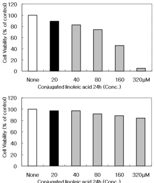
CLA에 의한 MCF-7 세포의 증식 억제
CLA가 MCF-7 세포의 증식에 미치는 영향을 알아보기 위
하여, 다양한 농도별로 CLA를 처리하여 배양한 후, MTT as-
say를 이용하여 조사하였다. Fig. 1A에 나타난 바와 같이,
(A)
(B)
Fig. 1. Growth inhibition of MCF-7 cells after treatment with CLA. (A) Cell viability of MCF-7 cells were seeded at 4×10³ cells/well in a 12-well culture plate and in- cubated for 24 hr. The cells were treated with variable concentrations of CLA for 24 hr. The growth inhibition was measured by the metabolic-dye-based MTT assay.
The data shown are means±SD of three independent experiments. (B) Under the same conditions, the cell viability of L6 skeletal muscle cells was measured by MTT assay.
CLA의 농도가 높아짐에 따라 MCF-7 세포의 증식 억제 정도 가 증가하여, 160 μM 농도 처리 시에는 50%, 320 μM 농도 처리 시에는 90%의 증식 억제 효과가 나타난 것을 관찰할 수 있었다. 반면에 Fig. 1B에 보여 지는 것과 같이, 정상인 L6 세포에서는 MCF-7 세포에서와 동일한 농도로 CLA를 처리 하였지만 암세포의 증식 억제에는 변화를 일으키지 않았다.
이상의 실험에 따르는 평균값과 표준오차는 Microsoft Exel program을 이용하여 분석하였다. 이와 같이 Fig. 1A에서처 럼 CLA를 MCF-7 세포에 처리하였을 경우에는 암세포의 증 식이 억제되었지만, Fig. 1B와 같이 L6 세포에서는 암세포의 증식 억제가 일어나지 않았다. 위와 같은 실험의 결과로부터, CLA 처리 시에 암세포의 증식이 억제되었으며, 아울러 CLA 농도가 높아짐에 따라 암세포의 증식이 더욱 더 억제되는 것 을 관찰할 수 있었다.
한편 Fig. 2에서 나타난 바와 같이, CLA 농도를 100 μM로 일정하게 MCF-7 세포에 처리하고, MTT assay를 이용하여 서로 다른 시간별로 암세포의 증식 억제를 조사한 결과, 시 간이 지남에 따라서 MCF-7 세포의 증식 억제 효과가 증가하 는 것을 관찰할 수 있었다. 즉 1 시간이 경과하였을 때, 10%
Fig. 2. Cell viability of MCF-7 cells treated with different time of same-dose CLA. The cells were seeded at 4×10³ cells/well in a 12-well plate and incubated for 24 hr. The cells were treated with a constant concentration of 100 μM CLA for 24 hr. The growth inhibition was measured by the metabolic-dye-based MTT assay. The data shown are means±SD of three independent experiments.
의 암세포의 증식 억제가 나타났고, 6 시간 후에는 30%, 12 시간 후에는 60%, 그리고 24 시간 후에는 90%의 암세포의 증식 억제 효과가 나타난 것을 확인할 수 있었다. 이러한 실 험 결과로부터 CLA 농도를 100 μM로 일정하게 처리했을 때, 시간이 경과함에 따라 MCF-7 세포의 억제 효과가 증가 하는 것으로 나타났다.
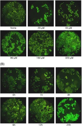
CLA에 의한 MCF-7 세포의 apoptosis
앞에서 실시한 MTT assay의 실험결과로부터 관찰된 암세 포의 증식 억제 효과가 apoptosis에 의한 것인지 아닌지를 확인하기 위하여 Hoechst 33342 염색시약을 이용한 chroma- tin staining 실험을 수행하였다. Fig. 3A에서 나타난 바와 같 이 CLA를 농도별로 MCF-7 세포에 처리했을 때, 그림에서 화살표가 가리키는 것처럼 apoptotic body를 관찰할 수 있었 다. 이러한 apoptotic body는 염색사의 응축으로부터 유발된 것이며, CLA 농도가 높아질수록 더욱 많은 apoptotic body 가 나타났는데, CLA를 농도를 160 μM로 처리했을 때, 가장 많은 apoptotic body를 확인할 수 있었다. 이와 같이 apop- totic body가 생성된 것은 CLA에 의해 MCF-7 세포에서 apoptosis가 일어난 것으로 보인다. 한편 Fig. 3B에서 보는 바와 같이, CLA 농도를 100 μM로 일정하게 MCF-7 세포에 처리하고 시간별로 apoptotic body의 발현을 관찰했을 때, 시간이 지남에 따라 apoptotic body가 증가하는 것을 관찰할 수 있었으며, 12 시간 후에 apoptotic body의 발현이 최고조 에 이르는 것으로 관찰되었다.
CLA가 reactive oxygen species (ROS) 생성에 미치는 영향
CLA 처리가 ROS의 생성에 어떤 영향을 미치는지를 알아
보기 위하여, MCF-7 세포 내의 활성산소와 반응하여 형광물
(A)
(B)
Fig. 3. Induction of apoptosis of MCF-7 cells treated with Hoechst 33342. (A) The MCF-7 cells were treated with variable concentrations of CLA. The arrows indicate cleaved nuclei in the MCF-7 cells. Cells were photo- graphed using a Nikon digital camera and a fluo- romicroscope after being treated with Hoechst 33342 staining. (B) The MCF-7 cells were treated with differ- ent time of same-dose CLA (100 μM). Under the same conditions, apoptotic bodies are shown with cells treated with Hoechst 33342.
질을 만들어내는 DCFH-DA를 이용하여 H
2O
2생성을 확인 하였다. Fig. 4A에서 보여지는 것과 같이, 배양한 MCF-7 세 포에 CLA를 다양한 농도로 6시간 동안 처리했을 때, CLA 농도가 증가할수록 ROS의 생성이 활발해지는 것을 확인할 수 있었다. 또한 Fig. 4B에서 나타난 것처럼, 시간별 실험군 에서는 CLA 농도를 100 μM로 일정하게 유지하고, 시간별로 24 시간 동안 ROS의 생성을 관찰했을 때, 시간이 경과함에 따라서 활성산소의 생성이 증가하는 것을 관찰하였다.
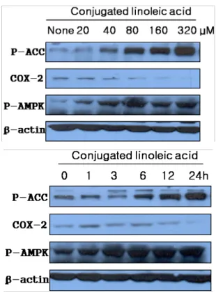
CLA 처리에 의한 AMPK 및 COX-2의 활성 변화
CLA 처리가 AMPK 및 COX-2 단백질의 활성에 어떠한
(A)(B)
Fig. 4. ROS generation by CLA in MCF-7 cells. (A) ROS gen- eration by CLA in MCF-7 cells was determined by DCFH-DA dye. Cells were treated with various con- centrations of CLA. After additional 30 min of in- cubation in the presence of 10
μ
M DCFH-DA, changes in fluorescence intensity were measured by fluo- rescence-activated cell scanning analysis. (B) Cells were treated at different time with CLA. Under the identical conditions, ROS generation by CLA in MCF-7 are shown by DCFH-DA dye.영향을 미치는지를 관찰하기 위하여 Western blot 분석을
실시한 결과, Fig. 5A에서 나타난 바와 같이 CLA를 다양
한 농도로 MCF-7에 처리하였을 때, AMPK의 기질인
ACC가 인산화 된 p-ACC 및 인산화 된 p-AMPK는 CLA
의 농도가 증가함에 따라 활성화 되었으나, 반면에 COX-2
의 발현은 감소되었다. 한편 Fig. 5B에서 제시한 바와 같
이 CLA의 농도를 100 μM로 일정하게 MCF-7 세포에 처
리하고, 시간별로 나누어 p-ACC, p-AMPK, COX-2의 활성
을 조사한 결과, 시간이 지남에 따라서 p-ACC와 p-AMPK
의 활성은 증가한 반면, COX-2의 발현은 감소된 것으로
관찰되었다.
(A)
(B)
Fig. 5. Effects of CLA on MCF-7 cells induced COX-2 expression via activating AMPK. (A) The MCF-7 cells were treated with variable concentrations of CLA for 6 hr, and then p-ACC, COX-2 and p-AMPK expression was measured by Western blot analysis. β-actin was used as an internal control. (B) The MCF-7 cells were treated with different time of same-dose CLA (100 μM). Under the same con- ditions, p-ACC, COX-2 and p-AMPK expression was measured by Western blot analysis. β-actin was used as an internal control.
고 찰
본 실험을 통하여 CLA를 다양한 농도와 시간별로 MCF-7 세포에 처리했을 때, MCF-7 세포의 증식이 억제되는 것을 관찰하였고, 이것이 apoptosis와 연관이 있는지를 알아보았 으며, 또한 이러한 apoptosis를 유도할 수 있는 AMPK 및 COX-2 단백질의 활성 변화를 확인하였다. 우선 CLA가 MCF-7 세포의 증식에 어떠한 영향을 미치는지를 조사한 MTT assay 결과는 Fig. 1A에 나타난 바와 같다. 이 그림에서 처럼 CLA의 농도가 증가 할수록 MCF-7 세포의 증식이 억제 되는 것을 관찰할 수 있었는데, 이는 Fig. 1B에서 보여 지는 것처럼 CLA를 정상인 L6 세포에 처리하였을 때에는 MCF-7 세포의 증식에 변화가 일어나지 않은 것과 비교하여 확인할 수 있었다. 또한 Fig. 2에 나타난 것처럼 CLA의 농도를 일정 하게 처리했을 때, 시간이 지남에 따라 MCF-7 세포의 증식 이 억제되는 것을 관찰할 수 있었다. 이렇게 CLA 처리에 의 해서 MCF-7 세포의 증식이 억제되는 것이 apoptosis와 연관
이 있는지를 확인하기 위하여 Hoechst 33342 염색을 이용하 여 apoptosis 발생여부를 관찰한 결과, Fig. 3A, B의 화살표 에서 보이는 바와 같이, apoptotic body 형태인 염색사의 응 축이 일어난 것을 확인할 수 있었다. 따라서 Fig. 1과 Fig. 2 에서 관찰된 MCF-7 세포의 증식 억제는 apoptosis에 의해서 유도된 것으로 보인다.
CLA에 의해서 apoptosis가 유도될 수 있다는 또 다른 실 험은 Fig. 4에서 밝힌 것처럼 ROS의 활성 측정실험을 통하여 이루어졌다. 최근의 연구보고에 의하면 대장암 세포주에서 ROS의 활성이 증가하게 되면 apoptosis가 유도되어 암세포 의 성장이 억제되는 것으로 알려져 있다[25]. Fig. 4A, B에서 나타난 것처럼, ROS 활성은 DCFH-DA를 이용한 H
2O
2의 생 성을 관찰함으로써 알 수 있는데. 이 그림에서 보듯이 농도 가 높아질수록, 또한 시간이 경과할수록 ROS의 활성이 강해 지는 것을 확인할 수 있었다. 이와 같은 결과는 ROS의 활성 이 증가함에 따라 세포 내의 미토콘드리아에서 apoptosis가 유도되는[19,26] 것으로 알려져 있는데, 결국 ROS 활성이 증 가되면 AMPK의 활성을 증가시켜 apoptosis를 유도하는 것 으로 보인다[25].
CLA 처리에 따른 apoptosis의 유도가 AMPK와 AMPK의 영향을 받는 COX-2의 발현과는 어떠한 연관성이 있는지를 밝히기 위해서 Western blot 실험을 실시하였다. 뇌의 시상 하부로부터 분비되는 AMPK는 세포 내에서 에너지 센서를 통하여 단백질의 합성을 조절하는데 중요한 역할을 한다[3].
AMPK는 우리 몸의 에너지 수준을 조절해 생체활동을 돕는 일종의 효소이며, 외부로부터의 스트레스에 대하여 세포 내 의 에너지 항상성을 유지시키는데 중요한 역할을 한다.
AMPK는 세포가 외부 환경으로부터 강한 스트레스를 받게 되었을 때, 세포 안에서 에너지 센서 역할을 하며, 이러한 AMPK의 활성도는 세포 내의 AMP:ATP의 비율에 의해 조 절된다고 알려져 있다[3]. 즉 세포 내에서 ATP양이 변화할 때, AMP 농도의 변화가 ADP 보다 훨씬 더 빠르고, 세포 내 의 에너지 상태를 인식하기 위해서는 ADP보다 AMP에 반응 하는 시스템이 훨씬 더 활발하다. 그러므로 AMPK는 세포 내에서 AMP에 의해 활성화되어 다양한 기질단백질을 조절 함으로써, 세포 내의 ATP 변화를 가장 민감하게 인식하는 단백질 키나제이다[10,11]. 최근에 AMPK가 종양억제유전자 와 연관되어 나타나는데, 암세포에 있어서 신호분자의 역할 을 통하여 암의 발달과 조절에 중요한 역할을 하는 것으로 보고되었다[11].
COX-2는 여러 자극을 통하여 체내에서 염증을 유발시키
는데, COX-2 효소를 선택적으로 억제하는 약물인 celecoxib,
아스피린, nonsteroidal anti-inflammatory drug (NSAID) 등
은 암 억제에 효과가 있는 것으로 밝혀졌으며, 이러한
COX-2는 AMPK에 의해 조절된다[12,14]. 또한 COX-2의 과
발현은 종양형성과 매우 밀접한 관계가 있는데, 암조직이 영
양분을 공급받기 위해 새로운 혈관을 만드는 angiogenesis의 대부분은 vascular endothelial growith factor (VEGF)에 의 해서 조절되며, VEGF는 COX-2의 영향아래서 angiogenesis 를 유도하는 것으로 보인다[21,27]. Fig. 5에서 나타난 바와 같이, AMPK의 활성이 증가하게 되면 COX-2의 발현이 감소 하게 되고, COX-2의 조절을 받는 VEGF가 결국 감소됨에 따 라 angiogenesis가 억제됨으로써 암의 전이가 억제되는 것으 로 알려져 있다[28]. 결론적으로, CLA를 MCF-7 세포에 처리 했을 때, AMPK가 활성화되면 COX-2의 억제효과가 나타나 암세포에서 apoptosis가 유도되는 것으로 여겨지는데, CLA 로부터 apoptosis가 유도될 수 있다는 연구는 이미 대장암 세포주를 비롯한 여러 연구들에서도 보고되고 있다[24].
CLA의 항암 효과 기전으로는 CLA가 항산화제로 작용하 거나[7,8], LA로부터 arachidonic acid가 생성되는 과정을 방 해함으로써 DNA의 합성을 억제하거나 또는 apoptosis를 유 도하여 일어나는 것으로 알려져 있다[15,16,23]. 한편, CLA는 LA와 마찬가지로 소장을 통해 흡수된 체지방에 결합되는데, 체지방 triglyceride에는 8개의 CLA 이성체 모두가 결합되지 만, 세포막의 인지질에는 cis, 9-trans, 11 CLA 이성체만이 결 합되며, 이러한 인지질과의 결합이 항암작용에 결정적인 역 할을 한다고 알려져 있다[17,22]. 또한 CLA는 체지방을 감소 시키고, 혈중 콜레스테롤을 낮추어 동맥 경화증의 발생을 억 제시키며, 식이효율을 증가시킴으로써 성장 인자로 작용한 다고 알려져 있다[4]. 이 외에도, 면역 증진의 효과가 있고, 인슐린 민감성을 증가시켜 당뇨병에도 효과가 있는 것으로 보고되었다[1].
이상의 결과들로부터, CLA를 MCF-7 세포에 처리했을 때, CLA의 농도 및 시간 의존적으로 암세포의 증식이 억제되는 것은, 암세포의 apoptosis에 의해 유도된 것으로 보인다.
CLA의 이러한 특성은 앞으로 VEGF의 연구와 함께, COX-2 와 연결된 mitogen activated protein kinase (MAPK)-신호경 로의 연구를 통하여 더욱 상세하게 밝혀질 것으로 여겨진다.
CLA와 마찬가지로, AMPK를 활성화시키는 물질들, 예를 들 어, 녹차에서 유래한 epigallocatechin-3-gallate (EGCG)는 암 세포의 전이에 중요한 유전자인 VEGF와 glucose trans- porter 1 (Glut1)을 감소시켜 암의 전이를 방해하며[13], 고추 의 성분인 capsaicin은 여러 단계의 신호경로를 통하여 대장 암세포의 증식을 억제하고 apoptosis를 유도하는 것으로 밝 혀져 있다[20]. 따라서 본 연구는 향후 식품 혹은 의약품속에 포함된 어떤 성분에서, 암세포의 증식 억제와 apoptosis의 유 도에 관련된 연구를 하고자 할 때, 과학적으로 도움이 될 수 있는 연구논문이라고 여겨진다.
요 약
본 연구는 쇠고기와 유제품에 들어 있는 CLA의 항암효과
를 조사하기 위하여 수행되었다. 이 실험을 위하여 MCF-7 인 체 유방암 세포주를 사용하였으며, CLA를 처리했을 때 MCF-7 세포의 증식은 CLA의 농도가 증가할수록, 또한 일정 한 농도에서는 시간이 경과함에 따라 의존적으로 억제되었다.
이와 같이 암세포의 증식이 억제되는 이유는 Hoechst 33342 염색을 이용한 chromatin staining 및 ROS의 활성 측정실험 결과, apoptosis와 연관이 있는 것으로 확인되었다. CLA 처리 에 의한 apoptosis가 AMPK 및 COX-2 단백질의 활성 발현과 는 어떤 연관성이 있는지를 조사하기 위하여 Western blot 실 험을 실시한 결과, CLA 처리에 따라 AMPK의 활성이 증가되 었고, COX-2의 발현은 감소됨으로써, MCF-7 세포에서 apop- tosis가 유도되었다는 것을 알 수 있었다. 본 연구를 통하여 조사한 CLA의 항암효과로부터, 향후 다른 식품에 포함된 성 분들에서도 암세포의 증식 억제와 apoptosis의 유도를 연구할 수 있는 기초 자료를 제시한 것이라고 할 수 있다.
감사의 글
본 논문은 2008학년도 한남대학교 교비학술연구비의 지원 으로 수행되었으며 이에 감사드립니다.
References
1. Brown, J. M. and M. K. Mclntosh, 2003. Conjugated lino- leic acid in humans: regulation of adiposity and insulin sensitivity. J. Nutr. 133, 3041-3046.
2. Campas, C., J. M. Lopez, A. F. Santidrian, M. Barragan, B.
Bellosillo, D. Colomer and J. Gil. 2003. Acadesine activates AMPK and induces apoptosis in B-cell chronic lympho- cytic leukemia cells but not in T lymphocytes. Blood 101, 3674-3680.
3. Carling, D. 2004. The AMP-activated protein kinase cas- cade a unifying system for energy control. Trends Biochem.
Sci. 29, 18-24.
4. Chin, S. F., J. M. Storkson, W. Liu, K. J. Albrigt, M. E.
Cook and M. W. Pariza. 1994. Conjugated linoleic acid is a growth factor for rats as shown by enhanced weight gain and improved feed efficency. J. Nutr. 24, 2344-2349.
5. Chujo, H., M. Chujo, S. Yamasaki, N. Nou, H. Koyanagi, K. Tachiban and Yamada. 2003. Effect of conjugated lino- leic acid isomers on growth factor-induced proliferation of human breast cancer cells. Cancer Lett. 202, 81-87.
6. Doll, R. and R. Peto, 1981. The cause of cancer: quantita- tive estimates of avoidable risks of cancer in the united states today. J. Natl. cancer Inst. 66, 1191-1308.
7. Ha, Y. L., N. K. Grimm and M. W. Pariza. 1987.
Anticarcinogens from fried ground beef: heat-altered de- rivatives of linoleic acid. Carcinogenesis 8, 1881-1887.
8. Ha, Y. L., N. K. Grimm and M. W. Pariza. 1989. Newly recognized anticarcinogenic fatty acid. Identification and quantification in natural and processed chesses. J. Agr.
Food Chem. 37, 75-81.
9. Ha, Y. L., J. Storkson and M. W. Pariza. 1990. Inhibition of benzo (a) pyrene-inudced mouse forestomach neoplasia by conjugated dienoic derivatives of linoleic acid. Cancer Res. 50, 1097-1101.
10. Hardie, D. G. 2003. The AMP-activated protein kinase cas- cade: The key sensor of cellular energy status. Endocrinology 144, 5179-5487.
11. Hardie, D. G. 2005. New roles for the LKB1/AMPK pathway. Curr. Opin. Cell Biol. 17, 167-173.
12. Hasegawa, K., Y. Ohashi, K. Ishikawa, A. Yasue, R. Kato and Y. Achiwa 2005. Expression of cyclooxygenase-2 in uterine endometrial cancer and anti-tumor effects of a se- lective COX-2 inhibitor. Int. J. Oncol. 26, 1419-1428.
13. Hwang, J. T., I. J. Park, J. I. Shin, Y. K. Lee, S. K. Lee, H.
W. Baik, J. Ha and O. J. Park 2005. Genistein, EGCG, and capsaicin inhibit adipocyte differentiation process via acti- vating AMP-activated protein kinase. Biochem. Biophys.
Res. Commun. 338. 694-699.
14. Hwang, J. T., Y. M. Kim, Y. J. Surh, H. W. Baik, S. K. Lee, J. Ha and O. J. Park. 2006. Selenium regulates COX-2 and ERK signaling pathways by activating AMPK in colon cancer cells. Cancer Res. 66, 10057-10063.
15. Ip, C. 1997. Review of the effects of trans fatty acids, oleic acid, n-3 polyunsaturated fatty acids, and conjugated lino- leic acid on mammary carcinogenesis in animals. Am. J.
Clin. Nutr. 66, 1523-1529.
16. Ip, C., S. F. Chin, J. A. Scimeca and M. W. Pariza. 1991.
Mammary cancer prevention by conjugated dienoic de- rivative of linoleic acid. Cancer Res. 51, 6118-6124.
17. Ip, C, M. Singh, H. J. Thompson and J. A. Scimeca. 1994.
Conjugated linoleic acid suppress mammary carcino- genesis and proliferative activity of the mammary gland in the rat. Cancer Res. 54, 1212-1215.
18. Kim, E. J., I. J. Kang, H. J. Cho, W. K. Kim, Y. L. Ha and J. H. Park. 2005. Conjugated linoleic acid downregulates insulin-like growth factor-I receptor levels in HT-29 hu- man colon cancer cells. J. Nutr. 133, 2675-2681.
19. Kim, K. H., K. J. Kang and H. S. Park. 2002. Effect of con- jugated linoleic acid on colon tumor incidence and anti-
oxidant enzymes and fecal excretion of secondary bile acids in DMH_treated rats. Kor. J. Nutr. 35, 1308-1044.
20. Kim, Y. M., J. T. Hwang, D. W. Kwak, Y. K. Lee and O.
J. Park. 2007. Involvement of AMPK signaling cascade in capsaicin-induced apoptosis of HT-29 colon cancer cells.
Ann. N.Y. Acad. Sci. 1095, 496-503.
21. Masso-Welch, P. A., D. Zangani, C. Ip, M. M. Vaughan, S.
F. Shoemaker, S. O. McGee and M. M. Ip. 2004. Isomers of conjugated linoleic acid differ in their effects on angio- genesis and survival of mouse mammary adipose vasculature. J. Nutr. 134, 299-307.
22. Ochoa, J. J., A. J. Farquharson, I. Grant, L. E. Moffat, S. D.
Heys and K. W. Wahle. 2004. Conjugated linoleic acids (CLA’s) decrease prostate cancer cell proliferation: differ- ent molecular mechanisms for cis-9, trans-11 and trans-10, cis-12 isomers. Carcinogenesis 25, 1185-1191.
23. Oh, Y. S., H. S. Lee, H. J. Cho, S. G. Lee, K. C. Jung and J. H. Park. 2003. Conjugated linoleic acid inhibits DNA synthesis and induces apoptosis in TSU-Pr1 human blad- der cancer cells. Anticancer Res. 23, 4765-4772.
24. Palombo, J. D., A. Ganguly, B. R. Bistrian and M. P.
Menard. 2002. The antiproliferative effects of biologically active isomers of conjugated linoleic acid on humanand prostatic cancer cells. Cancer Lett. 177, 163-172.
25. Park, I. J., J. T. Hwang, Y. M. Kim, J. H. Ha and O. J.
Park. 2006. Differential Modulation of AMPK Signaling Pathways by Low or High Levels of Exogenous Reactive Oxygen Species in Colon Cancer Cells. Ann. N.Y. Acad.
Sci. 1091, 102-109.
26. Skulachev, V. P. 2005. How to clean the direct place in the cell; cationic antioxidants as intramitochondrial ROS seavengers. IUBMB Life 57, 305-310.
27. Tjiu, J. W., Y. H. Liao, S. J. Lin, Y. L. Huang, W. L. Tsai, C. Y. Chu, M. L. Kuo and S. H. Jee. 2006. Cyclooxygenase-2 overexpression in human basal cell carcinoma cell line in- creases antiapoptosis, angiogenesis, and tumorigenesis. J.
Invest Dermatol. 126, 1143-1151.
28. Veikkola, T. and K. Alitalo. 1999. VEGF's Receptors and angiogenesis. Semin. Cancer Biol. 9, 211-220.