39(8), 1119~1125(2010) DOI: 10.3746/jkfn.2010.39.8.1119
Primary 인체 전립선 암세포에서 Resveratrol의 Apoptosis 유도 효과
강혜인
1
․김재용2
․조현동1
․박경욱3
․강점순4
․서권일1†
1
순천대학교 식품영양학과,
2순천대학교 기초과학연구소
3
(주)에스바이오푸드,
4부산대학교 원예생명과학과
Resveratrol Induces Apoptosis in Primary Human Prostate Cancer Cells
Hye-In Kang
1
, Jae-Yong Kim2
, Hyun-Dong Cho1
, Kyung-Wuk Park3
, Jum-Soon Kang4
, and Kwon-Il Seo1†
1
Dept. of Food and Nutrition and2
Research Institute of Basic Science, Sunchon National University, Jeonnam 540-742, Korea3
S-biofood, Jeonnam 540-742, Korea4
Dept. of Hortcultural Bioscience, Pusan National University, Gyeongnam 627-702, KoreaAbstract
To evaluate resveratrol as a prostate cancer preventive material, we investigated its anti-proliferative and apoptotic effects in RC-58T/h/SA#4 primary human prostate cancer cells. Resveratrol significantly decreased the number of viable RC-58T/h/SA#4 cells in a dose- and time-dependent manner. Resveratrol showed cytotox- icity against RC-58T/h/SA#4, LNCaP, PC-3 human prostate cancer cells with IC
50values of 245, 320 and 340 μ M, respectively. However the cytotoxic potential of resveratrol against normal RWPE-1 cells was lower (IC
50=982 μM). Resveratrol induced cell death as evidenced by the increased formation of apoptotic bodies, nuclear condensation, sub-G1 phase, and DNA fragmentation. Resveratrol activated initiator caspases 8, and 9 as well as effector caspase 3 in a dose-dependent manner. Furthermore, the general caspase inhibitor z-VAD-fmk sig- nificantly inhibited resveratrol-induced apoptosis compared to cells without treatment. These results clearly indicate that resveratrol-induced apoptosis was dependent on caspase activation. Further, resveratrol modulated the down regulation of Bcl-2 (anti-apoptotic), and Bid. However, the level of Bax (pro-apoptotic) remained unchanged. These results suggest that resveratrol induced apoptosis in RC-58T/h/SA#4 cells via a mitochon- drial-mediated caspase-dependent pathway, suggesting therapeutic potential against prostate cancer.
Key words: resveratrol, prostate cancer cell, RC-58T/h/SA#4, apoptosis
†
Corresponding author. E-mail: [email protected]
†
Phone: 82-61-750-3655, Fax: 82-61-752-3657
서 론
암은 정상세포가 외부 자극에 의해서 유전자의 형질전환 이 발생되면 변형세포가 무절제한 증식을 함으로써 형성된 악성 종양으로, 전 세계적으로 인간의 생명을 위협하는 가장 중요한 요소로 지목되고 있다(1). 암은 세계 사망원인 중 1위 를 차지하고 있는 질환이며(2), 우리나라에서도 암으로 인한 사망률이 계속 증가되고 있는 실정이다. 특히, 전립선암은 미국에서 남성암 중 가장 발생빈도가 높고, 암 관련 사망의 두 번째 원인이 되는 종양으로 국내에서도 서구화된 식습관, 고령화 등의 생활양식 변화로 그 발생빈도 및 사망률이 증가 하고 있는 추세이다(3,4). 기존에 이용되고 있는 항암제는 면역기능저하, 조혈장애, 탈모 및 유전자 손상 등의 부작용 을 유발한다. 최근 이러한 전립선암을 예방 및 치료하기 위 해 부작용을 감소시키면서 암세포에만 특이적으로 작용하
여 정상세포에 영향을 주지 않는 천연물 유래 항암치료제를 개발하는 연구들이 진행되고 있다(5). 이러한 항암치료제는 암세포의 사멸을 apoptosis를 유도하여 암의 발생 및 진행을 억제한다고 보고하고 있다(6).
Apoptosis는 세포가 정상적인 상태에서나 또는 병리학적 요인에 노출된 후 죽음에 이루게 되는 생리학적 과정이며, 대표적인 현상으로 세포질 및 염색질 응축, 세포막 수포화 현상, DNA 단편화 등이 수반되는데 이러한 현상은 세포내 부의 정교한 신호전달에 의해 조절함으로써 조직의 항상성, 감염이나 손상된 세포에 대한 방어를 통해 암을 억제할 수 있다(7,8).
Resveratrol은 많은 식물에서 박테리아를 포함한 다양한
외부 환경의 변화에 식물체 스스로가 자신을 보호하기 위한
수단으로 만들어내는 phytoalexin의 일종이며, 포도, 오디
및 땅콩을 포함한 약 70여종 이상의 식물체에서 발견되어지
고 있다(9). 특히 식물 종자들이 발아하면 유용한 생리활성 물질들이 최대가 된다고 보고되고 있다(10). 이러한 발아채 소의 주요 생리활성물질 중 하나가 resveratrol이라고 알려 져 있다(11).
Resveratrol은 다양한 암세포주의 사멸을 apoptosis를 유 도하여 억제한다고 보고하고 있다(12-14). 그러나 전립선암 세포에 대한 연구는 특정 세포에만 국한되어 진행되고 있다.
기존의 전립선 암 연구로 사용된 암세포인 LNCaP, PC-3 세포는 다른 조직으로 전이된 세포로 각각 상피 및 척추로 전이된 형태의 세포로 순수한 전립선암세포로 보기는 어렵 다(15). 하지만 본 연구에서 사용된 인체 전립선 암세포인 RC-58T/h/SA#4 세포는 전이되기 전 형태의 primary can- cer 조직으로부터 분리된 세포로서 앞으로
in vitro에서 전립 선암 연구를 위해 새로운 전립선 암세포 배양 모델로서 중요 한 역할을 할 것으로 생각된다.
따라서 본 연구에서는 resveratrol을 전립선 암 예방 치료 제로 사용하기 위한 기초 자료를 제공하기 위하여 resvera- trol에 의한 RC-58T/h/SA#4 인체 전립선 암세포의 사멸 및 그 기전에 대하여 조사하였다.
재료 및 방법
암세포 배양 및 resveratrol 처리
본 실험에 사용한 인체 전립선암세포주인 LNcaP, PC-3 및 DU145는 한국세포주은행(KCLB)에서 분양 받았으며, primary 인체 전립선 암세포인 RC-58T/h/SA#4 및 RWPE- 1 정상세포는 Center for Prostate Disease Reserch(CPDR, Washington, DC, USA)로부터 분양 받아 10% FBS(fetal bovine serum)를 첨가한 DMEM(Gibco, NY, USA) 배지를 첨가하여 37
oC, 5% CO
2incubator에서 계대배양하면서 실 험에 사용하였다. Resveratrol(Sigma Chemical Co., St.
Louis, MO, USA)은 구입하여 DMSO로 용해하여 본 연구에 사용하였다.
암세포 증식 억제효과
Monolayer로 자란 전립선 암세포주를 0.25% trypsin- EDTA용액으로 처리하여 single cell로 만든 후 최종농도가 1×10
5cells/mL가 되도록 희석하여 24 well plate에 분주한 다음 37
oC, 5% CO
2incubator(HERA cell 150, Heraeus, Hanau, Germany)에서 24시간 동안 배양한 후, resveratrol 를 50, 100, 300 및 500 μM 농도로 첨가하여 24, 48 및 72시간 반응시켜 암세포 증식 정도를 SRB(sulforhodamine B, Sigma Chemical Co.) 및 cell counting 방법에 의하여 측정 하였다(16).
암세포 형태의 관찰
SRB 정량 분석을 하기 전에 대조군과 실험군의 암세포 모양 변화를 200 배율의 위상차 현미경으로 관찰하고 사진
을 촬영하였다.
Hoechest 332580 염색을 통한 핵의 관찰
RC-58T/h/SA#4 세포를 1×10
6cells/mL 밀도로 분주한 다음 37
oC, 5% CO
2incubator에서 24시간 배양한 후 resvera- trol을 100, 300 μM 농도를 처리하여 24시간 배양하였다. 배 양이 종료된 well에서 회수한 세포를 PBS로 3회 세척하고 Hoechest 332580을 첨가하여 실온에서 20분 염색한 후 형광 현미경으로 관찰하였다(17).
Sub-G1 함량 분석
Resveratrol 처리에 의하여 apoptosis가 유도된 세포의 빈 도를 확인하기 위하여 flow cytometry 분석을 실시하였다.
Monolayer로 배양한 세포주를 0.25% trypsin-EDTA 용액 으로 처리하여 single cell로 만든 후 최종 농도가 1×10
6cells/mL가 되도록 희석하여 6 well plate에 분주한 후 24시 간 배양한 후 resveratrol을 50, 100, 300 및 500 μM 농도로 처리하여 24시간 반응시켰다. 암세포를 PBS(phosphate buf- fered saline)로 2회 세척한 후 70% 에탄올을 첨가하고 4
oC 에서 하루 동안 방치하였다. 고정된 암세포를 PBS로 세척하 고 RNase(0.1 mg/mL)를 첨가하여 37
oC 30분 반응시킨 후 1 mg/mL의 PI(propidium iodide) 용액으로 30분간 염색한 후 Flow cytometer(EPICS XL, Backman Coulter, California, USA)를 이용하여 분석하였다(16).
DNA 분절 측정
Resveratrol에 의한 DNA 분절이 일어나는지 알아보기 위 하여 Wan 등(18)의 방법에 따라 RC-58T/h/SA#4 세포를 10 mm dish에 5×10
6cells/10 mL로 분주하고 resveratrol을 300, 500 μM 농도로 처리한 후 2시간 배양하였다. 반응이 종료된 dish에서 회수한 세포를 PBS로 3회 세척 후 상등액 을 제거한 세포에 lysis buffer(NET buffer, Proteinase K, 20% SDS)를 첨가하여 50
oC에서 4시간 동안 반응시킨 후 phenol buffer를 이용하여 DNA를 추출하였다. 2% agarose gel에서 전기영동 하여 DNA ladder를 확인하였다.
Caspase 활성 측정
Caspase의 활성은 Colorimetric assay kit(BioVison, CA,
USA, 색도 분석법)로 제조사의 방법에 따라 측정하였다. 즉,
monolayer로 배양한 전립선 암세포주를 0.25% trypsin-
EDTA 용액으로 처리하여 단일 세포로 만든 후 배양액으로
최종농도가 1×10
7cells/mL가 되도록 희석하여 10 mm dish
에 seeding한 다음 37
oC, 5% CO
2incubator에서 24시간 배양
하였다. 24시간 후 각 dish의 배양액을 제거한 후 다양한 농
도로 준비한 시료가 포함된 새 배양액을 dish에 첨가하고
24시간 더 반응시켰다. 반응이 종료된 dish에서 회수한 세포
를 PBS로 3회 세척 후 상등액을 제거한 세포에 50 μL lysis
buffer를 첨가하여 용해하고, 각각의 caspase 기질과 함께
37
oC에서 2시간 간격으로 incubation하였다. 반응 종료 후
96 well plate용 microplate reader로 405 nm에서 흡광도를 측정하여 활성 정도를 확인하였다.
Caspase inhibitor 측정
Resveratrol에 의한 전립선암세포(RC-58T/h/SA#4)의 사멸이 caspase에 의해 의존하여 일어나는 지를 확인하기 위해 RC-58T/h/SA#4의 최종 농도가 1×10
5cells/mL가 되 도록 희석하여 24 well에 첨가한 후 24시간 배양하였다.
Resveratrol을 처리하기 전에 caspase-family inhibitor(z- VAD-fmk)를 처리하고 2시간 후에 resveratrol를 농도별로 첨가하여 24시간 반응 후 trypan blue 염색을 통하여 측정하 였다(19).
Western blot을 이용한 단백질 분석
Monolayer로 배양한 세포를 0.25% trypsin-EDTA용액 으로 처리하여 single cell로 만든 후 배양액으로 최종 농도 가 1×10
7cells/mL가 되도록 희석하여 10 mm dish에 첨가 한 다음 37
oC, 5% CO
2incubator에서 24시간 배양하였다.
24시간 후 각 dish의 배양액을 제거한 후 다양한 농도로 준비 한 시료가 포함된 새 배양액을 dish에 첨가하고 48시간 더 반응시켰다. 반응이 종료된 dish에서 회수한 세포를 PBS로 3회 세척 후 500 μL lysis buffer(50 mM Tris, 150 mM NaCl, 1 mM EDTA, 50 mM NaF, 30 mM Na
4P
2O
7, 1 mM PMSF, 2 μg/mL aprotinin)를 첨가하여 얼음에서 용해하였다. 세포 용해액과 2×sample buffer를 동량으로 섞어 65
oC에서 5분간 끓인 후 12% sodium dodesyl sulfate-polyacrylamide gel electrophoresis(SDS-PAGE)를 시행하였다. 전기영동이 끝 난 gel의 단백질을 nitrocellulose membrane으로 transfer한 후 blocking buffer(2.5%, 5% BSA)로 상온에서 1시간 반응 시킨 후 primary antibody(anti-Bax, anti-Bcl-2, anti-Bid) 를 희석하여 4
oC에서 하루 동안 반응시켰다. T-TBS로 1시 간 이상 세척하고 secondary anti-rabbit IgG conjugated HRP를 희석하여 4
oC에서 1시간 반응시켰다. 반응 종료 후 membrane을 T-TBS로 1시간 이상 세척하고 enhanced chemiluminescence kit(ECL kit)를 사용하여 필름에 노출시 켜 단백질을 확인하였다(18).
통계처리
실험결과는 평균 표준편차로 표시하였으며, 각 실험군을 대조군에 대한 백분율로 나타내었다. 대조군과 실험군간의 통계적 유의성에 대한 검증은 Student's
t-test를 이용하여, p value가 0.05 미만일 때 통계적으로 유의성이 있다고 판단 하였다.
결과 및 고찰
Resveratrol의 RC-58T/h/SA#4 암세포 성장 억제효과 전립선 암세포의 증식에 미치는 resveratrol의 영향을 정 량적으로 측정하기 위하여 LNCaP, PC-3 및 RC-58T/h/
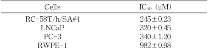
Table 1. IC
50values of resveratrol for 24 hr on prostate can- cer cells
Cells IC
50(μM)
RC-58T/h/SA#4 LNCaP RWPE-1 PC-3
245±0.23 320±0.45 340±1.20 982±0.98 Data values were expressed as mean±SD (n=3).
SA/#4의 3종의 전립선 암세포를 대상으로 IC
50값을 측정하 였다. 이들 전립선 암세포의 IC
50값의 범위는 245 μM에서 340 μM이었으며, 특히 RC-58T/h/SA#4 암세포에서 IC
50값 이 250 μM으로 가장 높았다. 그러나 전립선 정상세포인 RWPE-1의 저해 농도 IC
50값은 전립선 암세포보다 낮아 resveratrol은 암세포의 성장을 선택으로 억제하는 것을 알 수 있었다(Table 1).
Resveratrol의 IC
50의 활성이 가장 높은 RC-58T/h/SA#4 세포의 증식에 미치는 영향을 조사하기 위하여 50, 100, 300 및 500 μM 농도로 첨가하여 24, 48 및 72시간 동안 반응 후 암세포 증식을 측정한 결과는 Fig. 1과 같다. 즉 resvera- trol 처리 농도가 증가할수록 농도 의존적으로 유의적인 세 포 증식이 감소하였다. Resveratrol에 의한 세포 증식 감소 는 resveratrol 100 μM 농도로 처리 후 24시간부터 나타났으 며 시간이 경과함에 따라 세포 증식 감소는 더욱 현저히 나 타났다(Fig. 1A). 또한 trypan blue 염색을 통한 결과에서도 Fig. 1A 결과와 같이 농도 및 시간 의존적으로 암세포의 밀 도는 감소하였으며, 성장 억제율은 증가하는 것을 확인할 수 있었다(Fig. 1B). Resveratrol을 100, 300 μM 농도로 처리 하여 24시간 후 위상차 현미경으로 관찰한 결과 암세포의 증식이 감소하였으며, culture plate로부터 분리되어 배양액 에 부유한 세포는 일정한 모양을 잃고 크기가 줄어 사멸되는 것을 관찰할 수 있었다(Fig. 1C).
Aziz 등(20)은 resveratrol을 LNCaP 전립선 암세포와 HPEC 전립선 정상세포에 1, 2, 5, 25 및 50 μM 농도로 24시 간 처리하여 trypan blue exclusion 및 MTT 방법으로 세포 성장 억제효과를 비교 측정한 결과 LNCaP 전립선 암세포는 농도 의존적으로 성장이 억제하였으나, HPEC 세포의 성장 에는 영향을 미치지 않았다고 보고하였다. 본 연구 결과를 이전의 연구 결과와 비교하였을 때 resveratrol이 전립선 암 세포 성장을 억제하는 농도 차이는 있었으나, 전립선 암세포 의 성장을 선택적으로 억제하는 효과는 동일하였다. 따라서 resveratrol은 암세포에만 선택적으로 작용하는 전립선 항암 제로서 활용할 수 있을 것으로 생각된다.
Resveratrol이 RC-58T/h/SA#4의 apoptosis에 미치 는 영향
Resveratrol에 의한 전립선 암세포증식 억제효과가 apop-
tosis에 의한 것인 지를 확인하기 위하여 Hoechst 332580
염색을 통한 핵 관찰, 세포주기 중 sub-G1 함량 및 DNA
0 20 40 60 80 100 120
Control 50 100 300 500
Concentration (μM)
S u rvi va l ra te ( % ) .
24 hr 48 hr 72 hr
*
*
*
*
*
*
*
* *
* * (A)
0 10 20 30 40 50 60 70
Control 50 100 300 500
Concentration (μM) C e ll d e n si ty ( × 1 0
4) .
0 20 40 60 80 100 120
D e a d ce lls (% ) .
24 hr 48 hr 72 hr
24 hr 48 hr 72 hr
* *
* *
*
* * * * * * (B)
(C)
Fig. 1. Effect of resveratrol on the viability of RC-58T/h/
SA#4 human prostate cancer cells. (A) Cells were treated with resveratrol (0~500 μM) for 24, 48 and 72 hr after which cell growth was measured by SRB assay. Data values were expressed as mean±SD (n=3). Significant differences were compared with the control at
*p<0.05 using the Student's t-test. (B) Cells were treated with resveratrol (0~500 μM) for 24, 48 and 72 hr after which the viability of cells was determined by trypan blue ex- clusion assay. Data values were expressed as mean±SD (n=3).
Significant differences were compared with the control at
*p<0.05 using the Student's t-test. (C) After 24 hr incubation with 100 or 300 μM resveratrol, cell morphology was visualized by inverted microscopy. Magnification, ×200.
절편을 관찰하였다. Fig. 2A에 나타난 바와 같이 resveratrol 을 처리하지 않은 대조군은 핵의 모양이 정상적으로 염색된 반면에 resveratrol 100 및 300 μM 농도로 처리 시 세포 밀도 의 감소와 더불어 핵 응축 현상이 나타났다. 또한 resveratrol 에 의해 유도된 apoptosis 정도를 정량적으로 분석하기 위하 여 세포주기 중 sub-G1기에 속하는 세포의 함량을 flow cytometry를 이용하여 측정한 결과 resveratrol을 처리하지 않은 대조군의 sub-G1 함량이 약 1.54%였으나, resveratrol 처리 시 sub-G1 변화량이 농도 의존적으로 증가하였으며, 100 μM 농도에서 20% 이상 그 함량이 증가하였다(Fig. 2B).
(A)
0 10 20 30 40 50 60
Control 50 100 300
Concentration (µM)
S u b -G 1 p o p u la ti o n (% ) .
*
*
* (B)
(C) M Control 100 300 μM
Fig. 2. Effect of resveratrol on apoptosis in RC-58T/h/SA#4 human prostate cancer cells for 24 hr. (A) Apoptotic body for- mation was observed under a fluorescent microscope after Hoechst 332580 staining (magnification×400). (B) Sub-G1 con- tent was detected by flow cytometry after propidium iodide staining. Significant differences were compared with the control at
*p<0.05 using the Student's t-test. (C) DNA fragmentation was observed by 2% agarose gel electrophoresis. M refers to the 100 bp DNA marker.
또한 resveratrol을 100 및 300 μM 농도로 처리 시 DNA 분절 현상이 나타났다(Fig. 2C).
Wang 등(4)은 resveratrol를 5 μM 농도로 LNCap 전립선
암세포의 72시간 처리하여 세포주기를 측정한 결과 대조군
에 비하여 sub-G1 함량이 증가하였다고 보고하였다. 한편
Gill 등(21)은 전립선 암세포인 PC-3, DU145에 resveratrol
을 50 μM 농도를 24시간 동안 처리하여 Hoechst 염색을
통하여 핵의 관찰한 결과 핵 응축 및 절편이 나타났다고 보
고하였다. 따라서 resveratrol에 의한 RC-58T/h/SA#4 전립
선 암세포의 사멸은 apoptosis 유도와 연관이 있음을 알 수
있었다.
Resveratrol에 의한 caspase 활성
Caspase은 세포사멸을 조절하는 주요한 조절인자로 in- itiator caspase와 effector caspase로 구분되며, initiator caspase는 death 신호에 의해 활성화 되어 effector caspase 를 활성화 시킨다(22).
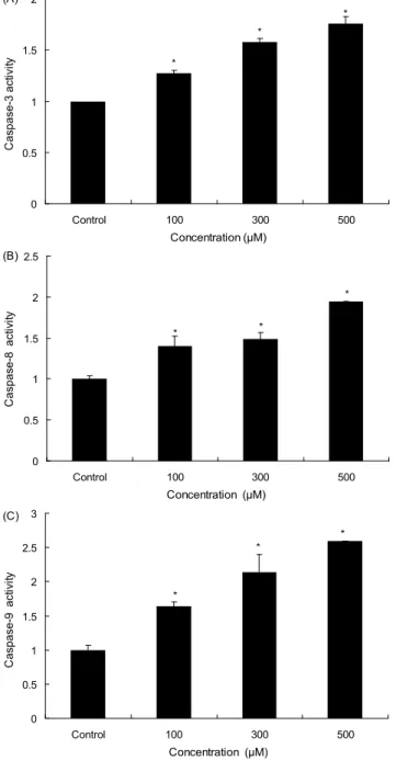
본 연구에서는 resveratrol이 caspase 활성에 미치는 영향 을 조사하기 위하여 initator caspase에 해당하는 caspase-8, -9와 effector caspase인 capase-3의 활성을 조사하였다.
*
*
*
0 0.5 1 1.5 2
Control 100 300 500
Concentration (μM)
C a sp a se -3 a ct ivi ty .
(A)
0 0.5 1 1.5 2 2.5
Control 100 300 500
Concentration (μM)
C a sp a se -8 a ct ivi ty . *
* *
(B)
0 0.5 1 1.5 2 2.5 3
Control 100 300 500
Concentration (μM)
C a sp a se -9 a ct ivi ty .
*
*
* (C)
Fig. 3. Caspase activity in RC58T/h/SA#4 cells treated with resveratrol. (A) caspase-3 activity, (B) caspase-8 activity, (C) caspase-9 activity. Data values were expressed as mean±SD (n=3). Significant differences were compared with the control at
*
p<0.05 by Student’s t-test.
Fig. 3에서 보는 바와 같이 resveratrol 첨가군은 농도 의존 적으로 caspase-3 활성이 증가되어, 100 μM 농도 이상에서 대조군에 비하여 약 1.5 배 정도 증가되었다. Caspase-8 및 -9의 경우도 resveratrol 처리 농도의 증가에 따라 활성의 정도가 증가되었다.
또한 resveratrol 처리에 의한 apoptosis 유도가 caspase 활성과 관련성이 있는 지를 재확인하기 위하여 caspase inhibitor, z-VAD-fmk를 처리하여 cell counting 방법을 통 하여 암세포 성장 억제효과를 조사하였다(Fig. 4). 그 결과 resveratrol 처리 전 z-VAD-fmk(10 μM)를 2시간 전 처리한 군은 resveratrol을 단독으로 처리한 군보다 살아 있는 세포 의 밀도가 증가하였다.
Gill 등(21)은 resveratrol 농도 50 μM을 전립선 암세포인 PC-3, DU145에 24시간 처리하여 capase-3, -8, -9 단백질 활성을 Western blot 방법에 의해 측정한 결과 이들 caspase 단백질들의 활성이 유도되었다고 보고하였다.
이전의 연구 결과와 본 연구 결과를 종합해 보면 resvera- trol에 의한 전립선 암세포의 apoptosis 유도에 caspase가 매우 중요한 역할을 하고 있음을 시사하고 있다.
Resveratrol에 의한 Bcl-2 family의 단백질 발현 변화 Apoptosis는 다양한 단백질들의 유기적인 작용으로 일어 나는 과정으로 세포 내․외 경로를 통해 조절된다. 즉 Bcl-2 family 단백질은 미토콘드리아 막 투과성을 조절하는 단백 질로 미토콘드리아 막에 존재하거나 세포사멸 유도 신호에 의해 미토콘드리아 막으로 이동하여 세포사멸을 조절하는 중요한 단백질이다(6). 그중 Bcl-2, Bcl-xL은 anti-apop- totic 인자로서 apoptosis의 유발을 억제하는 기능을 가지며, Bax, Bad 등은 pro-apoptotic 인자로 apoptosis 유도를 증가 시킨다(23). 또한 Bid는 cytosol에 비활성 상태로 존재하며 caspase-8에 의해 truncated Bid(
t-Bid)로 분절되어 미토콘
0 10 20 30 40 50
Control 100 300 500
Concentration (μM) C e ll d e n si ty ( × 1 0
4) .
(-) z-VAD-fmk (+) z-VAD-fmk
* *
*
*
*
*
Fig. 4. Effect of caspase inhibitor (Z-VAD-fmk) on cell
death induced by resveratrol. RC58T/h/SA#4 cells were pre-
incubated with 10 μM Z-VAD-fmk for 2 hr and then treated with
various concentrations of resveratrol. Data values were expressed
as mean±SD (n=3). Significant differences were compared with
the control at
*p<0.05 by Student’s t-test.
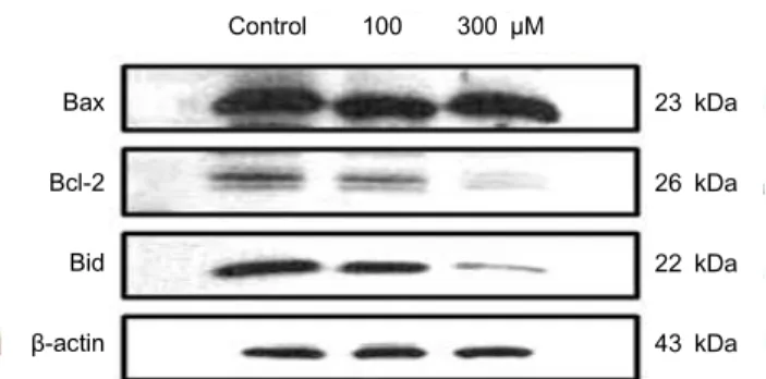
Control 100 300 μM Bax
Bcl-2
Bid β-actin
23 kDa
26 kDa
22 kDa
43 kDa Fig. 5. Effect of resveratrol on the levels of Bcl-2 family proteins in RC58T/h/SA#4 cells. RC58T/h/SA#4 cells were in- cubated with 100, 300 μM resveratrol for 24 hr after which cells were harvested and lysed. Protein lysates were subjected to 12%
SDS-PAGE and then immunoblotted with corresponding anti- bodies.
드리아 막으로 이동하여 미토콘드리아를 통한 내적 경로를 활성화시켜 death receptor를 통한 외적인 경로와 미토콘드 리아를 통한 내적 경로가 서로 연결된다고 알려져 있다(24).
본 연구에서는 resveratrol이 RC-58T/h/SA#4 세포의 apoptosis 유도 경로를 확인하기 위하여 Western blot를 통 하여 apoptosis를 조절하는 단백질 발현 양상을 조사하였다.
Fig. 5에 나타낸 바와 같이 resveratrol은 pro-apoptotic 인자 인 Bax 단백질 발현에는 영향을 미치지 않았으나, anti- apoptotic 단백질인 Bcl-2 발현은 감소하였다. 또한 resvera- trol에 의해
t-Bid의 발현을 확인할 수 없으나 분자량 22 kDa 인 full length Bid가 감소하였으므로 resveratrol에 의해 Bid 의 분절화가 유도되었을 것으로 생각된다.
Kim 등(23)은 후추의 성분인 piperine이 대장암세포의 사 멸을 유도하는 지를 확인하기 위하여 Bcl-2 family 단백질 발현의 조사한 결과, pro-apoptotic 인자인 Bax 발현에는 영 향을 미치지 않았으나, anti-apoptotic 인자인 Bcl-2 단백질 수준은 감소하였다고 보고하였다. 또한 Park 등(6)은 목향 헥산추출물이 LNCaP 전립선 암세포의 사멸의 Bcl-2 family 단백질과의 연관성을 조사한 결과 pro-apoptotic 단백질 중 Bax 단백질 수준은 변화하지 않았으나, Bak 단백질의 발현 은 유의적으로 증가하였다고 보고하였다. 본 연구에서도 resveratrol에 의한 Bcl-2 단백질의 상대적인 발현의 증가가 apoptosis 유도와 어느 정도 관련성이 있음을 알 수 있었다.
요 약
본 연구에서는 resveratrol을 전립선 암 치료제로의 활용 가능성을 조사하기 위하여 primary 인체 전립선 암세포에 대한 resveratrol의 성장억제 효과 및 그 기전에 대하여 조사 하였다. Resveratrol은 RC-58T/h/SA#4 세포에서 농도 및 시간에 의존적으로 세포의 증식을 억제하였으며, IC
50값은 암세포인 RC-58T/h/SA#4, LNCaP, PC-3에서는 각각 245, 320, 340 μM, 전립선 정상세포인 RWPE-1에서는 982 μM로 나타나 정상세포에서보다는 암세포에서 그 독성이 크게 나
타났다. 또한 resveratrol에 의해 유도된 세포 사멸은 핵 응 축, sub-G1 함량 증가 및 DNA 분절 현상이 나타나 apop- tosis를 유도함을 알 수 있었다. Resveatrol은 caspase-8, -9 및 effector casapse-3 활성을 농도 의존적으로 증가시켰으 며, caspase 저해제인 z-VAD-fmk로 caspase의 처리 시 resveratrol에 의한 apoptosis 유도 현상이 유의적으로 감소 되어 resveratrol에 의한 RC-58T/h/SA#4 세포의 apoptosis 유도에 caspase가 중요한 역할을 하고 있음을 확인하였다.
Resveratrol에 의해 anti-apoptotic 인자인 Bcl-2 및 Bid 단 백질의 발현은 감소하였으나, pro-apoptotic 인자인 Bax 단 백질 발현은 변화가 없었다. 따라서 본 연구는 resveratrol이 RC-58T/h/SA#4세포에서 caspase 의존형 미토콘드리아 경 로에 의해 유도되며, resveratrol은 전립선암 치료제로서 사 용 가능성을 시사한다.
감사의 글
본 논문은 2009년 농림수산식품부 농림기술개발사업의 연구비 지원으로 이루어진 결과이며 이에 감사드립니다.
문 헌