2002; 7(1): 19-26
서 론
혈관신생은 암화과정 뿐만 아니라 초기발생과
정, 상처치유 등 정상적인 상태에서도 중요한 역 할을 담당하고 있다. 특히, 악성암의 증식과 전이 및 여러 혈관신생 관련질환은 혈관신생과정과 밀 접한 관계가 있으며, 이 혈관신생과정은 많은 혈
계배융모막에서 인슐린-유사 성장인자 II (IGF-II)의 혈관신생 활성조절에 관한 연구
부산대학교 자연과학대학 분자생물학과, 1의과대학 피부과,
2서울대학교 약학대학 약학과 종합약학연구소
배명호․배수경․이옥희․권유욱․김문범1
오창근1․장호선1․권경술1․김규원2
Regulation of Angiogenic Activity of Insulin-like Growth Factor II (IGF-II) in Chick Chorioallantoic Membrane
Myung-Ho Bae, Soo-Kyung Bae, Ok-Hee Lee, Yoo-Wook Kwon, Moon-Bum Kim
1, Chang-Keun Oh
1, Ho-Sun Jang
1, Kyung-Sool Kwon
1and Kyu-Won Kim
2Departments of Molecular Biology and
1Dermatology, Pusan National University, Busan 609-735, Korea,
2Research Institute of Pharmaceutical Sciences, College of Pharmacy, Seoul National University, Seoul 151-742, Korea
Insulin-like growth factor II (IGF-II) is highly expressed during hepatocarcinogenesis and psoriasis. It has also angiogenic activity in quantitative chick chorioallantoic membrane (CAM) assay. However, the regulating factor of angiogenic activity of IGF-II is largely unknown. This study was undertaken to examine the control mechanism of angiogenic activity of IGF-II during the development of hepatocellular carcinoma and psoriasis. In this study, we examined angiogenic activity of IGF-II within HepG2 con- ditioned media by immunoprecipitaion and CAM assay. The EGF potentiated the angio- genic activity of IGF-II synergistically while heparin had no effect on the angiogenic activity of IGF-II. This effect of EGF may due to increased-activity of endothelial cell migration. However, EGF had no effect on the proliferating activity of endothelial cells.
Finally, we demonstrated that IGF-IIR induced the expression of IGF-II. These results may examine the development mechanism of the hepatocellular carcinoma and psoriasis.
ꠏꠏꠏꠏꠏꠏꠏꠏꠏꠏꠏꠏꠏꠏꠏꠏꠏꠏꠏꠏꠏꠏꠏꠏꠏꠏꠏꠏꠏꠏꠏꠏꠏꠏꠏꠏꠏꠏꠏꠏꠏꠏꠏꠏꠏꠏꠏꠏꠏꠏꠏꠏꠏꠏꠏꠏꠏꠏꠏꠏꠏꠏꠏꠏꠏꠏꠏꠏꠏꠏꠏꠏꠏ Key Words: Insulin like growth factor-II, Angiogenesis, Endothelial cell, Psoriasis
책임저자:김규원, ꂕ 151-742, 서울시 관악구 신림 9동 산 56-1, 서울대학교 약학대학 약학과 Tel: 02-880-6988, Fax: 02-872-1795, E-mail: [email protected]
접수일:2002년 2월 14일, 게재승인일:2002년 3월 9일
관신생인자 및 억제인자의 균형에 의해 조절된다.
이러한 인자들로는 vascular endothelial growth fac- tor (VEGF), basic fibroblast growth factor (bFGF), transfoming growth factor β1 (TGF β1) 등 여러 가지 성장인자들이 알려져 있다.1∼5) 이 중, 최근 혈관신생인자로 보고된 IGF-II는 간암에서 그 발 현이 증가하는 단백질로 알려져 있다.6)
Insulin-like growth factor II (IGF-II)는 proinsulin 과 구조적인 상동성을 가지며 여러 세포에서 증 식과 분화를 조절하는 인자로 알려져 있다.7) IGF- II는 insulin과는 달리 인체 대부분의 조직에서 생 산되고 있으며, 상당히 높은 혈중농도를 유지하고 있다. 이러한 IGF-II의 활성은 자가분비/주위분비 또는 내분비의 기전으로 조절되고 있다. 정확한 발생과정에서의 역할은 알 수 없지만 IGF-II가 태 생기의 somatomedin이란 사실은 쥐를 이용한 실 험을 통해 밝혀진 바 있고,8) 여러 세포주 실험을 통해 IGF-II가 DNA합성 및 세포증식을 유도하는 인자임이 알려져 있다.9∼14) 뿐만 아니라 IGF-II는 발생과정 중에 mesodermal origin 세포의 분화를 유도하고,2,3) 암화과정에서는 혈관신생인자로도 작용함이 알려져 있다.15,16)
이와 같이 암화과정과 발생과정 뿐만 아니라, IGF-II가 작용할 수 있는 혈관신생관련 질환으로 는 건선을 들 수 있다. 건선은 피부의 과형성, 다 양한 염증세포들의 유입 및 피부세포의 증식 등 으로 발생하는 피부병의 일종이다.17,18) 건선의 발 생에는 IGF-II를 비롯하여 transforming growth factor-α와 IL-6, IL-8과 같은 염증을 유발하는 인 자들이 관여하는 것이 알려져 있다.19∼21) 이 중 IGF-II는 인간의 각질세포의 증식을 촉진시키며,22) 건선 조직주변에 그 발현이 증가되어 있음이 밝 혀진 바 있다. 뿐만 아니라, IGF-II가 건선조직에 서 염증인자로 잘 알려진 IL-6의 발현증가를 통해 염증반응을 촉진시키는 사실은 본 연구진에 의해 증명되었다.23) 따라서 IGF-II는 대표적인 혈관신 생 관련질환인 건선을 촉진시키는 인자이며, 이러 한 IGF-II의 조절단백질은 건선의 발생과정을 이 해하는데 중요한 사실이 될 것이다.
한편, 간암은 한국인의 사망률에 있어서 높은 비율을 차지하고 있지만 아직까지 적절한 치료방 법이 없으며, 유전자 수준에서 국내외적으로 많은
연구가 이루어져 있지 않은 암종이다. 특히, 간암 은 조직주위로 과도한 혈관신생을 보이고 있으며, 이러한 간암의 혈관신생과정을 유도하는 혈관신 생인자로서 IGF-II가 작용할 뿐만 아니라 정상간 에 비해 간암조직에서 그 발현이 현저히 증가한 다는 보고가 있다.6) 따라서 간암발생과정에서 증 가하는 IGF-II의 발현은 간암의 악성화 및 전이에 있어서 필수적인 조건으로 생각된다. 이와 같이 암화과정에서 혈관신생과정은 존재하며, 매우 중 요한 단계임을 알 수 있다. 따라서 암화과정에서 IGF-II는 혈관신생과정의 중요한 인자로 사용될 가능성이 있으며 이러한 IGF-II의 생물학적 활성 조절기작은 암화과정을 이해하는 데 도움을 줄 것이다.
본 연구에서는 HepG2 인간 간암세포주로부터 추출한 conditioned media의 혈관신생활성을 CAM assay로 살펴보았고, 이 활성이 IGF-II에 의한 것 임을 immunoprecipitation assay 및 CAM assay를 통해 증명하였다. 또한 heparin과 EGF와 같은 보 조인자들이 IGF-II의 혈관신생활성에 미치는 영향 을 CAM assay를 통해 알아보았고, 혈관신생과정 의 특징적인 현상인 혈관내피세포의 증식 및 이 동에 관여하는지를 Thymidine incorporation assay, Wounding migration assay 등을 통해 살펴보았다.
뿐만 아니라 IGF-II가 유도하는 혈관신생활성의 정확한 기작을 알기 위해 IGF-II의 조절인자들 (IGFR, IGFBP-3)의 발현을 Northern blot analysis를 통해 살펴보았다.
재료 및 방법
1) HepG2 세포의 배양
American Type Culture Collection에서 구입한 HepG2 cell (HB-8065)을 Gibco BRL에서 구입한 fetal bovine serum (FBS)을 첨가한 Minimal Essen- tial Medium (MEM)에 배양하였다. 분말상태의 배 지를 2차 증류수 1 L로 녹인 후 sodium bicar- bonate 2.2 g을 첨가하여 HCl로 pH 7.2를 맞추었 다. 0.22μm Millipore filter로 이를 여과한 다음, 사용직전에 10% FBS와 1% penicillin-streptomycin (P-S)를 첨가하였다.
2) HepG2 세포의 conditioned medium 준비 HepG2 세포를 70∼80% confluent 한 상태로 배 양한 다음 10% FBS가 첨가된 배지로 세 번 세척 하였고, 세척한 세포에 serum-free 배지를 첨가하 고 24시간 동안 배양하였다. 세포의 배양이 끝나 면 배지를 수거한 다음 5,000 rpm, 4oC에서 10분 간 원심분리하여 바닥의 세포를 제거한다. 세포가 제거된 배지를 13,000 rpm, 4oC에서 5분간 원심분 리하여 세포의 불순물을 제거한다. 이렇게 준비된 conditioned media를 Millipore에서 구입한 한외여 과장치(Ultrafree-15)에 부은 뒤, 원심분리기에 넣 어 2,000 g에서 회전시키면 농축이 완료된다. 농축 된 conditioned media로 CAM assay를 수행하였다.
3) Immunoprecipitation (IP)
HepG2 conditioned media에 단클론 anti-rat IGF- II antibody (Upstate Biotechnology Inc., New York, USA)와 12시간 반응시킨 후 protein A-agarose (Sigma chemical Co., St Louis, USA)로 immuno- precipitate를 얻었다. 이렇게 얻은 immunoprecipi- tate와 상등액으로 CAM assay를 수행하였다.
4) Chorioallantoic membrane (CAM) assay
수정란을 구입하여 45시간 동안 18oC에 놓아둔 다음 90% 습도가 유지되는 37oC 배양기에 넣어 이를 0일배로 하여 배양한다. 3일배가 되면 계란 의 끝 부분에 구멍을 내어 주사기로 알부민을 3 ml 뽑아낸다. 4일배가 되면 계란의 공기주머니가 있는 쪽을 요오드 팅크로 소독한 후 메스를 이용 하여 지름 3 cm 크기의 원형 창문을 만든다. 공기 주머니 아래에 있는 막은 핀셋으로 제거한 후 유 리테이프로 구멍을 막는다. 혈관형성촉진제의 검 색을 위하여 이것을 계속 배양기에서 키워 9일배 가 되면 thermanox coverslip에 혈관형성을 촉진할 것이라 여겨지는 농축된 HepG2 conditioned media 를 도포한 후 이를 40분 동안 말린다. 다 마른 것 을 확인하고 9일배의 유리테이프를 떼어낸 후, 이 coverslip을 발생 중의 embryo CAM 표면에 놓고 다시 유리테이프로 창문을 막는다. 이를 배양기에 서 3일 동안 배양시킨 후 10% fat emulsion (Intra- lipid, 녹십자)을 CAM막 안쪽에 주입하여 해부현미경(magnification×8)으로 혈관형성이 유도되었 는지 관찰하고 CAM의 사진을 찍는다.
5) 혈관내피세포의 증식조사(3H-Thymidine
incorporation assay)
HUVEC을 gelatin으로 코팅된 24 well multiplate 에 1×10⁴ cells/well의 밀도로 분주하고 1 ml의 배 지를 첨가하여 37oC에서 배양한다. 세포가 안정화 되면 IGF-II를 처리한다. 증식을 조사하고자 하고 자 하는 시간보다 4시간 전에 1μCi/ml의 농도로
3H-thymidine을 첨가한다. 4시간 동안 37oC 배양기 에서 배양한 후 배지를 버리고 100%의 methanol 을 첨가하여 4oC에서 30분 동안 고정시킨다. 그 후 PBS로 세포를 2번 씻고 10%의 TCA로 한 번 씻은 후 10%의 TCA를 300μl를 첨가하여 12시간 동안 처리한다. 12시간이 지난 후 TCA를 버리고 0.2 M의 NaOH와 0.1%의 SDS가 포함된 solubiliza- tion buffer를 150μl를 첨가하여 37oC에서 30분간 처리하여 세포를 lysis시킨다. 세포를 lysis시킨 후 scintillation solution을 첨가한 후 Liquid Scintilla- tion Counter (Packard)를 사용하여 방사능동위원소 의 활성을 측정한다.
6) Wounding migration assay
HUVEC을 60 mm culture dish에 바닥이 보이지 않는 정도가 될 때까지 배양한 후 면도날로 세포 에 상처를 내어 reference line을 긋고 일부를 긁어 낸다. Serum free medium으로 3번 씻어내고 10%
FBS가 포함된 M199 배지를 3 ml을 첨가한다. 그 리고 동시에 1mM thymidin과 IGF-II를 첨가하여 24시간 동안 37oC, 포화 습도로 유지되는 5% CO2
배양기에서 배양한다. 배지를 버리고 PBS로 씻어 내고 methanol로 1분간 고정시키고 Giemsa로 5분 간 염색한 후 물로 씻어낸다. Inverted microscopy 를 이용하여 40배로 관찰하여 reference line을 넘 어 이동한 세포 수를 센다.
7) Northern blot analysis
HUVEC 세포로부터 전체 RNA를 분리하고 Nor- thern hybridization 분석을 수행한다. 전체 RNA를 분리하기 위하여 4oC phosphate-buffered saline (PBS) 으로 3번 씻은 후, Acid-Guanidine-Phenol-Chloro-
form방법으로 전체 RNA를 분리한다. 분리한 전 체 RNA를 1% formaldehyde-agarose gel 상에서 electrophoresis한 다음 nylon membrane에 transfer하 여 random primer labeling 방법으로 [32P]α-dCTP 를 이용해 labelling한 cDNA probe로 유전자 발현 에 대한 Northern analysis를 한다. 유전자 발현에 대한 control로는 GAPDH (glyceraldehyde-3-phosphate dehydrogenase) 유전자 혹은 β-actin 유전자의 발 현을 사용한다.
결 과
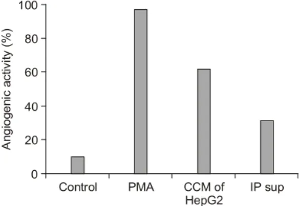
1) IGF-II의 혈관신생 촉진효과 검색 인간간암세포주인 HepG2 cell의 conditioned me- dia 내에 다량 존재하는 IGF-II의 혈관신생활성을 확인하기 위해 Immunoprecipitation에 의해 HepG2 conditioned media에 존재하는 IGF-II를 제거한 뒤, IGF-II가 제거된 conditioned media의 혈관신생활 성을 살펴보았다(Fig. 1). HepG2 conditioned media 의 혈관신생활성은 60% 정도 유도되는 것이 관찰 되었고, 정상적인 conditioned media에 비해 IGF-II 가 제거된 conditioned media는 절반 이상 현저히 줄어든 혈관신생활성을 나타내었다(Fig. 1). 따라 서 HepG2 conditioned media 내의 IGF-II가 간암진 행과정에 있어 강력한 혈관신생유도인자로 작용 함을 확인할 수 있었다.
2) IGF-II의 혈관신생활성에 미치는 heparin의 역
할조사여러 가지 다른 혈관신생인자의 활성 조절단백 질로 알려진 heparin의 역할을 알아보기 위해, CAM assay를 수행하였다. Fig. 2에서 보는 바와 같이 heparin은 IGF-II의 혈관신생활성에 큰 영향을 주 지 않았다. 따라서 IGF-II의 혈관신생활성은 hepa- rin과는 무관함을 알 수 있다.
3) IGF-II의 혈관신생활성에 미치는 EGF의 역할조사
IGFBP3의 활성을 저해하는 것으로 알려진 EGF
Fig. 1. Decreased angiogenic activity of HepG2 condi-
tioned media by immunoprecipitation. PMA was used as positive control (CCM: concentrated conditioned me- dia, Sup: supernatant).Angiogenic activity (%)
Control PMA CCM of
HepG2 IP sup 0
100
40 80
60
20
Fig. 3. Enhanced angiogenic activity of IGF-II by the
cotreatment with EGF. In case of cotreatment with IGF-II and EGF, considerable angiogenic activity was shown on the CAM, while IGF-II or EGF alone did not induce angiogenesis.Angiogenic activity (%)
Control PMA IGF-II EGF IGF-II+
EGF 0
100
40 80
60
20
Fig. 2. Effect of heparin on the IGF-II action. Heparin
had no effect on IGF-II action in angiogensis.Angiogenic activity (%)
Control PMA IGF-II Heparin IGF-II+
heparin 0
100
40 80
60
20
Fig. 4. Effects of IGF-II and EGF on the proliferation
of HepG2 cells. Following the dispension and stabiliz- ing incubation of 1×10⁵ cells per well, 1μCi methyl tritiated thymidine and each samples was treated simul- taneously. After 4 hours of incubation, incorporated thy- midines were measured by liquid scintillation counter.With IGF-II Without IGF-II
DNA Synthesis, cpm
12000
0.0 .5 1.0 5.0
Concentration of EGF (ng/ml) 0
14000
2000 10000 8000 6000 4000
의 역할을 알아보기 위하여 IGF-II와 EGF를 계배 융모막에 동시 처리하여 각각 처리한 군과의 차 이를 알아보았다. Fig. 3에서 보는 바와 같이, EGF 가 IGF-II의 혈관신생활성을 상승적으로 촉진함을 관찰할 수 있었고, 이것은 EGF 단독이 나타내는 혈관신생작용의 2.5배 가량이 되는 것을 관찰할 수 있었다. 따라서, EGF는 IGF-II의 활성을 저해 하는 IGFBP3의 발현을 억제하여 IGF-II가 일으키 는 혈관신생활성에 영향을 끼친다는 사실을 알 수 있다.
4) EGF가 촉진하는 IGF-II의 혈관신생작용 의 검증
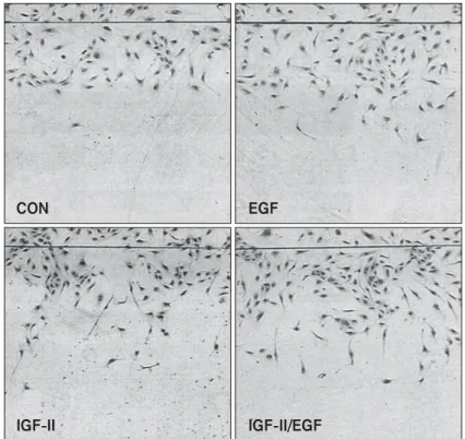
상승적으로 IGF-II의 혈관신생활성을 촉진하는 EGF의 역할에 초점을 맞추어, 혈관내피세포를 이 용한 angiogenesis assay를 수행하였다. 먼저 thy-
Fig. 5. Effect of IGF-II and/or EGF on migration of HUVECs. Each of EGF (5 ng/ml) and
IGF-II (100 ng/ml) stimulated the migration of HUVECs. When EGF and IGF-II were added in combination (IGF-II/EGF), the migration stimulatory effect was greater than the additive effects of each growth factor added alone. Phase-contrast photomicrographs show HUVECs that mi- grated from edge of the wound.CON EGF
IGF-II IGF-II/EGF
midine incorporation assay에 의해 혈관내피세포의 증식에 미치는 영향을 살펴본 결과, 5 ng/ml까지 EGF농도를 증가시켜 보았음에도 불구하고 IGF-II 의 혈관내피세포 증식효과에 별다른 영향을 주지 못함을 확인하였다(Fig. 4). 그러나, wounding migra- tion assay를 통한 혈관내피세포의 이동에는 IGF- II의 단독효과보다 EGF를 함께 처리했을 때 약 3 배 가량의 혈관내피세포가 이동이 촉진됨을 관찰 하였다(Fig. 5). 따라서 EGF가 일으키는 IGF-II에 대한 상승적인 혈관신생 촉진활성은 혈관내피세 포의 증식이 아니라, 이동을 촉진함으로서 일어난 다는 것을 알 수 있다. 뿐만 아니라, EGF가 IGF-II 의 조절인자인 IGFIIR과 IGFBP3의 발현에 미치는 영향을 Northern blot analysis로 살펴보았다(Fig.
6). 20 ng/ml의 EGF를 6시간 처리하였을 때 2.3배, 12시간 처리시 약 2배 가량의 IGFIIR 발현증가가 관찰되었다. 반면 IGFBP3의 발현에는 다소 저해 효과가 있음을 알 수 있었다. 따라서, EGF는 IGF- II의 활성을 저해하는 IGFBP3의 발현을 억제하고, IGFIIR의 발현을 촉진함으로서 IGF-II가 일으키는 혈관신생활성에 영향을 끼친다는 사실을 알 수 있다.
고 찰
국내 호발암인 간암은 대표적인 hypervascular
tumor이다.24,25) 암조직에서 혈관생성은 암의 증식 과 전이에 직접적으로 관련되어 있다고 알려져 있다.25) 그러나 간암에 대해서는 이러한 연구가 국외에서도 매우 미약한 상황이므로 혈관생성억 제에 의한 간암의 증식과 전이 억제방법의 개발 은 국내에서 앞으로 필히 수행되어야 할 과제이 다. 또한 만성피부염인 건선은 피부세포의 증식과 염증반응을 촉진시키는 질환이다.17,18) 이러한 건 선은 난치병으로서 그 발생기작에 관여하는 인자 를 연구하는 것은 질병의 발생과정을 이해하는 데 도움을 줄 것이다.
IGF-II는 간암으로의 이행과정과 건선의 발생과 정에서 그 발현이 급격히 증가되는 것으로 알려 져 있다.23,26) 이러한 IGF-II의 활성을 직접적으로 조절할 수 있는 인자로는 IGF binding proteins (IGFBPs)와 IGF receptors (IGFR) 등을 들 수 있다.
특히 IGFBP-3는 혈청 내에 존재하는 IGFBP 중 가 장 많은 양을 차지하고 있고, 강한 affinity로 IGF- II에 결합할 수 있으며, IGF-II의 활성을 저해하는 작용을 가지고 있다. 이러한 IGFBP-3에 의한 IGF- II 활성저해는 IGF receptor에 IGF-II가 결합하는 것을 IGFBP-3가 방해함으로서 일어난다.27,28) 그러 므로 이들 IGFBP-3와 IGFR의 발현은 IGF-II의 활 성과 직접적으로 연결되어 있으며 이들의 발현을 조절하는 인자 역시 IGF-II의 활성과 연관되어 있 다고 말할 수 있다. 따라서 IGFBP-3를 전사활성 을 억제하는 EGF29)는 IGF-II의 혈관신생활성과 직접적으로 연계되어 있을 가능성이 높다.
또한 VEGF, bFGF와 같이 강력한 혈관신생인자 로 알려진 단백질은 heparin결합단백질로 알려져 있으며, 특히 IGF-I의 경우는 heparin에 의해 조절 받는다는 사실이 알려져 있다.30,31) 즉, VEGF, bFGF의 혈관신생활성은 각각 단독으로 작용할 때보다 heparin과의 결합상태에서 더욱 증가한다 하겠다. 따라서 heparin은 이들 성장인자들과 결합 함으로써 성장인자들의 혈관신생활성을 직접적으 로 조절할 수 있는 보조인자로서의 역할을 수행 할 것으로 사료된다.
본 연구에서는 위의 사실을 바탕으로 하여 HepG2 인간 간암세포주의 conditioned media 내에 다량 존재하는 IGF-II의 혈관신생촉진활성을 확인 하기 위해 IGF-II antibody를 이용한 immunopre-
Fig. 6. Induction of IGFIIR expression by EGF. Con-
fluent HUVECs culture was maintained for 12 hr in M199 containing with 1% FBS and then treated with or without EGF (20 ng/ml) for indicated times. Nor- thern blot analysis was performed with 20μg of total RNA/lane using human IGFIIR and IGFBP3 cDNA probe. Hybridization with the human β-actin control probe is shown below.
EGF
IGFIIR Time (hr)
- + - + - + - +
6 6 12 12 24 24 48 48
fold 2.3 1.9 1.1 1.3
IGFBP3
fold 5 0.6 0.1 1.5
β-actin
cipitation 방법에 의해 HepG2 conditioned media 내 에 존재하는 IGF-II를 제거한 뒤, IGF-II가 제거된 conditioned media의 혈관신생활성이 기존의 con- ditioned media보다 혈관신생활성이 저해됨을 CAM assay를 통해 관찰하였다(Fig. 1). 또한 IGFBP-3의 전사활성을 저해하는 EGF가 IGF-II의 혈관신생활 성을 상승적으로 촉진함을CAM assay를 통해 조 사하였다(Fig. 2, 3). 이와 같이 EGF가 촉진하는 IGF-II의 혈관신생작용은 혈관내피세포의 이동을 촉진함으로써(Fig. 5) 일어나며, 이러한 현상은 IGFBP3와 IGFR의 발현변화에 기인한 것임(Fig.
6)을 알 수 있었다.
이와 같은 사실은 간암이행과정과 건선의 발생 과정에서 급격한 증가를 보이면서, 혈관신생활성 인자로서 작용하는 IGF-II의 새로운 조절 단백질 로서 EGF가 작용할 수 있음을 직접적으로 밝힌 사실이라 하겠다. 즉, 간암발생과정에서 급격히 발현이 증가되는 IGF-II는 간암의 형성과정중 간 암세포로부터 분비되어 간암의 성장을 유도하는 혈관신생활성을 가지고, EGF는 이러한 IGF-II의 혈관신생작용에 상승효과를 가지게 하는 단백질 로서 악성종양으로의 유도를 촉진하여 암전이를 일으키고, IL-6와 같은 염증반응에 관여하는 인자 들의 발현촉진을 통해 건선을 일으킴을 추측할 수 있다.
결 론
IGF-II가 암조직에서 발현되는 혈관신생 촉진인 자이며, 이 IGF-II의 혈관신생작용을 EGF 단백질 이 상승적으로 촉진함을 확인하였다. 또한 이러한 상승효과는 EGF가 혈관내피세포의 이동을 촉진 하는 데에서 비롯됨을 증명하였다. 뿐만 아니라, EGF의 혈관내피세포 이동촉진은 IGFBP3와 IGFR 의 발현변화에 기인한 것임을 알 수 있었다. 이상 의 결과에 의해 암화과정 및 건선발생과정 중에 서 IGF-II가 EGF에 의해 혈관신생이 촉진되는 방 향으로 조절되고 있음을 시사하고 있다. 향후 암 조직과 건선조직에서의 IGF-II 작용에 관한 연구 와 발현 기작에 관한 연구가 진행되면 악성암과 건선에서의 IGF-II의 역할을 이해할 수 있을 것 이다.
감사의 글
본 연구는 2001년도 부산대학교병원 의학연구 소 연구비의 지원으로 이루어진 것으로 이에 감 사드립니다.
참고 문헌
1) Shweiki D, Itin A, Soffer D, Keshet E. Vascular end- othelial growth factor induced by hypoxia may med- iate hypoxia-initiated angiogenesis. Nature 1992; 359:
843-845.
2) Plate KH, Breier G, Weich HA, Risau W. Vascular endothelial growth factor is a potential factor in hu- man gliomas in vivo. Nature 1992; 359: 845-848.
3) Nabel EG, Yang Z, Plautz, G, Forough R, Zhan X, Haudenschild DD, Maciag T, Nabel GJ. Recombinant fibroblast growth factor-1 promotes intimal hyper- plasia and angiogenesis in arteries in vivo. Nature 1993; 362: 844-846.
4) Yang EY, Moses HL. Transforming growth factor β 1-induced changes in cell migration, proliferation, and angiogenesis in the chicken chorioallantoic membrane.
J Cell Biol 1990; 111: 731-741.
5) Pepper MS, Montesano, R, Orci L, Vassalli JD.
Transforming growth factor beta-1 modulates basic fibroblast growth factor-induced proteolytic and an- giogenic properties of endothelial cells in vitro. J Cell
Biol 1990; 111: 743-754.
6) Park BC, Huh MH, Seo JH. Differential expression of transforming growth factor alpha and insulin-like growth factor II in chronic active hepatitis B, cirrhosis and hepatocellualr carcinoma. J Hepatol 1995; 22:
286-294.
7) Cohick WS, Clemmons DR. The insulin-like growth factors. Annu Rev Physiol 1993; 55: 131-153.
8) Moses AC, Nissley SP, Short PA, Rechler MM, White RM, Knight AB, Higa OZ. Elevated levels of insulin- like growth factor, multiplication stimulatory activity in fetal rat serum. Proc Natl Acad Sci USA 1980; 77:
3649-3653.
9) Hill DJ, Milner RDG. Somatomedins and fetal growth.
In the fetus and independent life, Ciba foundation
symposium 1981; 86: 124-151. London: Pitman.
10) Milner RDG, Hill DJ. Fetal growth: The role of insulin and related peptides. Clin Endocrinol 1984;
21: 415-433.
11) Underwood LE, D'Ercole AJ. Insulin and somato- medins/insulin-like growth factors in fetal and neo- natal development. In: ed, by Daughaday WH Clinics in Endocrinology and Metabolism vol. 13, Tissue Growth Factors. pp 68-89, East Essex, England:
Saunders WB Co, Ltd, 1984.
12) Froesch ER, Schmid C, Schwander J, Zapf J. Actions of insulin-like growth factors. A Rev Phsiol 1985; 47:
443-467.
13) Zapf J, Froesch ER. Insulin-like growth factors/ soma- tomedins: Struture, secretion, biological actions and physiological role. Homone Res 1986; 24: 121-130.
14) Baxter RC. The somatomedins: Insulin-like growth factors. Adv Clin Chem 1986; 25: 49-115.
15) Kim KW, Bae SK, Lee OK, Bae MH, Lee MJ, Park BC. Insulin-like growth factor II induced by hypoxia may contribute to angiogenesis of human hepato- cellular carcinoma. Cancer Res 1998; 58: 348-351.
16) Bae MH, Lee MJ, Bae SK, Lee OK, Lee YM, Park BC, Kim KW. Insulin-like growth factor II (IGF-II) secreted from HepG2 human hepatocellular carcinoma cells shows angiogenic activity. Cancer Lett 1998;
128: 41-46.
17) Christophers E, Parzefall R, Braun-Falco O. Initial events in psoriasis: quantitative assessment. Br J Der-
matol 1973; 89: 327-334.
18) Braverman IM, Keh-Yen A. Three-dimensional recon- struction of endothelial cell gaps in psoriatic vessels and their morphologic identity with gaps produced by the intradermal injection of histamine. J Invest Der-
matol 1986; 86: 577-581.
19) Xu S, Cwyfan-Hughes SC, van der Stappen JW, San- som J, Burton JL, Donnelly M, Holly JM. Altered insulin-like growth factor-II (IGF-II) level and IGF- binding protein-3 (IGFBP-3) protease activity in inter- stitial fluid taken from the skin lesion of psoriasis. J
Invest Dermatol 1996; 106:109-112.
20) Elder JT, Fisher GJ, Lindquist PB, Bennett GL, Pit- telkow MR, Coffey RJ Jr, Ellingsworth L, Derynck R, Voorhees JJ. Overexpression of transforming growth factor alpha in psoriatic epidermis. Science 1989; 243:
811-814.
21) Gillitzer R, Berger R, Mielke V, Muller C, Wolff K, Stingl G. Upper keratinocytes of psoriatic skin lesions
express high levels of NAP-1/IL-8 mRNA in situ. J
Invest Dermatol 1991; 97: 73-79.
22) Neely EK, Morhenn VB, Hintz RL, Wilson DM, Ro- senfeld RG. Insulin-like growth factors are mitogenic for human keratinocytes and a squamous cell carci- noma. J Invest Dermatol 1991; 96: 104-110.
23) Kwon YW, Jang ER, Lee YM, Kim YS, Kwon KS, Jang HS, Oh CK, Kim KW. Insulin-like growth factor II induces interleukin-6 expression via NFkappaB activation in psoriasis. Biochem Biophys Res Commun 2000; 278: 312-317.
24) Liotta LA, Steeg PA, Stetler-Stevenson WG. Cancer metastasis and angiogenesis: an imbalance of positive and negative regulation. Cell 1991; 64: 327-336.
25) Folkman J, Klagsbrun M. Angiogenic factors. Science 1987; 235: 442-447.
26) Yang DY, Rogler CE. Analysis of Insulin-like Growth Factor II (IGF-II) expression in neoplastic nodules and hepatocellular carcinomas of the woodchuck utilizing in situ hybridization and immunohistochemistry. Car-
cinogenesis 1991; 12: 1893-1901.
27) Mohan S, Bautista CM, Wergedat J, Baylink DJ. Iso- lation of an inhibitory Insulin-like Growth Factor (IGF) binding protein from bone cell-conditioned medium: a potentia; local regulator of IGF action.
Proc Natl Acad Sci U.S.A. 1989; 86: 8338-8342.
28) Rose M, Francis GL, Szabo L, Wallace JC, Ballard, FJ. Insulin-like Growth Factor (IGF)-binding proteins inhibit the biological activities of IGF-1 and IGF-2 but des-(1-3)-IGF-1. Biochem J 1989; 258: 267-272.
29) Hembree JR, Agarwal C, Eckert RL. Epidermal growth factor suppresses insulin-like growth factor binding protein 3 levels in human papillomavirus type 16- immortalized cervical epithelial cells and thereby potentiates the effects of insulin-like growth factor 1.
Cancer Res 1994; 54:3160-3166.
30) Folkman J, Shing Y. Control of angiogenesis by he- parin and other sulfated polysaccharides. Adv Exp
Med Biol 1992; 313: 355-364.
31) Clemmons DR, Underwood LE, Chatelain PG, Van Wyk JJ. Liberation of immunoreactive somatomedin-C from its binding proteins by proteolytic enzymes and heparin. J Clin Endocrinol Metab 1983; 56: 384-389.